. 2013 Nov 1;188(9):1126-36.
doi: 10.1164/rccm.201302-0403OC.

Bronchus-associated lymphoid tissue in pulmonary hypertension produces pathologic autoantibodies

Affiliations

Bronchus-associated lymphoid tissue in pulmonary hypertension produces pathologic autoantibodies

Kelley L Colvin et al. Am J Respir Crit Care Med. .

Abstract

Rationale: Autoimmunity has long been associated with pulmonary hypertension. Bronchus-associated lymphoid tissue plays important roles in antigen sampling and self-tolerance during infection and inflammation.

Objectives: We reasoned that activated bronchus-associated lymphoid tissue would be evident in rats with pulmonary hypertension, and that loss of self-tolerance would result in production of pathologic autoantibodies that drive vascular remodeling.

Methods: We used animal models, histology, and gene expression assays to evaluate the role of bronchus-associated lymphoid tissue in pulmonary hypertension.

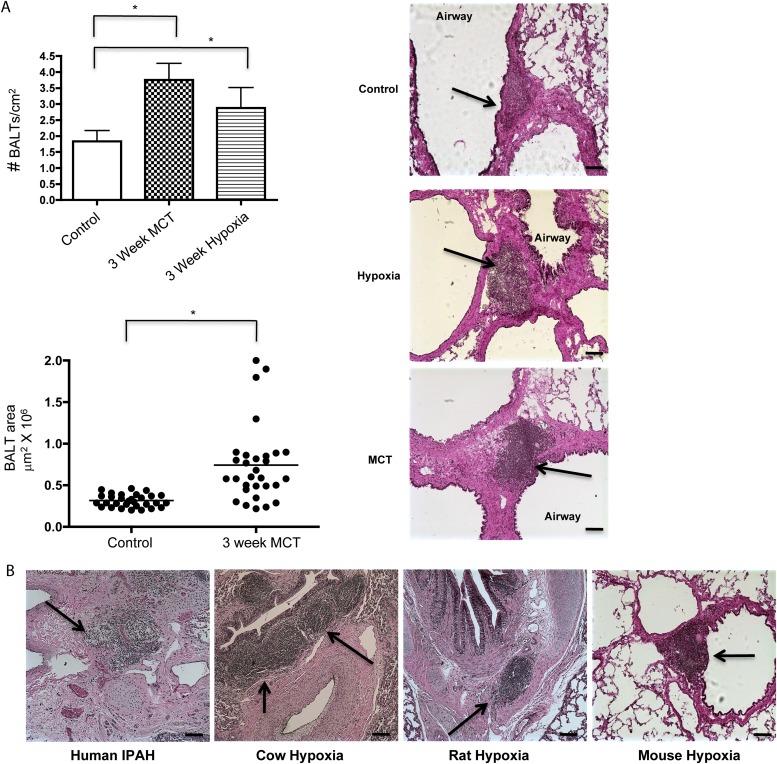
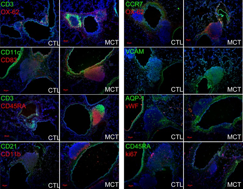
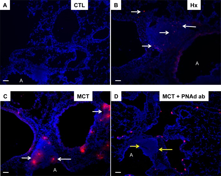
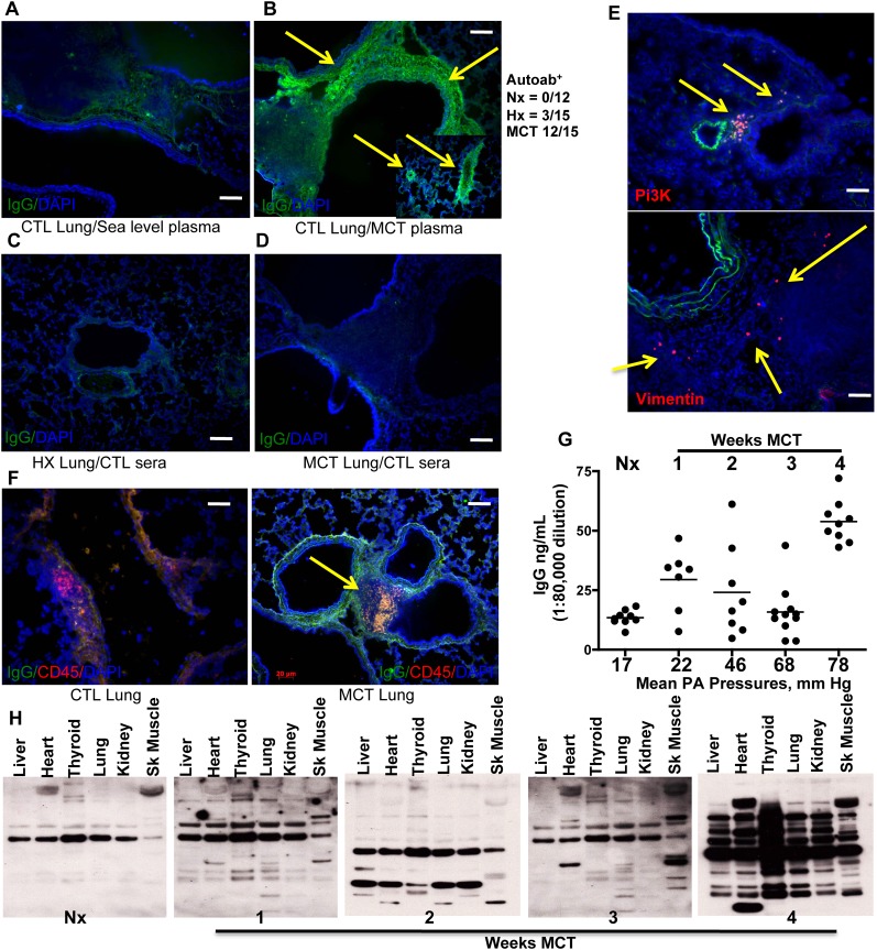
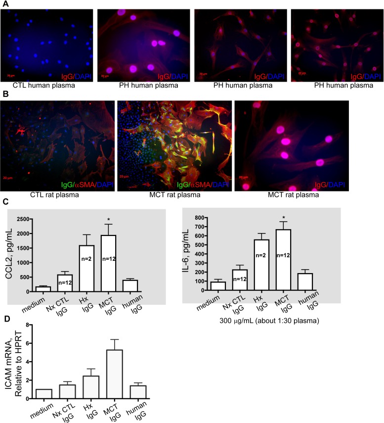
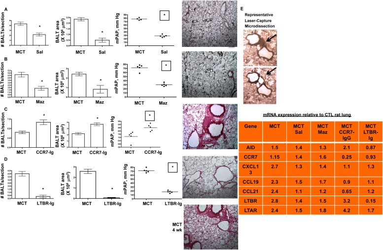
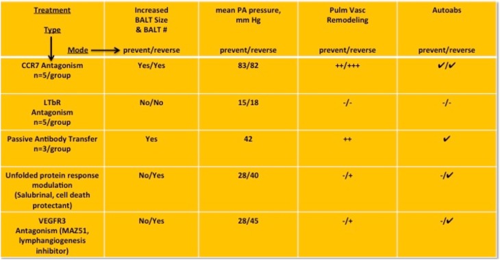
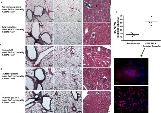
Measurements and main results: Bronchus-associated lymphoid tissue was more numerous, larger, and more active in pulmonary hypertension compared with control animals. We found dendritic cells in and around lymphoid tissue, which were composed of CD3(+) T cells over a core of CD45RA(+) B cells. Antirat IgG and plasma from rats with pulmonary hypertension decorated B cells in lymphoid tissue, resistance vessels, and adventitia of large vessels. Lymphoid tissue in diseased rats was vascularized by aquaporin-1(+) high endothelial venules and vascular cell adhesion molecule-positive vessels. Autoantibodies are produced in bronchus-associated lymphoid tissue and, when bound to pulmonary adventitial fibroblasts, change their phenotype to one that may promote inflammation. Passive transfer of autoantibodies into rats caused pulmonary vascular remodeling and pulmonary hypertension. Diminution of lymphoid tissue reversed pulmonary hypertension, whereas immunologic blockade of CCR7 worsened pulmonary hypertension and hastened its onset.

Conclusions: Bronchus-associated lymphoid tissue expands in pulmonary hypertension and is autoimmunologically active. Loss of self-tolerance contributes to pulmonary vascular remodeling and pulmonary hypertension. Lymphoid tissue-directed therapies may be beneficial in treating pulmonary hypertension.


Figures

<i>Figure 1.</i>
Figure 1.
Bronchus-associated lymphoid tissues (BALT) are more conspicuous in pulmonary hypertension (PH). BALTs are increased in number (A, top left, and hematoxylin and eosin sections, top right) in rats with PH compared with control animals. (B) BALTs are readily apparent in humans and in bovine (representative images from cows, rats, and mice with PH). Representative images and measurements in A taken from lung sections from n = 6 animals per group, with two to three sections per rat from three independent experiments. Bars = 20 μm. Representative image using human section (B, left) taken from analysis of n = 3 patient samples, three sections per sample. *P less than 0.05 compared with control, Student t test. IPAH = idiopathic pulmonary arterial hypertension; MCT = monocrotaline.
<i>Figure 2.</i>
Figure 2.
Bronchus-associated lymphoid tissues (BALTs) in rats with pulmonary hypertension (PH) are highly organized and more vascularized compared with control animals. In PH versus control animals, CD3+ T cells and CD45RA+ B cells adopt distinct segregational localizations in PH BALT, and are encircled by OX-62+ dendritic cells, and CD11b+, CD11c+, and CD83+ cells (left column of images and top right images). VCAM+ structures are more evident in PH versus control animals, whereas aquaporin (AQP)-1–positive structures seemed to be proportional to BALT size in both PH and control animals (right column of images, middle). CD45RA+ cells did not colocalize with ki-67 immunoreactivity in either control animals or PH, even when control BALT sizes were large (lowest control left image, right column). Note robust ki-67–positive cells (red, lowest right image) in adventitial of bronchovascular space. Representative images and measurements taken from lung sections from n = 6 animals per group, with two to three sections per rat from three independent experiments. All images, original magnification: ×100; bars = 20 μm where applicable. CTL = control; MCT = monocrotaline; VCAM = vascular cell adhesion molecule.
<i>Figure 3.</i>
Figure 3.
Pulmonary hypertension (PH) lung bronchus-associated lymphoid tissues (BALTs) are bound by T cells by peripheral node addressin (PNAd). BALTs in experimental PH bind dye-labeled immunomagnetic bead sorted spleen and thymic rat CD3+ T cells (B and C), but control lung sections (A) do not. The T-cell adhesion may be largely attributed to binding to PNAd in BALTs (white arrows) because preincubation of lung sections with PNAd antibody attenuated T-cell adhesion to the BALT but not the lung parenchyma (D, yellow arrows). Representative images from tissue-cell incubations taken from lung sections from n = 6 animals per group, with two to three sections per rat from three independent experiments. Bars = 20 μm. All binding experiments were performed in triplicate. A = airway; MCT = monocrotaline.
<i>Figure 4.</i>
Figure 4.
Rats with pulmonary hypertension (PH) circulate autoantibodies. (A) Control (CTL) lung sections incubated with plasma from normal (no PH) rats is not bound by sequentially applied Alexa 488–conjugated antirat IgG. In contrast, control lung sections incubated with plasma from rats with monocrotaline (MCT) induced PH can be bound by antirat IgG (B, green signal indicated by yellow arrows) indicating that the PH plasma contains IgG that bind self-antigens (autoantibodies). The inset in B shows resistance sized vessels (50–500 μm internal diameter) are also labeled with autoantibodies in PH plasma. (C and D) Lung sections from rats with hypoxic PH or MCT-PH incubated with control plasmas lack autoantibody binding. MCT-PH lung sections incubated with fluor-labeled phosphatidyl inositol 3 kinase (Pi3K) or vimentin (E, red signal, yellow arrows) indicate the presence of autoantibodies in situ. (F) Lung sections from MCT rats, but not control rats, demonstrate the presence of rat IgGs (green) localized to regions with CD45RA+ B cells (red). (G) Plasma IgG titers increase in MCT-PH rats compared with control rats, peaking at Week 4 and correlating with increasing mean pulmonary artery (PA) pressure. (H) Immunoblots using rat plasma as the primary antibody reveal autoimmune epitope spreading. Compared with the lack of autoantibodies in control rat plasma, MCT-PH plasma contains autoantibodies that increasingly recognize a wider array of tissue lysates, peaking at 4 weeks post-MCT. Representative images taken from lung sections from n = 6 animals per group, with two to three sections per rat from three independent experiments. Bars = 20 μm. ELISAs were performed in three separate experiments in triplicate on rat plasmas from n = 3 rats per group. Immunoblots shown are representative of tissue lysates run in three separate experiments. Blotting used pools (n = 3) of indicated rat plasmas, performed independently using three separate pools.
<i>Figure 5.</i>
Figure 5.
Human and monocrotaline (MCT) rat autoantibodies label pulmonary artery fibroblasts. (A) Compared with control (CTL) human plasma, plasma from patients with pulmonary hypertension (PH) (verified in clinical laboratory workup to have autoantibodies) label human lung fibroblasts in nuclear, punctate, and filamentous patterns. (B) Similarly, rat plasmas label rat lung fibroblasts in nuclear, punctate, and filamentous patterns. IgG from PH rats, but not human IgG or preimmune plasma IgG, stimulate PA fibs toward a phenotype that is proinflammatory (C, increased IL-6 and CCL2 secretion) and proadhesive (D, increased expression of intercellular adhesion molecule [ICAM]). Cell cultures and ELISAs were performed in three separate experiments in triplicate with rat plasmas from the indicated rats per group, compared with n = 3 control media experiments. Quantitative polymerase chain reaction was performed in triplicate using mRNA isolated from fibroblasts used in three independent experiments. All images, original magnification: ×100; bars = 20 μm where applicable. *P less than 0.05 compared with control, Student t test.
<i>Figure 6.</i>
Figure 6.
Strategies aimed at diminution of bronchus-associated lymphoid tissues (BALTs) biology prevent and reverse pulmonary hypertension (PH). (A) Preventing lung cell apoptosis (and presumably self-antigen burden) by salubrinal (Sal), an unfolded protein response modulator, prevents and reverses pulmonary vascular remodeling and PH in a manner associated with decreased BALT size and number. (B) Pharmacologic VEGFR3 antagonism prevents and reverses monocrotaline (MCT)-PH and is associated with decreased macrophage influx, decreased blood and lymphatic vascularization, and smaller BALT size and numbers. (C) CCR7 controls migration of lung CCR7+ dendritic cells (DC) and CCR7+ T cells into BALT; pharmacologic CCR7 antagonism worsens MCT-PH and is associated with larger BALT size and numbers. (D) Lymphotoxin β receptor (LTBR) control animals BALT formation and maintenance; pharmacologic LTBR antagonism prevents and reverses MCT-PH and is associated with decreased blood and lymphatic vascularization, and smaller BALT size and numbers. In AD, pressure measurement, BALT counts, and representative lung section images were taken from n = 6 animals per group, with two to three sections per rat from three independent experiments. All images, original magnification: ×100. *P less than 0.05 compared with control, Student t test. (E) Representative laser capture microdissection of BALT from rat lung section. Black arrows show the microdissected BALT region. The table lists the mRNAs isolated from the rat groups and their respective mean fold changes in expression relative to the control lung BALT. n = 2–3 BALT per section, 3–5 sections per rat, n = 6 animals per group. For each fold change equal to or greater than ± 1.2 fold, P less than 0.05 compared with control using Student t test. CTL = control; Maz = Maz51 VEGFR3 inhibitor.
<i>Figure 7.</i>
Figure 7.
Summary of in vivo data from experiments aimed at bronchus-associated lymphoid tissues (BALT) biology. LTBR = lymphotoxin β receptor; PA = pulmonary artery.
<i>Figure 8.</i>
Figure 8.
Pulmonary hypertension (PH) can be caused by autoantibody passive transfer from monocrotaline (MCT) rats into naive rats. (A) Naive rats injected with either preimmune sera, adjuvant (saline) alone, or adjuvant mixed with control human IgG (B) do not develop pulmonary vascular remodeling or PH. Lung (left and middle images) and heart (right images) histology is normal, as is pulmonary artery pressure (PAP) in these rats. (C) Lung remodeling and heart fibrosis with elevated PAP in rats injected with plasma (C) or purified IgG (D) from MCT rats. Note the more intense blue staining in heart sections from rats receiving autoantibody transfer (far right images, C and D). (E) High titers of IgG in rats 4 weeks after cell immunizations or passive antibody transfers have ceased. Note low titers in control rat groups. IgG from rats, 4 weeks after apoptotic lung cell immunizations or autoantibodies have ceased, label nuclei and perinuclear compartments of cultured pulmonary artery fibroblasts. Representative images and measurements taken from lung sections from n = 6 animals per group, with two to three sections per rat from three independent experiments. Cell cultures and ELISAs were performed in three separate experiments in triplicate with rat plasmas from the indicated rats per group, compared with n = 3 control media experiments. All images, original magnification: ×100; bars = 20 μm where applicable. BALT = bronchus-associated lymphoid tissues.

References

    1. Simonneau G, Robbins IM, Beghetti M, Channick RN, Delcroix M, Denton CP, Elliott CG, Gaine SP, Gladwin MT, Jing ZC, et al. Updated clinical classification of pulmonary hypertension. J Am Coll Cardiol. 2009;54:S43–S54. - PubMed
    1. Tuder RM, Abman SH, Braun T, Capron F, Stevens T, Thistlethwaite PA, Haworth SG. Development and pathology of pulmonary hypertension. J Am Coll Cardiol. 2009;54:S3–S9. - PubMed
    1. Hassoun PM, Mouthon L, Barberà JA, Eddahibi S, Flores SC, Grimminger F, Jones PL, Maitland ML, Michelakis ED, Morrell NW, et al. Inflammation, growth factors, and pulmonary vascular remodeling. J Am Coll Cardiol. 2009;54:S10–19. - PubMed
    1. Randall TD. Bronchus-associated lymphoid tissue (BALT) structure and function. Adv Immunol. 2010;107:187–241. - PMC - PubMed
    1. Foo SY, Phipps S. Regulation of inducible BALT formation and contribution to immunity and pathology. Mucosal Immunol. 2010;3:537–544. - PubMed

Publication types