. 2013 Oct 1;18(4):588-95.
doi: 10.1016/j.cmet.2013.09.009.

Rapid versus delayed stimulation of feeding by the endogenously released AgRP neuron mediators GABA, NPY, and AgRP

Affiliations

Rapid versus delayed stimulation of feeding by the endogenously released AgRP neuron mediators GABA, NPY, and AgRP

Michael J Krashes et al. Cell Metab. .

Abstract

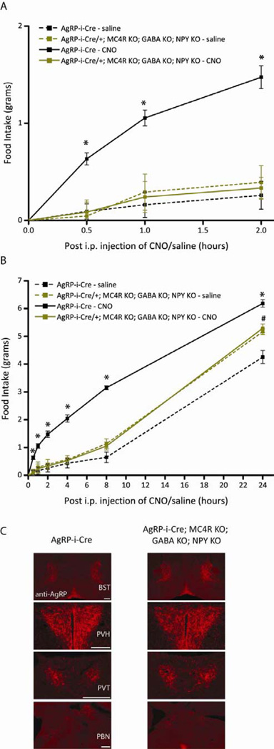
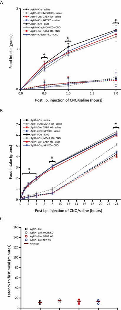
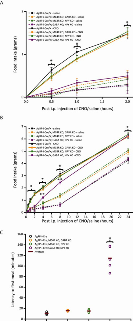
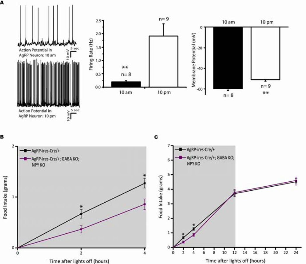
Agouti-related peptide (AgRP) neurons of the hypothalamus release a fast transmitter (GABA) in addition to neuropeptides (neuropeptide Y [NPY] and Agouti-related peptide [AgRP]). This raises questions as to their respective functions. The acute activation of AgRP neurons robustly promotes food intake, while central injections of AgRP, NPY, or GABA agonist results in the marked escalation of food consumption with temporal variance. Given the orexigenic capability of all three of these neuroactive substances in conjunction with their coexpression in AgRP neurons, we looked to unravel their relative temporal role in driving food intake. After the acute stimulation of AgRP neurons with DREADD technology, we found that either GABA or NPY is required for the rapid stimulation of feeding, and the neuropeptide AgRP, through action on MC4 receptors, is sufficient to induce feeding over a delayed yet prolonged period. These studies help to elucidate the neurochemical mechanisms of AgRP neurons in controlling temporally distinct phases of eating.


Figures

Figure 1
Figure 1
Acute pharmaco-genetic activation of AgRP neurons in mice without release of GABA, NPY and AgRP signaling via MC4Rs, collectively, lack DREADD-mediated increases in food intake (Pooled data across multiple trials). All mice in these studies were bilaterally injected with AAV8-DIO-hM3Dq-mCherry in the ARC. (A-B) Activation of AgRP neurons increases food intake in AgRP-ires-Cre mice (black line), but has no effect on food intake in AgRP-ires-Cre; Mc4r−/−; Vgatflox/flox; Npy−/− triple KO mice (light green line). CNO (solid line; 0.3 mg/kg of body weight, i.p.) or saline (dotted line) was injected 3 hr after the start of the “lights on” cycle and food intake was assessed (A) 1-, 2- , (B) 4-, 8- and 24-hr post-injection (PI) over three trials of each treatment. Data shown is from male mice (Error bars indicate mean +/− SEM, n=8 AgRP-ires-Cre mice; n=5 AgRP-ires-Cre; Mc4r−/−; Vgatflox/flox; Npy−/− mice; *P<0.05 AgRP-ires-Cre CNO group versus all other groups; #P<0.05 AgRP-ires-Cre; Mc4r−/−; Vgatflox/flox; Npy−/− groups versus AgRP-ires-Cre saline group). (C) Immunohistochemical analysis of AgRP projections in AgRP-ires-Cre mice (left) and AgRP-ires-Cre; Mc4r−/−; Vgatflox/flox; Npy−/− triple KO mice (right) to (Top-Bottom) the bed nucleus of the stria terminalis (BST), paraventricular hypothalamus (PVH), paraventricular thalamus (PVT) and lateral parabrachial nucleus (PBNl) reveals no gross differences in morphology and/or density (200 μm). See also Figure S1,S4 and Table S1.
Figure 2
Figure 2
Acute pharmaco-genetic activation of AgRP neurons in mice without release of GABA, NPY or AgRP signaling via MC4Rs, individually, display intact DREADD-mediated increases in food intake (Pooled data across multiple trials). All mice in these studies were bilaterally injected with AAV8-DIO-hM3Dq-mCherry in the ARC. (A-C) Activation of AgRP neurons increases comparable levels of food intake and latency to first meal in AgRP-ires-Cre mice (black line), AgRP-ires-Cre; Mc4r−/− KO mice (grey line), AgRP-ires-Cre; Vgatflox/flox KO mice (red line) and AgRP-ires-Cre; Npy−/− KO mice (blue line). CNO (solid line; 0.3 mg/kg of body weight, i.p.) or saline (dotted line) was injected 3 hr after the start of the “lights on” cycle and food intake was assessed (A) 1-, 2-, (B) 4-, 8- and 24- hr post-injection (PI) over three trials of each treatment. Data shown is from male mice (Error bars indicate mean +/− SEM, n=8 AgRP-ires-Cre mice; n=5 AgRP-ires-Cre; Mc4r−/− mice; n=7 AgRP-ires-Cre; Vgatflox/flox mice; n=8 AgRP-ires-Cre; Npy−/− mice; *P<0.05 CNO groups versus all saline groups; #P<0.05 AgRP-ires-Cre; Mc4r−/− saline group versus all other saline groups). (C) Latency to first meal following acute pharmaco-genetic activation of AgRP neurons. Each circle represents the average of two trials for each mouse; horizontal bar represents average of all mice. Data shown is from male mice (mean ± SEM, n=7 AgRP-ires-Cre mice; n=5 AgRP-ires-Cre; Mc4r−/− mice; n=5 AgRP-ires-Cre; Vgatflox/flox mice; n=5 AgRP-ires-Cre; Npy−/− KO mice). See also Figure S2 and Table S1.
Figure 3
Figure 3
Acute pharmaco-genetic activation of AgRP neurons in mice without release of GABA and NPY, collectively, display delayed DREADD-mediated increases in food intake (Pooled data across multiple trials). All mice in these studies were bilaterally injected with AAV8-DIO-hM3Dq-mCherry in the ARC. (A-C) Activation of AgRP neurons increases comparable levels of food intake (A,B) and latency to first meal (C) in AgRP-ires-Cre mice (black line), AgRP-ires-Cre; Mc4r−/−; Vgatflox/flox double KO mice (orange line) and AgRP-ires-Cre; Mc4r−/−; Npy−/− double KO mice (green line). In contrast, activation of AgRP neurons in AgRP-ires-Cre; Vgatflox/flox; Npy−/− double KO mice (purple line) resulted in highly attenuated short-term feeding (A), increased latency to first meal (C) and late-onset hyperphagia (B). CNO (solid line; 0.3 mg/kg of body weight, i.p.) or saline (dotted line) was injected 3 hr after the start of the “lights on” cycle and food intake was assessed (A) 1-, 2-, (B) 4-, 8- and 24-hr post-injection (PI) over three trials of each treatment. Data shown is from male mice (Error bars indicate mean +/− SEM, n=8 AgRP-ires-Cre mice; n=5 AgRP-ires-Cre; Mc4r−/−; Vgatflox/flox mice; n=5 AgRP-ires-Cre; Mc4r−/−; Npy−/− mice; n=8 AgRP-ires-Cre; Vgatflox/flox; Npy−/− mice; *P<0.05 AgRP-ires-Cre, AgRP-ires-Cre; Mc4r−/−; Vgatflox/flox and AgRP-ires-Cre; Mc4r−/−; Npy−/− CNO groups versus AgRP-ires-Cre; Vgatflox/flox; Npy−/− CNO and all saline groups; #P<0.05 AgRP-ires-Cre; Mc4r−/−; Vgatflox/flox and AgRP-ires-Cre; Mc4r−/−; Npy−/− saline groups versus all other saline groups; &P<0.05 AgRP-ires-Cre; Vgatflox/flox; Npy−/− CNO group versus all saline groups). (C) Latency to first meal following acute pharmaco-genetic activation of AgRP neurons. Each circle represents the average of two trials for each mouse; horizontal bar represents average of all mice. Data shown is from male mice (mean ± SEM, n=7 AgRP-ires-Cre mice; n=5 AgRP-ires-Cre; Mc4r−/−; Vgatflox/flox mice; n=5 AgRP-ires-Cre; Mc4r−/−; Npy−/− mice; n=5 AgRP-ires-Cre; Vgatflox/flox; Npy−/− mice; *P<0.05 AgRP-ires-Cre; Vgatflox/flox; Npy−/− group versus all other groups). See also Figure S3 and Table S1.
Figure 4
Figure 4
Mice with compromised release of both GABA and NPY from AgRP neurons display a delayed physiological increase in dark cycle food intake. (A) Light cycle (10 am; n=8) versus dark cycle (10 pm; n=9) electrophysiological properties of AgRP neurons (Error bars indicate mean + SEM; *P<0.05). Top, representative trace of AgRP neuron in the light cycle. Bottom, representative trace of AgRP neuron in the dark cycle. Right, quantitative analyses of firing rate and membrane potential of AgRP neurons in the light cycle (black bar) versus dark cycle (white bar). (B-C) Dark cycle food intake measurements assessed at (B) 2-, 4-, (C) 12-and 24-hr after lights are shut off. Grey background indicates dark cycle period. Data shown is from male mice (Error bars indicate mean +/− SEM, n=9 AgRP-ires-Cre mice; n=9 AgRP-ires-Cre; Vgatflox/flox; Npy−/− mice; *P<0.05).

References

    1. Alexander GM, Rogan SC, Abbas AI, Armbruster BN, Pei Y, Allen JA, Nonneman RJ, Hartmann J, Moy SS, Nicolelis MA, et al. Remote control of neuronal activity in transgenic mice expressing evolved G protein-coupled receptors. Neuron. 2009;63:27–39. - PMC - PubMed
    1. Aponte Y, Atasoy D, Sternson SM. AGRP neurons are sufficient to orchestrate feeding behavior rapidly and without training. Nature neuroscience. 2011;14:351–355. - PMC - PubMed
    1. Atasoy D, Betley JN, Su HH, Sternson SM. Deconstruction of a neural circuit for hunger. Nature. 2012;488:172–177. - PMC - PubMed
    1. Balthasar N, Dalgaard LT, Lee CE, Yu J, Funahashi H, Williams T, Ferreira M, Tang V, McGovern RA, Kenny CD, et al. Divergence of melanocortin pathways in the control of food intake and energy expenditure. Cell. 2005;123:493–505. - PubMed
    1. Bouret SG, Bates SH, Chen S, Myers MG, Jr, Simerly RB. Distinct roles for specific leptin receptor signals in the development of hypothalamic feeding circuits. J Neurosci. 2012;32:1244–1252. - PMC - PubMed

Publication types

MeSH terms

LinkOut - more resources