Review
. 2013 Oct;38(10):417-55.
doi: 10.1016/j.cpcardiol.2013.08.001.

Genotype- and phenotype-guided management of congenital long QT syndrome

Review

Genotype- and phenotype-guided management of congenital long QT syndrome

John R Giudicessi et al. Curr Probl Cardiol. 2013 Oct.

Abstract

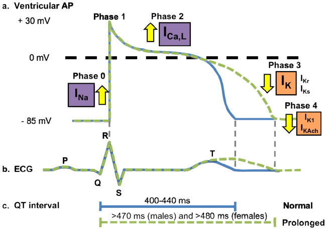
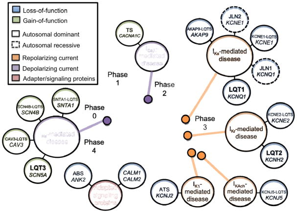
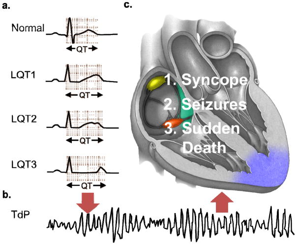
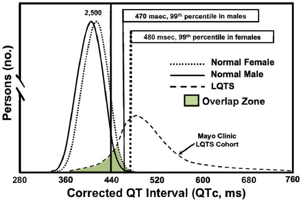
Congenital long QT syndrome (LQTS) is a genetically heterogeneous group of heritable disorders of myocardial repolarization linked by the shared clinical phenotype of QT prolongation on electrocardiogram and an increased risk of potentially life-threatening cardiac arrhythmias. At the molecular level, mutations in 15 distinct LQTS-susceptibility genes that encode ion channel pore-forming α-subunits and accessory β-subunits central to the electromechanical function of the heart have been implicated in its pathogenesis. Over the past 2 decades, our evolving understanding of the electrophysiological mechanisms by which specific genetic substrates perturb the cardiac action potential has translated into vastly improved approaches to the diagnosis, risk stratification, and treatment of patients with LQTS. In this review, we describe how our understanding of the molecular underpinnings of LQTS has yielded numerous clinically meaningful genotype-phenotype correlations and how these insights have translated into genotype- and phenotype-guided approaches to the clinical management of LQTS.


Figures

Figure 1
Figure 1. Electrophysiological basis of long QT syndrome
a. Tracings of the normal cardiac ventricular action potential (blue) observed in health and prolonged cardiac ventricular action potential (green) observed in long-QT syndrome. b. Schematic representation of a normal ECG (blue) and QT interval prolongation (green). c. Schematic depiction of normal (blue) and prolonged (green) QT intervals. Abbreviations: LQTS, long QT syndrome; ICa,L, L-type calcium current; IK1, inwardly rectifying current; IKr, rapid component of the delayed rectifier potassium current; IKs, slow-component of the delayed rectifier potassium current; INa, cardiac sodium current.
Figure 2
Figure 2. Current-centric classification of long QT syndrome (LQTS)-susceptibility genes
Perturbation of ventricular cardiac action potential depolarization (purple), repolarization (orange), or adapter/signaling proteins that influence depolarization/repolarization (maroon) by mutations in long QT syndrome-susceptibility genes are grouped according to the specific current perturbed by the underlying genetic defect. Blue circles represent loss-of-function mutations to the specified current, whereas green circles represent a gain-of-function. Solid lines indicate those disorders that are autosomal dominant, whereas dashed lines indicate those disorders that are autosomal recessive. Abbreviations: ABS, ankyrin-B syndrome; ATS, Andersen-Tawil syndrome; LQT, long-QT syndrome; ICa,L, L-type calcium current; IK1, inwardly rectifying current; IKr, rapid component of the delayed rectifier potassium current; IKs, slow-component of the delayed rectifier potassium current; INa, cardiac sodium current; and TS, Timothy syndrome. Adapted from Giudicessi, J.R., and Ackerman, M.J. Determinants of incomplete penetrance and variable expressivity in heritable cardiac arrhythmia syndromes. Translational Research 161(1), 1–14 (2013) with permission from Elsevier.
Figure 3
Figure 3. Electrocardiographic and clinical hallmarks of long QT syndrome
a. Schematic ECGs displaying the broad-based T wave pattern associated with long-QT syndrome type 1 (LQT1), notched T wave pattern associated with long-QT syndrome type 2 (LQT2), and long isoelectric segment with normal symmetrical T wave pattern associated with long-QT syndrome type 3 (LQT3) and QT prolongation observed in LQT1–LQT3 compared to normal. b. Triggers such as strenuous exercise (LQT1), sudden noises (LQT2), or sleep/rest (LQT3) can cause the stable, but prolonged myocardial repolarization to degenerate into torsades de pointes (TdP), the characteristic form of polymorphic ventricular fibrillation observed in long QT syndrome, depicted in this schematic ECG. c. The clinical manifestations of TdP in long QT syndrome are dependent on whether (syncope/seizures) or not (sudden death) order is restored to cardiac rhythm, either spontaneously or by a defibrillator.
Figure 4
Figure 4. QTc distribution in health and disease
The distribution of QTc values in health was derived from nearly 80,000 healthy adult males and females. [81] The distribution of QTc values in LQTS were derived from all patients with genetically proven LQTS evaluated in the Mayo Clinic’s Long QT Syndrome Clinic. Permission obtained from the American Heart Association © Taggart, N.W. et al. Diagnostic miscues in congenital long-QT syndrome. Circulation 115(20), 2613–2620 (2007).
Figure 5
Figure 5. Evidence-based algorithm designed to aid in the interpretation of a long QT syndrome (LQTS) genetic test result
Algorithm for interpreting a “positive” LQTS genetic test. Radical mutations that significantly alter/truncate Kv7.1 or Kv11.1 such as insertions/deletions, alteration of intronic/exonic splice site boundaries, and nonsense mutations, are probably LQTS-associated. Those raremissense mutations that localize to Kv7.1 (TM/pore, SA, or C terminal domains), Kv11.1 (C terminal when ≥ 3 tools are in agreement, TM/Pore, PAS/PAC, or cNBD), or Nav1.5 (TM/pore/linker or C terminus) are probably or possibly pathogenic. Variants outside these topological structure-function domains are truly ambiguous variants or variants of uncertain significance (VUS) without the aid of additional evidence (e.g. co-segregation with disease, LQTS-like electrophysiological phenotype etc.). Abbreviations: cNBD, cyclic nucleotide binding domain; EPV, estimated predictive value; IDL, interdomain linker; PAC, per-arnt-sim C-terminal associated; PAS, per-arnt-sim; SA, subunit assembly; TM, transmembrane. Adapted from Giudicessi, J.R. and Ackerman, M.J. Genetic testing in heritable cardiac arrhythmia syndromes; differentiating pathogenic mutations from background genetic noise. Current Opinion in Cardiology 28(1), 63–71 (2013) with permission from Wolters Kluwer Health.
Figure 6
Figure 6. Genotype- and phenotype-guided risk classification of long QT syndrome patients
Risk groups have been defined based on the previously published probability of suffering a first or recurrent cardiac event (syncope, seizure, sudden cardiac arrest, or sudden cardiac death) before 40 years of age without appropriate therapeutic interventions. A probability of suffering a first cardiac event > 80% defines the extremely high-risk group, > 50% the high-risk group, between 30% and 49% as the intermediate-risk group, and below 30% as the lowest-risk group. Genotype-guided recommendations are indicated by purple text, phenotype-guided recommendations are indicated by orange text, and a combination of genotype- and phenotype-guided recommendations is indicated by black text within the figure.
Figure 7
Figure 7. Left cardiac sympathetic denervation
a. Anatomical drawing depicting the extrapleural exposure of the left cardiac sympathetic chain during video assisted thoracic surgery left cardiac sympathetic denervation (VATS-LCSD). The stellate ganglion is located under the superior edge of the incision. The dashed line indicates the resection of the lower half of the left stellate ganglion occurring just above the major lower branches. b. Videoscopic stillframe from a VATS-LCSD depicting the left cardiac sympathetic chain before dissection of the pleura. c. Videoscopic stillframe from a VATS-LCSD depicting the left cardiac sympathetic chain after dissection of the pleura. Permission obtained from Elsevier © Collura et al. Left cardiac sympathetic denervation for the treatment of long QT syndrome and catecholaminergic polymorphic ventricular tachycardia using video-assisted thoracic surgery. Heart Rhythm 6(6), 752–759 (2009).
Figure 8
Figure 8. An integrated approach to the diagnosis, risk stratification, and genotype- and phenotype-guided management of patients with long QT syndrome
Blue boxes denote the recommended steps in assessing the index of clinical suspicion for long QT syndrome based on personal and family history, thorough cardiac evaluation including a 12-lead electrocardiogram, and if appropriate clinical long QT syndrome genetic testing. For those individuals where a bona fide long QT syndrome-causative mutation is identified, purple boxes indicate the recommended genotype-guided management of specific long QT syndrome genetic substrates based in part on established genotype-phenotype correlations and our current understanding of the pathogenesis/electrophysiological mechanisms of these disorders. Lastly, orange boxes indicate the recommended phenotype-guided management of individuals where a specific long QT syndrome genotype remains elusive or cannot be established as well as genotype-positive individuals already receiving gene-specific treatment. Importantly, it should be noted that for genotype-positive individuals that both genotype- and phenotype-guided management strategies are utilizedconcurrently. Abbreviations: ACA, aborted cardiac arrest; AR LQT1, autosomal recessive long-QT syndrome type 1; ICD, implantable cardioverter-defibrillator; JLNS, Jervell and Lange-Nielsen syndrome; LCSD, left cardiac sympathetic denervation; LQTS, long QT syndrome; QTc, heart-rate corrected QT interval; SCD, sudden cardiac death; TS, Timothy syndrome; and VUS, variant of unknown significance.

References

    1. Moss AJ. Long QT Syndrome. JAMA. 2003;289:2041–4. - PubMed
    1. Schwartz PJ, Crotti L, Insolia R. Long-QT syndrome: from genetics to management. Circ Arrhythm Electrophysiol. 2012;5:868–77. - PMC - PubMed
    1. Romano C, Gemme G, Pongiglione R. Rare Cardiac Arrythmias of the Pediatric Age. Ii. Syncopal Attacks Due to Paroxysmal Ventricular Fibrillation. (Presentation of 1st Case in Italian Pediatric Literature) Clin Pediatr (Bologna) 1963;45:656–83. - PubMed
    1. Ward OC. A New Familial Cardiac Syndrome in Children. J Ir Med Assoc. 1964;54:103–6. - PubMed
    1. Schwartz PJ, Stramba-Badiale M, Crotti L, Pedrazzini M, Besana A, Bosi G, et al. Prevalence of the congenital long-QT syndrome. Circulation. 2009;120:1761–7. - PMC - PubMed