. 2013 Oct 1;105(7):1562-8.
doi: 10.1016/j.bpj.2013.08.037.

The role of vimentin intermediate filaments in cortical and cytoplasmic mechanics

Affiliations

The role of vimentin intermediate filaments in cortical and cytoplasmic mechanics

Ming Guo et al. Biophys J. .

Abstract

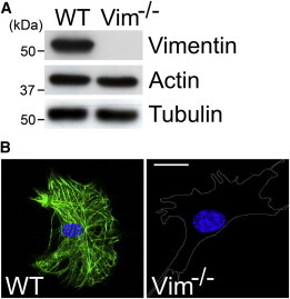
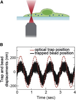
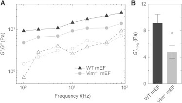
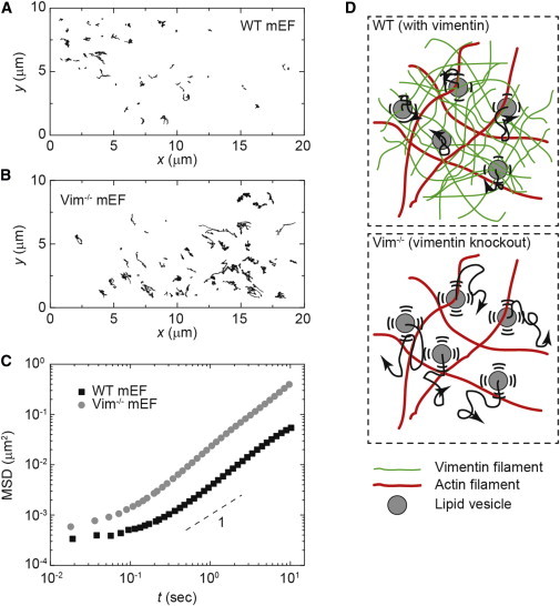
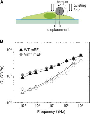
The mechanical properties of a cell determine many aspects of its behavior, and these mechanics are largely determined by the cytoskeleton. Although the contribution of actin filaments and microtubules to the mechanics of cells has been investigated in great detail, relatively little is known about the contribution of the third major cytoskeletal component, intermediate filaments (IFs). To determine the role of vimentin IF (VIF) in modulating intracellular and cortical mechanics, we carried out studies using mouse embryonic fibroblasts (mEFs) derived from wild-type or vimentin(-/-) mice. The VIFs contribute little to cortical stiffness but are critical for regulating intracellular mechanics. Active microrheology measurements using optical tweezers in living cells reveal that the presence of VIFs doubles the value of the cytoplasmic shear modulus to ∼10 Pa. The higher levels of cytoplasmic stiffness appear to stabilize organelles in the cell, as measured by tracking endogenous vesicle movement. These studies show that VIFs both increase the mechanical integrity of cells and localize intracellular components.


Figures

Figure 1
Figure 1
Analysis of control (WT) and VIM−/− mEF cells. (A) Immunoblot analyses of cell lysates from WT and VIM−/− mEFs using antibodies to vimentin, actin, and tubulin. Representative blots from three experiments are shown. (B) Immunofluorescence using antibodies against vimentin in control (WT, left) and VIM−/− (right) mEFs. The cell boundary in VIM−/− mEFs is represented by the yellow line. Representative images from three experiments are shown. Scale: 10 μm. To see this figure in color, go online.
Figure 2
Figure 2
Optical-tweezers measurement of intracellular mechanics. (A) Schematic of the optical tweezer experiment. PEG-coated inert particles (500 nm) are endocytosed into mEF cells and are then trapped and manipulated by a spatially sinusoidal oscillating optical trap, which generates a force F at frequency ω. The frequency-dependent complex spring constant is calculated by measuring the resultant displacement x of the bead in the trap oscillation, as F/x. (B) Typical displacements of the trapped bead and the optical trap oscillating at 1 Hz. To see this figure in color, go online.
Figure 3
Figure 3
Active microrheology with optical tweezers controlling 500 nm endocytosed beads in the cytoplasm of mEFs. (A) Frequency-dependent cytoplasmic elastic moduli G′ (solid symbols) and loss moduli G″ (open symbols) of the WT and Vim−/− mEFs. The cytoplasm of the WT mEFs (triangles) is stiffer than that of the Vim−/− mEFs (circles). (B) Cytoplasmic elastic moduli in the WT and Vim−/− mEFs at 1 Hz. Error bars: SEM (p < 0.05).
Figure 4
Figure 4
Intracellular movement of endogenous vesicles inside WT and Vim−/− mEFs. (A and B) Ten-second trajectories of endogenous vesicles in the cytoplasm of (A) WT mEFs and (B) Vim−/− mEFs. (C) Calculation of the MSD of vesicles shows that vesicles move faster in the Vim−/− mEFs than in the WT mEFs. (D) Illustration of random vesicle movement in networks with and without vimentin. In the WT cells, the vimentin network constrains the diffusive-like movement of organelles; in the VIM−/− cells, organelles move more freely. To see this figure in color, go online.
Figure 5
Figure 5
Cell cortical material properties of WT and VIM−/− mEFs measured with OMTC. (A) Schematic of the OMTC measurement. A magnetic field introduces a torque that causes the 4.5-μm ferromagnetic bead to rotate and to deform the cell cortex to which it is bound. (B) Elastic (G′, solid symbol), and loss (G″, open symbol) moduli for WT (black triangles) and Vim−/− (gray circles) mEFs cultured overnight on collagen-I-coated rigid plastic dishes, as measured by OMTC. Approximately 100 single cells are measured for each cell type. To see this figure in color, go online.

Comment in

References

    1. Alberts B., Johnson A., Lewis J., Raff M., Roberts K., Walter P. Garland Science; New York: 2007. Molecular Biology of the Cell.
    1. Carlier M.F. Role of nucleotide hydrolysis in the dynamics of actin filaments and microtubules. Int. Rev. Cytol. 1989;115:139–170. - PubMed
    1. Herrmann H., Bär H., Aebi U. Intermediate filaments: from cell architecture to nanomechanics. Nat. Rev. Mol. Cell Biol. 2007;8:562–573. - PubMed
    1. Janmey P.A., Euteneuer U., Schliwa M. Viscoelastic properties of vimentin compared with other filamentous biopolymer networks. J. Cell Biol. 1991;113:155–160. - PMC - PubMed
    1. Sivaramakrishnan S., DeGiulio J.V., Ridge K.M. Micromechanical properties of keratin intermediate filament networks. Proc. Natl. Acad. Sci. USA. 2008;105:889–894. - PMC - PubMed

Publication types