. 2013 Oct 10;155(2):285-95.
doi: 10.1016/j.cell.2013.08.057. Epub 2013 Oct 3.

The epithelial cell-derived atopic dermatitis cytokine TSLP activates neurons to induce itch

Affiliations

The epithelial cell-derived atopic dermatitis cytokine TSLP activates neurons to induce itch

Sarah R Wilson et al. Cell. .

Abstract

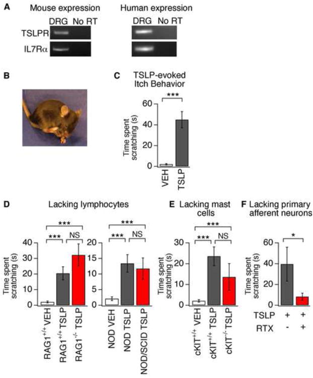
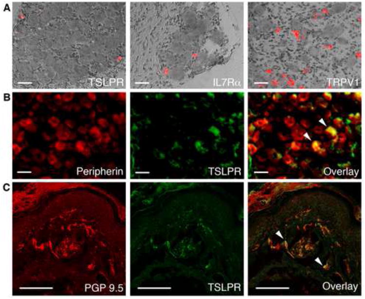
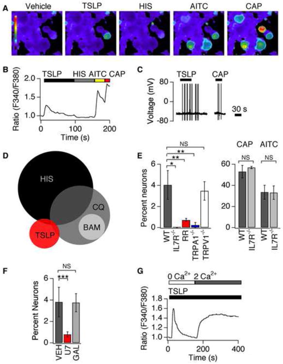
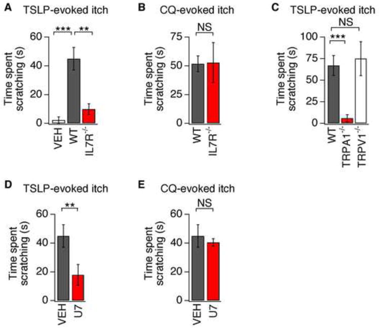
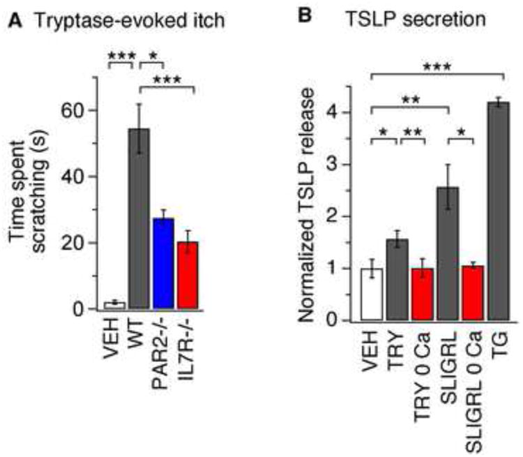
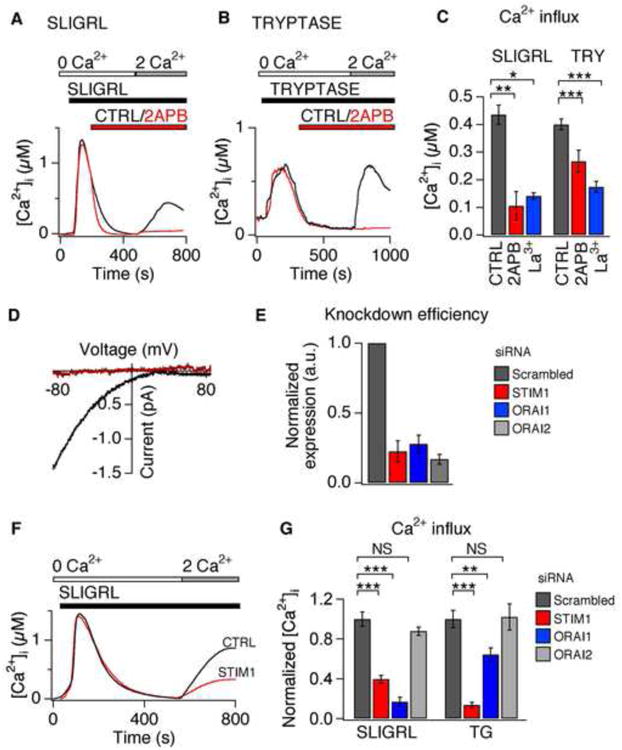
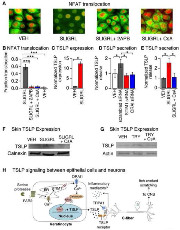
Atopic dermatitis (AD) is a chronic itch and inflammatory disorder of the skin that affects one in ten people. Patients suffering from severe AD eventually progress to develop asthma and allergic rhinitis, in a process known as the "atopic march." Signaling between epithelial cells and innate immune cells via the cytokine thymic stromal lymphopoietin (TSLP) is thought to drive AD and the atopic march. Here, we report that epithelial cells directly communicate to cutaneous sensory neurons via TSLP to promote itch. We identify the ORAI1/NFAT calcium signaling pathway as an essential regulator of TSLP release from keratinocytes, the primary epithelial cells of the skin. TSLP then acts directly on a subset of TRPA1-positive sensory neurons to trigger robust itch behaviors. Our results support a model whereby calcium-dependent TSLP release by keratinocytes activates both primary afferent neurons and immune cells to promote inflammatory responses in the skin and airways.


Figures

Figure 1
Figure 1. TSLP triggers robust itch behaviors in mice by activating sensory neurons
(A) PCR analysis of TSLPR and IL7Rα in mouse (left) and human (right) dorsal root ganglia (DRG). No product was amplified from the “no RT” control. (B) Image of itch-evoked scratching following intradermal injection of TSLP (2.5 μg/20 μl) into the cheek. (C) Quantification of scratching following TSLP injection in the cheek. TSLP (black) induced robust scratching compared to vehicle (white). n≥18 per group. (D) Itch behavior in RAG+/+, RAG-/-, NOD, and NOD/SCID mice following vehicle (PBS) or TSLP cheek injection. n≥8 per group. (E) Itch behavior in cKIT+/+ and cKIT-/- mice following vehicle (PBS) or TSLP injection. n≥8 per group. (F) TSLP-evoked scratching following neuronal ablation by RTX (red) versus control (black). n≥6 per group. *P<0.05; **P<0.01; ***P<0.001. Error bars represent s.e.m.
Figure 2
Figure 2. TSLP receptor components are expressed in sensory neurons
(A) DIC overlay images of in situ hybridization with cDNA probes detecting TSLPR, IL7Rα and TRPV1 in mouse DRG. Scale bar = 400μm. (B) Immunostaining of DRG sections with antibodies against peripherin and TSLPR in DRG sections. White arrows (right) mark peripherin- and TSLPR-positive neurons. Scale bar = 400μm. n≥4 mice/condition. (C) Immunostaining of PGP 9.5 and TSLPR in glabrous hind paw skin. The white arrows (right) mark PGP 9.5- and TSLPR-positive neurons. Scale bar = 200μm. n≥3 mice per condition.
Figure 3
Figure 3. TSLP directly activates a subset of sensory neurons
(A) Representative images of Fura-2 loaded DRG neurons treated with vehicle, TSLP (2 ng/mL), histamine (HIS, 1mM), AITC (200μM) and capsaicin (CAP, 1μM). (B) Representative trace shows a neuron that responds to TSLP, AITC and CAP, but not HIS. (C) Current-clamp recording showing TSLP- and CAP-evoked action potential firing in a DRG neuron. n≥60 cells. (D) A small percentage of the TSLP-sensitive population overlaps with the population of histamine- (HIS, 6%) or chloroquine-sensitive neurons (CQ, 6%), but not the BAM8-22 population (BAM, 0%). (E) Left: Prevalence of TSLP sensitivity in wild-type neurons (black), IL7Rα-deficient (grey) neurons, neurons treated with 20μM ruthenium red (RR; red), TRPA1-deficient neurons (blue) and TRPV1-deficient neurons (white). Right: prevalence of AITC and CAP sensitivity in wild-type (black) and IL7Rα-deficient (grey) neurons n≥1000 cells. (F) Prevalence of TSLP sensitivity in neurons pre-treated with vehicle (black), a PLC blocker, U73122 (red) and the Gβγ blocker, gallein (grey) n≥600 cells. (G) Representative response to TSLP in the absence (0mM Ca2+) and presence (2mM Ca2+) of extracellular Ca2+ n≥200 cells. *P<0.05; **P<0.01; ***P<0.001. Error bars represent s.e.m.
Figure 4
Figure 4. TSLP induces robust TSLPR- and TRPA1-dependent itch behaviors
(A) Itch behaviors following intradermal cheek injection of vehicle (10μL PBS, white) or TSLP (2.5μg/10 μL) into wild type (WT; black) or IL7Rα-deficient (red) mice. (B) Scratching in WT (black) and IL7Rα-deficient (red) mice following chloroquine (CQ) injection in the cheek. (C) Scratching in WT (black), TRPA1-deficient (red) and TRPV1-deficient (white) mice following TSLP injection (2.5 μg/10 μL). (D) Attenuation of TSLP-evoked scratching by 30 min preinjection with the PLC blocker, U73122 (U7) compared to vehicle (VEH). (E) CQ-evoked scratching in mice preinjected with U73122 or vehicle. The time spent scratching was quantified for 20 min after injection. n≥7 mice/condition. **P<0.01; ***P<0.001. Error bars represent s.e.m.
Figure 5
Figure 5. PAR2 activation promotes itch behaviors and Ca2+-dependent release of TSLP
(A) Itch-evoked scratching following injection of tryptase into the cheek (100 pg/20 μL) of wild type (WT; black), PAR2-deficient (blue) or IL7Rα-deficient mice (red), or PBS (white, 20μL) injection into WT mice, n≥8 mice per condition. The time spent scratching was quantified for 1 h after injection. (B) TSLP secretion evoked by 24 h treatment with vehicle (VEH), tryptase (TRY, 100nM), tryptase in the absence of extracellular Ca2+ (TRY 0Ca), SLIGRL (100μM), SLIGRL in the absence of extracellular Ca2+ (SLIGRL 0Ca), or TG (1μM). n≥4 replicates/condition *P<0.05; **P<0.01;, ***P<0.001. Error bars represent s.e.m.
Figure 6
Figure 6. ORAI1 and STIM1 are required for PAR2- and TG-evoked Ca2+ influx
(A) Representative response to SLIGRL (100μM) following pretreatment with vehicle (black) or 2-aminoethoxydiphenyl borate (50 μM 2-APB; red). (B) Representative response to tryptase (100nM) following pretreatment with vehicle (black) or 2-APB (red). (C) Average steady state Ca2+ level following SLIGRL- or tryptase (TRY)-evoked Ca2+ influx (2 mM Ca2+), in the presence of 2-APB (red), lanthanum (50nM La3+, blue), or vehicle (CTRL, black). n≥1000 cells. (D) Representative current-voltage trace in the presence of SLIGRL (100μM) in perforated-patch, whole-cell voltage-clamp recordings. Representative baseline subtracted currents before (red) and during application of SLIGRL (black). n≥3 cells/condition. (E) siRNA-induced silencing of STIM1 (red), ORAI1 (blue), and ORAI2 (grey) mRNA in keratinocytes. Expression was normalized to scrambled-siRNA control (black). n≥1000 cells. (F) Representative traces of SLIGRL-evoked (100μM) Ca2+ signals following treatment with siRNA targeting STIM1 (red) or scrambled control (CTRL, black). (G) Average steady state Ca2+ concentration after treatment with SLIGRL (100μM) or TG (1μM) in cells treated with scrambled siRNA (black), STIM1 (red), ORAI1 (blue), or ORAI2 (grey) siRNA. n≥500 cells. *P<0.05; **P<0.01, ***P<0.001. Error bars represent s.e.m.
Figure 7
Figure 7. PAR2 activation promotes Ca2+-dependent NFAT translocation and TSLP secretion
(A) Representative images displaying cytosolic and nuclear localization of NFAT (green) and DAPI (red) in keratinocytes after a 30 min incubation with vehicle (VEH), SLIGRL (100μM), SLIGRL + 2APB (50μM) or SLIGRL + CsA (1μM). Pretreatment with 2APB or CsA prevented SLIGRL-induced NFAT nuclear translocation. n≥300 cells. (B) Fraction of HaCaT keratinocytes displaying nuclear localization of NFAT-GFP following treatment with SLIGRL (100μM; black), SLIGRL and 2APB (50 μM; red), SLIGRL + CsA (1μM; blue) or vehicle (VEH; white). n≥1000 cells. (C) TSLP expression in human keratinocytes following a 3h treatment with vehicle (VEH, black) or SLIGRL (100μM, red). n≥3. (D) SLIGRL-evoked TSLP release in cells treated with scrambled (black), STIM1 (red) or ORAI1 siRNA (blue). Secretion was normalized to vehicle-treated cells (white). n≥3. (E) TSLP release in response to treatment with vehicle (VEH, black), SLIGRL (100 μM, red) or SLIGRL + CsA (1μM, blue). (F) Western blot of skin lysates from mice following intradermal injection with vehicle (VEH), SLIGRL, or SLIGRL+CsA. Samples were probed with antibodies against TSLP and calnexin (loading control). n≥3 mice. (G) Western blot of skin lysates isolated from mice following intradermal injection with vehicle (VEH), tryptase (TRY; 100pg/20μL), or tryptase+CsA (TRY + CsA). Samples were probed with antibodies against TSLP, and actin (loading control). n≥3 mice. *P<0.05; **P<0.01, ***P<0.001. Error bars represent s.e.m. (H) Schematic diagram depicting the ORAI1 signaling pathway in keratinocytes that links PAR2 to TSLP secretion and activation of itch neurons. Activation of PAR2 triggers release of Ca2+ from the ER and activation of STIM1, which opens ORAI1 channels to promote Ca2+ influx. Ca2+ activates the phosphatase calcineurin, which dephosphorylates NFAT and causes nuclear translocation, thus inducing transcription of TSLP. Secreted TSLP depolarizes a subset of C-fibers to evoke itch, in a TSLPR- and TRPA1-dependent manner. Activation of TRPA1-expressing sensory neurons can then lead to release of neuropeptides in the skin in a process known as neurogenic inflammation.

Comment in

References

    1. Al-Daraji WI, Grant KR, Ryan K, Saxton A, Reynolds NJ. Localization of calcineurin/NFAT in human skin and psoriasis and inhibition of calcineurin/NFAT activation in human keratinocytes by cyclosporin A. J Invest Dermatol. 2002;118:779–788. - PubMed
    1. Almers N. The Ca signal from fura-2 loaded mast cells depends strongly on the method of dye-loading. FEBS Letters. 1985;192:13–18. - PubMed
    1. Andoh T, Katsube N, Maruyama M, Kuraishi Y. Involvement of leukotriene B(4) in substance P-induced itch-associated response in mice. J Invest Dermatol. 2001;117:1621–1626. - PubMed
    1. Basbaum AI, Bautista DM, Scherrer G, Julius D. Cellular and molecular mechanisms of pain. Cell. 2009;139:267–284. - PMC - PubMed
    1. Bautista DM, Pellegrino M, Tsunozaki M. TRPA1: A gatekeeper for inflammation. Annu Rev Physiol. 2013;75:181–200. - PMC - PubMed

Publication types