Review
. 2013 Dec;51(4):206-12.
doi: 10.1016/j.bcmd.2013.09.005. Epub 2013 Oct 4.

A short history of hemogenic endothelium

Affiliations
Review

A short history of hemogenic endothelium

Gemma Swiers et al. Blood Cells Mol Dis. 2013 Dec.

Abstract

Definitive hematopoietic cells are generated de novo during ontogeny from a specialized subset of endothelium, the so-called hemogenic endothelium. In this review we give a brief overview of the identification of hemogenic endothelium, explore its links with the HSC lineage, and summarize recent insights into the nature of hemogenic endothelium and the microenvironmental and intrinsic regulators contributing to its transition into blood. Ultimately, a better understanding of the processes controlling the transition of endothelium into blood will advance the generation and expansion of hematopoietic stem cells for therapeutic purposes.

Keywords: AGM; E; EHT; Endothelial to hematopoietic transition; HSC; HSPC; Hematopoiesis; Hematopoietic stem cell; Hemogenic endothelium; Microenvironment; PAS; aorta-gonad mesonephros; embryonic day; endothelial-to-hematopoietic transition; hematopoietic stem and/or progenitor cells; hematopoietic stem cell; para-aortic splanchnopleura.


Figures

Figure 1
Figure 1. Hematopoietic cell generation occurs asynchronously during development
A During development hematopoietic cell generation occurs in three separate, but overlapping waves. The first wave is transient, initiating around embryonic day (E) 7 in the yolk sac blood islands producing primitive erythrocytes and macrophages. The second wave is also transient, initiating around E8.5 and produces multi-potent and lineage-restricted hematopoietic cells with an adult-like morphology. The third wave is responsible for the generation of the definitive HSC from E10.5 in development. B Schematic of the mouse embryo at the timepoints highlighted as important for hematopoietic cell generation in (A). The tissues responsible for hematopoietic cell generation are labelled; see main text for detailed description. Al = allantois, YS = yolk sac, YS BI = yolk sac blood island, PSp = para-aortic splanchnopleura, Ht = heart endothelium, AGM = Aorta-gonad-mesonephros, Hd = head vasculature, VA = vitelline artery, UA = umbilical artery.
Figure 2
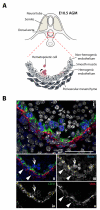
Figure 2. Hematopoietic cell generation in the E10.5 AGM region
A Schematic of a cross section through an E10.5 AGM with the ventral wall of the dorsal aorta boxed and magnified to reveal the different cell types. Clusters of hematopoietic cells (red) bud out from the hemogenic endothelium (pink) of the dorsal aorta. Non-hemogenic endothelium is labelled in light grey. Surrounding the endothelium is a layer of smooth muscle cells (mid grey) and underlying that are mesenchymal cells (dark grey). B Expression of Runx1, the smooth muscle marker SMA (smooth muscle actin), and the endothelial marker CD31 in the E10.5 dorsal aorta. Sections were immunolabeled with the appropriate antibodies, and images pseudo-coloured. Runx1 expression can be detected in endothelial cells (asterisk), hematopoietic cluster cells (arrow) and the abluminal cell layers of the dorsal aorta, here identified as smooth muscle (arrowhead). Runx1 (blue; Alexa Fluor 594), SMA (red; Cy3) and CD31 (green; Alexa Fluor 488), nuclear stain (Dapi; white). Merged channel image with nuclear stain (i) and without nuclear stain (ii). Single channel images for Runx1 (iii), CD31 (iv) and SMA (v) are shown for clarity. Scale bar is 50 μm. Image was taken on a Zeiss LSM780 inverted microscope, processed using ZEN lite 2011, adobe photoshop and illustrator.

References

    1. Palis J, et al. Development of erythroid and myeloid progenitors in the yolk sac and embryo proper of the mouse. Development. 1999;126(22):5073–84. - PubMed
    1. Ferkowicz MJ, Yoder MC. Blood island formation: longstanding observations and modern interpretations. Exp Hematol. 2005;33(9):1041–7. - PubMed
    1. McGrath K, Palis J. Ontogeny of erythropoiesis in the mammalian embryo. Curr Top Dev Biol. 2008;82:1–22. - PubMed
    1. Medvinsky A, Dzierzak E. Definitive hematopoiesis is autonomously initiated by the AGM region. Cell. 1996;86(6):897–906. - PubMed
    1. Muller AM, et al. Development of hematopoietic stem cell activity in the mouse embryo. Immunity. 1994;1(4):291–301. - PubMed

Substances