Review
. 2015 Jan;61(1):203-16.
doi: 10.1016/j.jvs.2013.08.019. Epub 2013 Oct 3.

Vein graft failure

Affiliations
Review

Vein graft failure

Christopher D Owens et al. J Vasc Surg. 2015 Jan.

Abstract

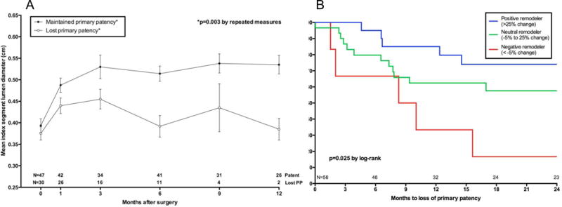
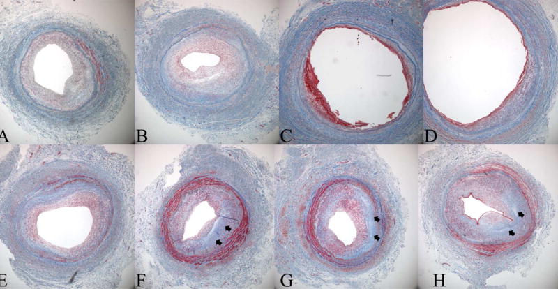
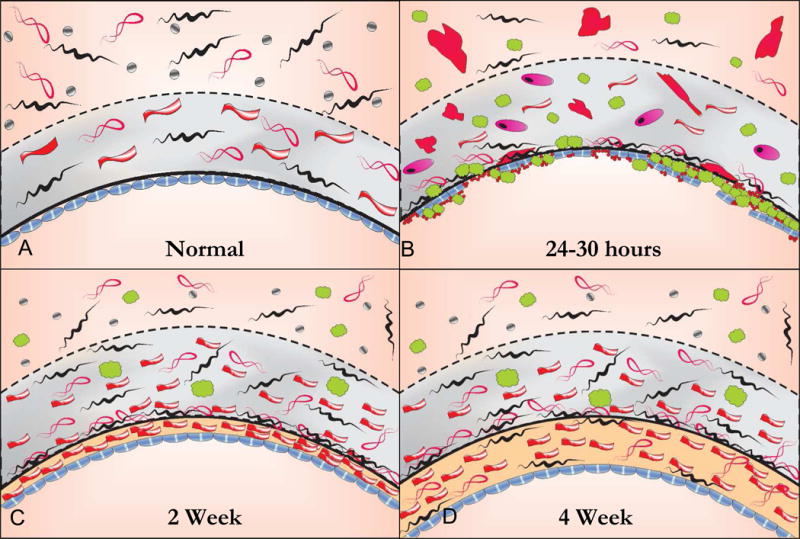
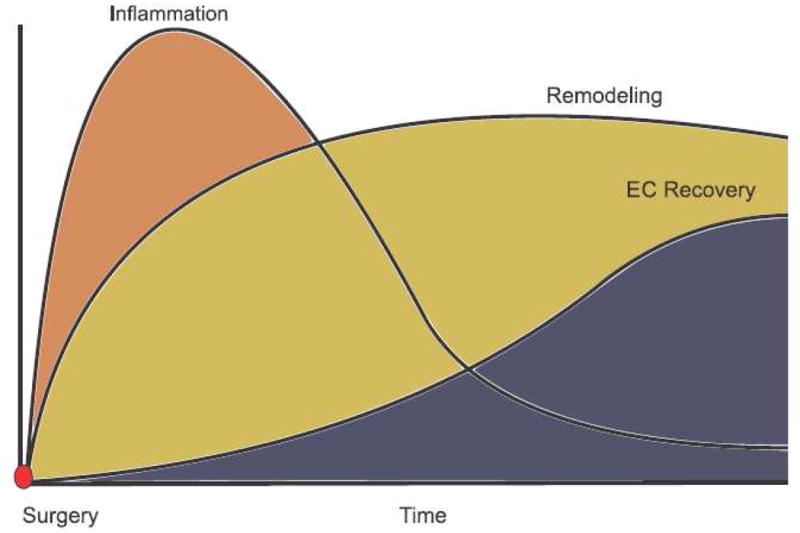
After the creation of an autogenous lower extremity bypass graft, the vein must undergo a series of dynamic structural changes to stabilize the arterial hemodynamic forces. These changes, which are commonly referred to as remodeling, include an inflammatory response, the development of a neointima, matrix turnover, and cellular proliferation and apoptosis. The sum total of these processes results in dramatic alterations in the physical and biomechanical attributes of the arterialized vein. The most clinically obvious and easily measured of these is lumen remodeling of the graft. However, although somewhat less precise, wall thickness, matrix composition, and endothelial changes can be measured in vivo within the healing vein graft. Recent translational work has demonstrated the clinical relevance of remodeling as it relates to vein graft patency and the systemic factors influencing it. By correlating histologic and molecular changes in the vein, insights into potential therapeutic strategies to prevent bypass failure and areas for future investigation are explored.


Conflict of interest statement

Author conflict of interest: none

Figures

Figure 1
Figure 1
Index segment diameter based on eventual loss of primary patency. Values shown represent the mean diameter ± standard error of the mean at each interval. Although the starting diameter was at a similar size as those that remained patent, vein grafts that eventually lost primary patency failed to dilate and had a significantly smaller lumen size over time, A. Early (30 day) remodeling is associated with primary patency, P=.02. Adapted from Gasper et al.
Figure 2
Figure 2
Histological evidence for negative remodeling and intimal hyperplasia as a cause of late lumen loss in a great saphenous vein bypass graft. Masson's Trichrome sections are from an 8 month old femoro-posterior tibial artery vein bypass graft that was explantated due to hemodynamically significant stenosis identified by surveillance duplex ultrasound. The repair was constructed by an interposition graft and an 8 cm piece of diseased segment was explanted, registered, and serially sectioned from A (proximal graft) to H (distal graft). Note two areas of significant stenosis, sections A and B, and sections F-H with an intervening area of relatively normal vein. While the vein was uniform size and luminal caliber at the time of original surgery the stenotic areas demonstrate loss of total vessel area indicating lumen loss is due not only to initimal hyperplasia but also negative remodeling. Note the amount of fibrous protein, blue stain (arrows), in the stenotic segments of the graft.
Figure 3
Figure 3
The histology of the healing autogenous vein graft. In the normal vein, the intima is lined by large flat endothelial cells that are more permeable than those in arteries. The intima is separated from the media by a fenestrated internal elastic laminae. The tunica media is thin compared to an artery with 2 or more layers of smooth muscle cells (SMC) while the adventitia is relatively thick consisting of a loose collagenous network interspersed with fibroblasts and vaso vasorum and small autonomic nerves, A. Within 24 hours following implantation the vein grafts exhibit significant endothelial cell loss and sub-endothelial edema. Inflammatory cells, platelets and fibrin are seen adherent to the surface and infiltrating underneath the attenuated endothelial cell monolayer. There is edema in the tunica media with extensive SMC necrosis or swelling and hypertrophy of the remaining SMCs with infiltration of inflammatory cells, B. By 2 to 4 weeks there is re-endothelialization of the luminal surface and a developing neointima. While the endothelium is continuous, it remains dysfunctional as evidenced by organelle hypertrophy and adhesion molecule expression. The medial edema and inflammation is reduced and there is increased collagen content. Surviving medial SMCs appear hypertrophic with increased rough endoplasmic reticulum and Golgi apparatus indicating synthetic transformation. Over time the adventitia becomes incorporated in surrounding tissue and vaso vasorum and adrenergic nerve fibers grow in from adjacent arteries and connective tissue, C. By 4 weeks there is a predominant layer of intimal thickening characterized by SMCs embedded in a matrix of collagen and ground substance. While early medial thickening is caused predominantly by edema and inflammation, fibrous transformation is responsible for late medial thickening. Areas of the medial wall are devoid of cells and entirely replaced by fibrosis. Clinically stiffening of the vein graft is likely from from the increase in fibrous protein as well as increased cross linking of extracellular matrix proteins.
Figure 4
Figure 4
Hypothetical sequence of vein graft healing. Inflammation peaks early following implantation and then subsides. The critical period of vein graft luminal remodeling is largely complete by the first 30 days. Functional endothelial recovery is temporally delayed by several months. Mature vein grafts exhibit an endothelial layer overlying a stable neointima. Endothelium-dependent relaxation in mature grafts is mediated by nitric oxide.

References

    1. Bhasin M, Huang Z, Pradhan-Nabzdyk L, Malek JY, LoGerfo PJ, Contreras M, et al. Temporal network based analysis of cell specific vein graft transcriptome defines key pathways and hub genes in implantation injury. PLoS One. 2012;7(6):e39123. - PMC - PubMed
    1. Owens CD, Wake N, Conte MS, Gerhard-Herman M, Beckman JA. In vivo human lower extremity saphenous vein bypass grafts manifest flow mediated vasodilation. J Vasc Surg. 2009;50(5):1063–70. - PMC - PubMed
    1. Alexander JH, Hafley G, Harrington RA, Peterson ED, Ferguson TB, Jr, Lorenz TJ, et al. Efficacy and safety of edifoligide, an E2F transcription factor decoy, for prevention of vein graft failure following coronary artery bypass graft surgery: PREVENT IV: a randomized controlled trial. Jama. 2005;294(19):2446–54. - PubMed
    1. Conte MS, Bandyk DF, Clowes AW, Moneta GL, Seely L, Lorenz TJ, et al. Results of PREVENT III: a multicenter, randomized trial of edifoligide for the prevention of vein graft failure in lower extremity bypass surgery. J Vasc Surg. 2006;43(4):742–751. discussion 751. - PubMed
    1. Conte MS, Belkin M, Upchurch GR, Mannick JA, Whittemore AD, Donaldson MC. Impact of increasing comorbidity on infrainguinal reconstruction: a 20-year perspective. Ann Surg. 2001;233(3):445–52. - PMC - PubMed

MeSH terms