Review
. 2013 Oct 5;382(9899):1214-23.
doi: 10.1016/S0140-6736(13)61897-8.

Allogeneic blood and bone marrow cells for the treatment of severe epidermolysis bullosa: repair of the extracellular matrix

Affiliations
Review

Allogeneic blood and bone marrow cells for the treatment of severe epidermolysis bullosa: repair of the extracellular matrix

Jakub Tolar et al. Lancet. .

Abstract

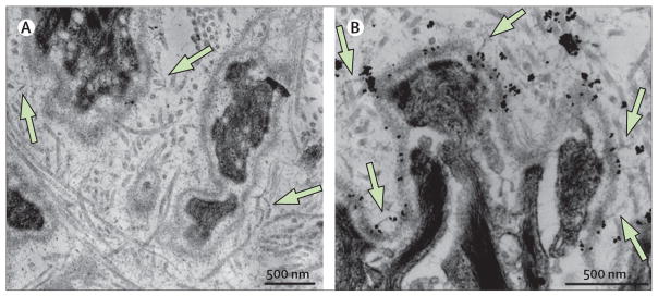
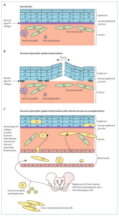
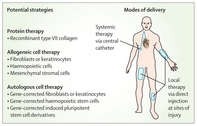
Contrary to the prevailing professional opinion of the past few decades, recent experimental and clinical data support the fact that protein replacement therapy by allogeneic blood and marrow transplantation is not limited to freely diffusible molecules such as enzymes, but also large structural proteins such as collagens. A prime example is the cross-correction of type VII collagen deficiency in generalised severe recessive dystrophic epidermolysis bullosa, in which blood and marrow transplantation can attenuate the mucocutaneous manifestations of the disease and improve patients' quality of life. Although allogeneic blood and marrow transplantation can improve the integrity of the skin and mucous membranes, today's accomplishments are only the first steps on the long pathway to cure. Future strategies will be built on the lessons learned from these first transplant studies.


Conflict of interest statement

Conflicts of interest

We declare that we have no conflicts of interest.

Figures

Figure 1
Figure 1. Structure of the dermal-epidermal junction and phenotype of generalised severe recessive dystrophic epidermolysis bullosa
(A) Ultrastructural features of a healthy human skin basement membrane zone with anchoring fibrils (arrows) extending from the basement membrane to the papillary dermis. (B) Immune fluorescence-aided visualisation of healthy human type VII collagen (in red) at the dermal-epidermal junction. Nuclei are blue (DAPI stain). (C) Wounds in recessive dystrophic epidermolysis bullosa. (D) Disfiguring scarring in recessive dystrophic epidermolysis bullosa.
Figure 2
Figure 2. Increased C7 expression and clinical benefit after blood and marrow transplantation for severe recessive dystrophic epidermolysis bullosa
(A) Immune fluorescence-aided visualisation of human type VII collagen (in red) at the dermal epidermal junction before blood and marrow transplantation (top panel), 200 days after blood and marrow transplantation (middle panel), and 360 days after blood and marrow transplantation (bottom panel). Nuclei are blue (DAPI stain). (B) Clinical comparison of extremity wounds before (left panels) and after (right panels) blood and marrow transplantation. Reprinted with permission from reference .
Figure 3
Figure 3. Evolution of the conditioning regimen for the treatment of severe recessive dystrophic epidermolysis bullosa
Day 0=day of stem cell infusion. HSCT=haemopoietic stem-cell transplantation. GVHD=graft-versus-host disease. MMF=mycophenolate mofetil. MSC=mesenchymal stromal cell. BU=busulfan. Flu=fludarabine. CY=cyclophosphamide. TBI=total body irradiation.
Figure 4
Figure 4. Engraftment of both haemopoietic and non-haemopoietic cells in recessive dystrophic epidermolysis bullosa skin after bone marrow transplantation
Y chromosome (red dot and arrow) and X chromosome (green dot and arrow) of male donor cells in skin of female recipient after sex-mismatched bone marrow transplantation. Some donor cells express pan-haemopoietic antigen CD45 (blue rectangles); other donor cells (non-haemopoietic cells in both epidermis and dermis) do not (white ovals). As would be expected in a recipient with an engrafted lymphohaemopoietic system, occasional donor haemopoietic cells were detected inside a dermal blood vessel (endothelium stained green with anti-CD31 antibody). A linear band (fuchsia) at the dermal-epidermal junction (dotted line) shows type VII collagen. Reprinted with permission from reference .
Figure 5
Figure 5. Anchoring fibrils in recessive dystrophic epidermolysis bullosa skin after bone marrow transplantation
Anchoring fibrils (homopolymers of type VII collagen) more than 3 years after bone marrow transplantation on ultrastructural examination by transmission electronmicroscopy (A) and anti-type VII collagen antibody-aided immune electronmicroscopy (B). The antibody is mAb185 (from Lynn Sakai, Microimaging Center, Shriner’s Hospital for Children, Portland, OR, USA) and is conjugated to gold particles (black dots).
Figure 6
Figure 6. Cross-correction for recessive dystrophic epidermolysis bullosa after blood and marrow transplantation
Models of (A) healthy skin and (B) skin in recessive dystrophic epidermolysis bullosa. Presence of patches of mutant type VII collagen at the dermal-epidermal junction does not prevent easy separation of the epidermis from the dermis upon minimal trauma. (C) Model of skin repair in recessive dystrophic epidermolysis bullosa after allogeneic blood and marrow transplantation. Presence of donor cells (yellow) in the dermis and epidermis, indicating the presence of haemopoietic (neutrophils and lymphocytes) and non-haemopoietic cells (stromal cells and possibly keratinocytes) of donor origin that have migrated from the blood or bone marrow graft, homed to the injured skin, and secreted wild-type (normal) type VII collagen above and below the dermal-epidermal junction, leading to enhanced integrity of the skin and resistance to trauma.
Figure 7
Figure 7. Therapeutic opportunities for recessive dystrophic epidermolysis bullosa
Several strategies can be used locally or systemically, and alone or in combination. Existing treatments include injection of third-party fibroblasts adjacent to wounds, direct injection of recombinant type VII collagen, and infusion of mesenchymal stromal cells or HLA-matched haemopoietic stem cells. New treatments in development include the use of genetically corrected keratinocytes and haemopoietic stem cells, and cellular reprogramming.

References

    1. Bruckner-Tuderman L. Dystrophic epidermolysis bullosa: pathogenesis and clinical features. Dermatol Clin. 2010;28:107–14. - PubMed
    1. Fine JD, Eady RA, Bauer EA, et al. The classification of inherited epidermolysis bullosa (EB): report of the Third International Consensus Meeting on Diagnosis and Classification of EB. J Am Acad Dermatol. 2008;58:931–50. - PubMed
    1. Uitto J, McGrath JA, Rodeck U, Bruckner-Tuderman L, Robinson EC. Progress in epidermolysis bullosa research: toward treatment and cure. J Invest Dermatol. 2010;130:1778–84. - PubMed
    1. Mellerio JE. Infection and colonization in epidermolysis bullosa. Dermatol Clin. 2010;28:267–69. - PubMed
    1. Pope E, Lara-Corrales I, Mellerio JE, Martinez AE, Sibbald C, Sibbald RG. Epidermolysis bullosa and chronic wounds: a model for wound bed preparation of fragile skin. Adv Skin Wound Care. 2013;26:177–88. - PubMed

Publication types

MeSH terms