. 2013 Dec;34(38):10043-55.
doi: 10.1016/j.biomaterials.2013.09.055. Epub 2013 Oct 1.

Automated procedure for biomimetic de-cellularized lung scaffold supporting alveolar epithelial transdifferentiation

Affiliations

Automated procedure for biomimetic de-cellularized lung scaffold supporting alveolar epithelial transdifferentiation

Eric D Girard et al. Biomaterials. 2013 Dec.

Abstract

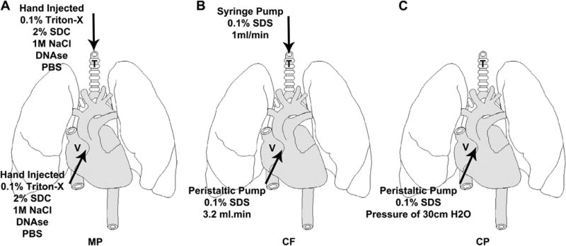
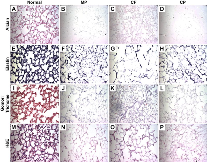
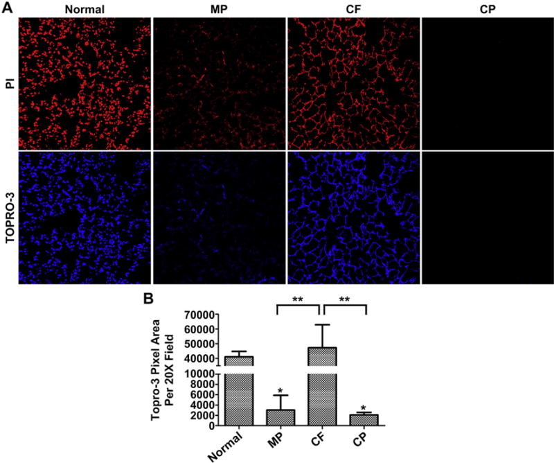
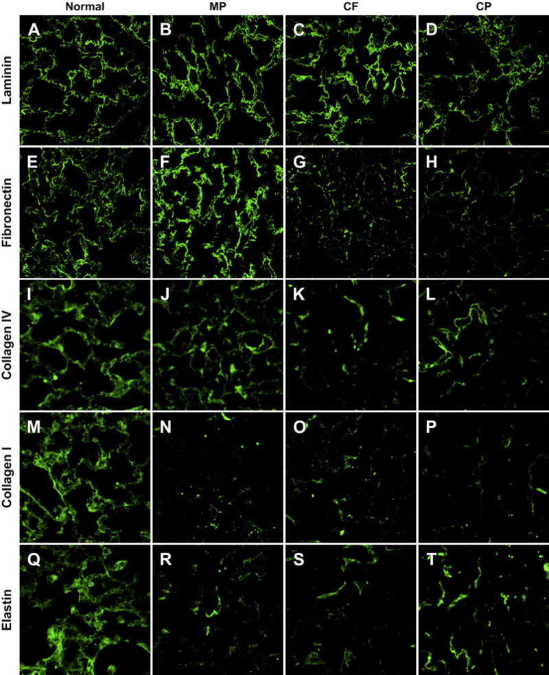
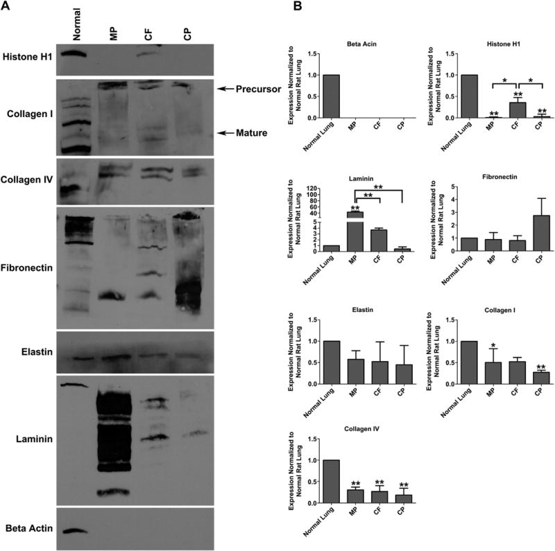
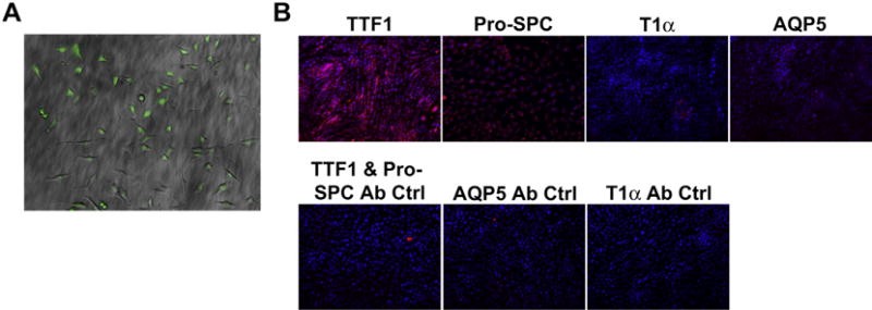
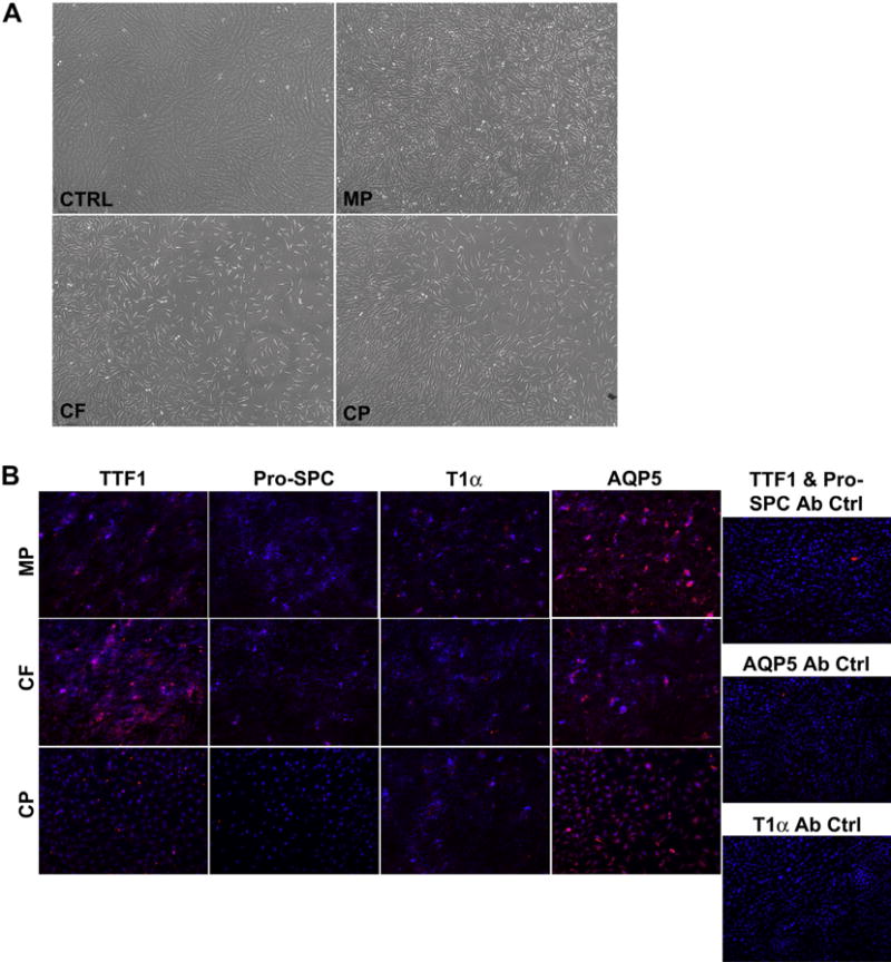
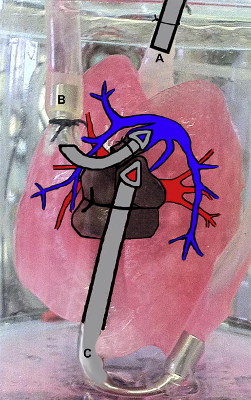
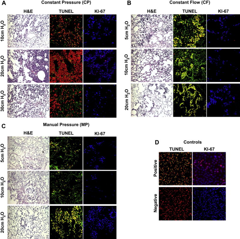
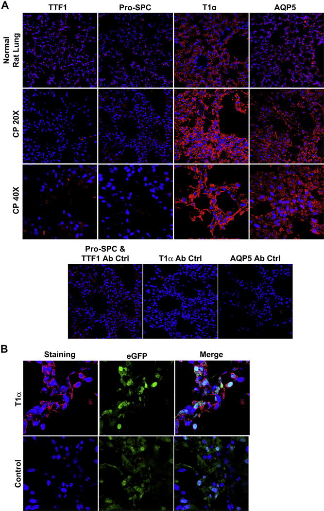
The optimal method for creating a de-cellularized lung scaffold that is devoid of cells and cell debris, immunologically inert, and retains necessary extracellular matrix (ECM) has yet to be identified. Herein, we compare automated detergent-based de-cellularization approaches utilizing either constant pressure (CP) or constant flow (CF), to previously published protocols utilizing manual pressure (MP) to instill and rinse out the de-cellularization agents. De-cellularized lungs resulting from each method were evaluated for presence of remaining ECM proteins and immunostimulatory material such as nucleic acids and intracellular material. Our results demonstrate that the CP and MP approaches more effectively remove cellular materials but differentially retain ECM proteins. The CP method has the added benefit of being a faster, reproducible de-cellularization process. To assess the functional ability of the de-cellularized scaffolds to maintain epithelial cells, intra-tracheal inoculation with GFP expressing C10 alveolar epithelial cells (AEC) was performed. Notably, the CP de-cellularized lungs were able to support growth and spontaneous differentiation of C10-GFP cells from a type II-like phenotype to a type I-like phenotype.

Keywords: Alveolar epithelial cell; C10 alveolar cells; De-cellularization; Extracellular matrix; Lung; Scaffold.


Figures

Fig. 1
Fig. 1
Schematic of De-cellularization Process MP de-cellularization (A) and CF de-cellularization (B) are achieved by infusing detergent through the trachea (T) and vasculature (V), while CP de-cellularization (C) infuses the detergent only through the vasculature (V).
Fig. 2
Fig. 2
Histologic Evaluation of De-cellularized Scaffolds Representative images for Alcian Blue, a glycosaminoglycan's stain, demonstrate a loss across all three de-cellularization methods (blue) (A–D). Verhoeff's elastin stain, which outlines elastin filaments (black) (E–H), also demonstrates a loss across all three methods. Gomori Trichrome, a collagen stain (blue) (I–L) also indicates a loss across all three methods. Lastly, H&E staining (M–P) confirms loss of nuclei, while maintain structural integrity in all three methods. Representative images are shown from triplicate lungs de-cellularized using each method (All Images 40× Magnification). (For interpretation of the references to colour in this figure legend, the reader is referred to the web version of this article.)
Fig. 3
Fig. 3
Presence of Nuclear Material Following De-cellularization (A) CP de-cellularization was most effective at removing nucleic acids. The MP protocol was also effective, while CF de-cellularization was very inefficient. Representative images are shown from triplicate lungs de-cellularized using each method (20× Magnification). (B) Quantification of TOPRO-3 signal using Image J and analyzed via one-tailed t-test demonstrates the effectiveness and reproducibility of CP de-cellularization on removing nucleic acids (*P ≤ 0.05 compared to normal lung, **P ≤ 0.005 compared to CF lung). Means + St Dev. from three representative lungs for each method are shown.
Fig. 4
Fig. 4
Qualitative Analysis of ECM Proteins by Immunofluorescence Representative images from triplicate lungs de-cellularized using each method and immunostained for laminin (A–D), fibronectin (E–H), collagen IV (I–L), collagen I (M–P), elastin (Q–T) qualitatively demonstrate loss of ECM proteins from the de-cellularization process. The MP protocol appears to retain the most laminin, fibronectin and Collagen IV compared to the CF and CP protocols. The CP method appears to retain the most elastin. (All Images 63× Oil Magnification).
Fig. 5
Fig. 5
Quantitative Analysis of ECM Proteins by Western Blot (A) Western blot analysis of 50 μg whole cell lysate from MP, CF or CP de-cellularized lung scaffolds relative to normal rat lung for nuclear (histone H1), intracellular (beta actin) and extracellular (collagen I, collagen IV, fibronectin, elastin, and laminin) proteins. (B) Densitometry was performed using Image J and analyzed via one-way ANOVA (* = P ≤ 0.05 ** = P ≤ 0.005 compared to normal lung). There is significant loss of nuclear proteins (Histone H1) in the MP and CP protocols. Laminin is significantly retained in the MP protocol, when compared to the CF and CP protocol. Fibronectin appears to be greatest in the CP protocol; however, it is not statistically significant. There is loss of elastin, collagen I and collagen IV in all three protocols. De-cellularized scaffolds reproduced and analyzed in triplicate.
Fig. 6
Fig. 6
Characterization of C10 Cells After GFP Transfection (A) C10 alveolar epithelial cells express GFP following viral transduction and FACS sorting. C10 cells following flow-based sorting were over 95% GFP positive (10× Magnification). (B) C10-GFP cells cultured on untreated plastic express type II alveolar epithelial markers TTF-1 and Pro-SPC, but do not express type I alveolar epithelial markers such as AQP5 or T1α (20× Magnification).
Fig. 7
Fig. 7
Comparison of C10 Cells Cultured on Homogenized Scaffolds (A) Culture wells coated with homogenized scaffolds and seeded with C10-GFP cells displayed a change in morphology after 3 days compared to those cultured on uncoated plastic (control) after three days (10× Magnification). (B) C10-GFP cells cultured for 3 days in wells coated with homogenized scaffolds and seeded with C10-GFP cells demonstrated loss of pro-SPC expression but increased that of T1α and AQP5. Variable TTF-1 expression was observed. Secondary antibody only controls were used to assess non-specific binding (20× magnification).
Fig. 8
Fig. 8
Schematic of Re-cellularization of De-cellularized Scaffolds in Bioreactor System A physiologic heart lung bioreactor system from Harvard Apparatus was utilized to re-cellularize and culture C10-GFP cells on MP CF, and CP de-cellularized lung scaffolds. Following de-cellularization, cells were inoculated via the trachea (A) and allowed to adhere for 4 h. After that time, maintenance media was perfused through the pulmonary artery (B) and exited via the pulmonary vein (C) for 3 days.
Fig. 9
Fig. 9
Histology, Apoptosis and Cell Proliferation of Re-cellularized Scaffolds After 3 Days In Culture Delivery pressure of maintenance medium was examined in range of 5–30 cm H2O (A) CP Seeded lungs appear to repopulate the scaffolds by H&E, but only yield proliferative cells when maintenance medium is delivered at a pressure of 20 cm H2O. (B) CF seeded lungs also appear to repopulate the scaffolds by H&E; however, all cells display an apoptotic, non-proliferative phenotype under all perfusion pressure conditions assessed. (C) MP seeded lungs were also apoptotic and non-proliferative under all perfusion pressure conditions assessed. (D) TUNEL negative control was normal lung and TUNEL Positive control was normal lung treated with DNAse (20× magnification). Ki67 negative control was no primary; positive control was C10-GFP cells dividing in culture (20× magnification). Ki67 staining (TOPRO3 = BLUE, Ki67 = RED), TUNEL Staining (PI (Nuclear Counterstain) = RED, TUNEL = Green, CELL DEATH = Yellow). (For interpretation of the references to colour in this figure legend, the reader is referred to the web version of this article.)
Fig. 10
Fig. 10
Immunofluorescence Staining of Re-cellularized CP Scaffolds (A) Characterization of CP re-seeded scaffolds indicates the expression of both AQP5 and T1α as well as the loss of pro-SPC expression (40× magnification). Low magnification images help demonstrate the widespread expression of this type I phenotype following re-seeding (20× magnification). No primary controls were used to assess non-specific binding of the secondary antibodies. (B) High magnification images demonstrate GFP positive cells on the scaffold that co-express T1α (Magnification 63× oil). Scaffolds reproduced and analyzed in triplicate.

References

    1. American Lung A. American lung association: lung disease data: 2008; improving life, one breath at a time. New York, N.Y.: American Lung Association; 2008.
    1. Organ procurement and transplantation network and scientific registry of transplant recipients 2010 data report. Am J Transplant. 2012;12(Suppl 1):1–156. - PubMed
    1. Badylak SF, Weiss DJ, Caplan A, Macchiarini P. Engineered whole organs and complex tissues. Lancet. 2012;379:943–52. - PubMed
    1. Vacanti JP. Tissue engineering and the road to whole organs. Br J Surg. 2012;99:451–3. - PubMed
    1. Warburton D, El-Hashash A, Carraro G, Tiozzo C, Sala F, Rogers O, et al. Lung organogenesis. Curr Top Dev Biol. 2010;90:73–158. - PMC - PubMed

Publication types