. 2013 Oct 17;5(1):194-206.
doi: 10.1016/j.celrep.2013.08.040. Epub 2013 Oct 3.

TRF2 interaction with Ku heterotetramerization interface gives insight into c-NHEJ prevention at human telomeres

Affiliations

TRF2 interaction with Ku heterotetramerization interface gives insight into c-NHEJ prevention at human telomeres

Albert Ribes-Zamora et al. Cell Rep. .

Abstract

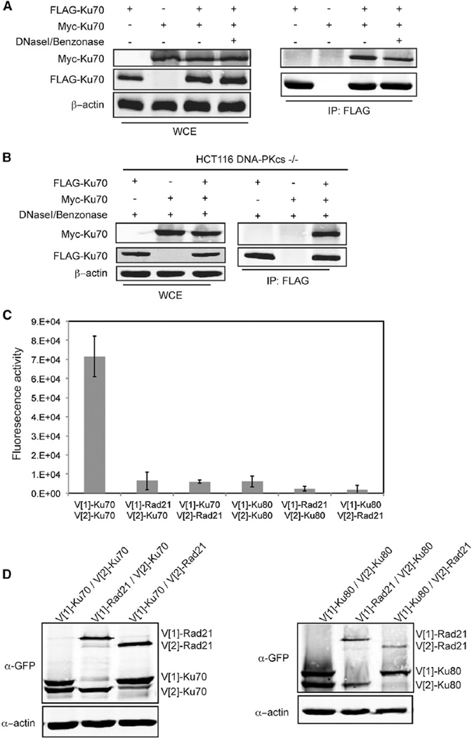
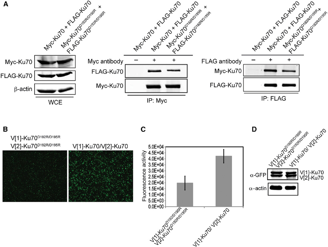
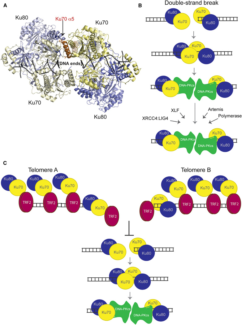
Telomeres are protected from nonhomologous end-joining (NHEJ) to avoid deleterious chromosome fusions, yet they associate with the Ku heterodimer that is principal in the classical NHEJ (c-NHEJ) pathway. T-loops have been proposed to inhibit Ku's association with telomeric ends, thus inhibiting c-NHEJ; however, deficiencies in the t-loop model suggest additional mechanisms are in effect. We demonstrate that TRF2 interacts with Ku at telomeres and via residues in Ku70 helix 5 (α5), which are vital for NHEJ. We show that Ku's interaction with a TRF2 mutant that induces telomeric fusions is significantly impaired. Additionally, we demonstrate that Ku70 α5 is required for Ku self-association in live cells, which can bridge DNA ends. Together, these findings lead us to propose a model in which telomeres are directly protected from c-NHEJ via TRF2 impeding Ku's ability to synapse telomere ends.


Figures

Figure 1
Figure 1. Ku70 Interacts with TRF1 and TRF2 at Telomeres
(A) Fluorescence quantification of PCA for Ku70 or Rad21 with either TRF1 or TRF2. Shown are the averages for three independent transfections. Error bars represent SD of the mean. (B) Colocalization of PCA for Ku70 with TRF1 or TRF2, YFP channel, with the DsRed-TRF2 telomeric marker, RFP channel, and (merge) in HEK293T cells. (C) Fluorescence quantification of PCA for Ku70 and TRF1 or Ku70 and TRF2 with coexpression of either FLAG-tagged empty vector (EV), TRF1 or TRF2, and TRF1ΔAΔM or TRF2ΔBΔM. p values were determined by Student’s two-tailed unpaired t test with *, p < 0.05; **, p < 0.01; and ns, not significant. For V[1]-TRF1/V[2]-Ku70: p = 0.0345 for EV versus TRF2ΔBΔ Mand p = 0.0011 for TRF2 versus TRF2ΔBΔM. For V[1]-TRF2/V[2]-Ku70: p = 0.8566 for EV versus TRF1ΔAΔM and p = 0.0343 for TRF1 versus TRF1ΔAΔM. The error bars represent SD of the mean. (D) Colocalization of PCA for Ku70 with TRF1 with coexpression of either FLAG-EV or FLAG-TRF2ΔBΔM (merge) in HEK293T cells. See also Figure S1.
Figure 2
Figure 2. TRF2, but Not Rap1 or TRF1, Interacts with the Ku70 NHEJ Domain
(A) Left, frontal view of Ku’s outward face. Ku70 and Ku80 are depicted in yellow and blue, respectively; Ku70 α5 in orange; and DNA in black. Right, alignment of human and budding yeast Ku70 residues located in α5. Residues mutagenized in Ku70D192R/D195R are depicted in red. (B) Fluorescence quantification of PCA for the designated Ku70 mutants or Rad21 with either TRF2 or Rap1. Shown are the averages for three independent transfections. Error bars represent SD of the mean. p values were obtained using the Student’s two-tailed unpaired t test. p values for pairs including TRF2 are as follows: p = 0.0072 for Ku70 versus Ku70D192R/D195R, p = 0.0026 for Ku70 versus Ku70D192R/R194D/D195R, and p = 0.0092 for Ku70 versus Ku70R185D/R187D. p values for pairs including Rap1 are as follows: p = 0.3540 for Ku70 versus Ku70D192R/D195R and p = 0.3162 for Ku70 versus Ku70D192R/R194D/D195R. (C) Yeast two-hybrid analysis for ADH1pLexA-DBD-TRF1 or the designated ADH1pLexA-DBD-Ku70 1–386 alleles (bait) with either GAL1pB42-AD-TRF2 or GAL1pB42-AD -TRF1 (prey). Serial dilutions were grown on Gal-His-Trp-Ura media without (−) or with (+) of leucine. Positive interaction is determined by growth in the absence of leucine. See also Figure S2.
Figure 3
Figure 3. Ku70 Has Decreased Association with TRF2ΔBΔM
(A) Fluorescence quantification of PCA for Ku70 and either TRF2 or TRF2ΔBΔM. Rad21 was used as a nonspecific control for interaction. The error bars represent SD of the mean. (B) Protein levels resulting from transfections in (A) detected by immunoblot analysis of WCE using a GFP antibody. β-actin was used as a loading control.
Figure 4
Figure 4. Ku70 α5 Is Not Required for Ku’s Association with XRCC4 or XLF
(A) Visualization of PCA using Ku70 or Ku70D192R/D195R with XLF or XRCC4. Images are at 4× magnification. (B) Fluorescence quantification of PCA shown in (A). The error bars represent SD of the mean. (C) Protein levels resulting from transfections in (A) detected by immunoblot analysis of WCE using a GFP antibody. β-actin was used as a loading control.
Figure 5
Figure 5. Ku Heterodimers Self-Associate via the Ku70 Subunit
(A) Coimmunoprecipitation of Myc-Ku70 with FLAG-Ku70. Immunoprecipitations with FLAG antibody were performed using WCEs untreated (−) or treated (+) with DNaseI/Benzonase. FLAG and Myc immunoblots were performed on the WCEs (left) and immunoprecipitates (IP) (right, IP:FLAG). β-actin represents a loading control. (B) Coimmunoprecipitation of Myc-Ku70 with FLAG-Ku70 in DNaseI/Benzonase treated WCEs from HCT116 DNA-PKcs−/− cells. (C) Fluorescence quantification of PCA using the indicated combinations of Ku70, Ku80, or Rad21. The error bars represent SD of the mean. (D) Protein levels resulting from the transfections in (C) detected by immunoblot analysis of WCEs using a GFP antibody. β-actin was used as a loading control. See also Figure S3.
Figure 6
Figure 6. Ku70 α5 Is Required for Heterotetramerization of Ku
(A) Differentially tagged Ku70 or Ku70D192R/D192 were transiently coexpressed in 293T cells as indicated. Immunoprecipitations with anti-FLAG, anti-Myc, or no antibody (−) were performed. FLAG and Myc immunoblots were performed on the WCEs (left) and the Myc (middle) and FLAG (right) immunoprecipitates (IP). β-actin immunoblot of the WCEs was performed as a loading control. (B) Visualization of PCA for Ku self-association using either Ku70D192R/D195R or Ku70. Images are at 4× magnification. (C) Fluorescence quantitation of PCA shown in (B). The error bars represent SD of the mean. (D) Protein levels resulting from the transient transfections in (A) detected by immunoblot analysis of WCE using a GFP antibody. β-actin was used as a loading control. See also Figure S4.
Figure 7
Figure 7. Models for Ku Heterotetramerization and Its Inhibition at Telomeres by TRF2
(A) Top view of a hypothetical Ku tetramer obtained via protein docking using HADDOCK web server. Yellow: Ku70. Blue: Ku80. Orange: Ku70 α5. Residues R185, D192, and D195 from Ku70 are shown in stick representation. The vWA domain from Ku70 would have to move to accommodate this model of heterotetramerization. Its original position with respect to its native heterodimer is shown in “ghost” (semitransparent) representation. (B) Mechanistic model for the role of Ku’s heterotetramerization during NHEJ. We propose that Ku heterotetramerization is required in the initial NHEJ steps to synapse the DNA ends of a DSB prior to the DNA-PKcs recruitment. Formation of the DNA-PK complex displaces Ku away from the end, which effectively disassociates the Ku-Ku interaction and allows DNA-PKcs to replace Ku in synapsing the two ends. Later roles of Ku in recruiting NHEJ factors to DSBs would not require heterotetramer formation. (C) Mechanistic model for the inhibition of Ku’s heterotetramerization by TRF2 at telomeres. We propose that TRF2’s interaction with the Ku70 α5 helix effectively inhibits Ku heterotetramerization at telomeres and the synapsis of telomeric ends, thereby blocking telomeric NHEJ.

References

    1. Bae NS, Baumann P. A RAP1/TRF2 complex inhibits nonhomologous end-joining at human telomeric DNA ends. Mol. Cell. 2007;26:323–334. - PubMed
    1. Barrientos KS, Kendellen MF, Freibaum BD, Armbruster BN, Etheridge KT, Counter CM. Distinct functions of POT1 at telomeres. Mol. Cell. Biol. 2008;28:5251–5264. - PMC - PubMed
    1. Bombarde O, Boby C, Gomez D, Frit P, Giraud-Panis MJ, Gilson E, Salles B, Calsou P. TRF2/RAP1 and DNA-PK mediate a double protection against joining at telomeric ends. EMBO J. 2010;29:1573–1584. - PMC - PubMed
    1. Cary RB, Peterson SR, Wang J, Bear DG, Bradbury EM, Chen DJ. DNA looping by Ku and the DNA-dependent protein kinase. Proc. Natl. Acad. Sci. USA. 1997;94:4267–4272. - PMC - PubMed
    1. Celli GB, de Lange T. DNA processing is not required for ATM-mediated telomere damage response after TRF2 deletion. Nat. Cell Biol. 2005;7:712–718. - PubMed

Publication types

MeSH terms