. 2013 Dec:65:9-18.
doi: 10.1016/j.yjmcc.2013.09.015. Epub 2013 Oct 2.

Angiotensin II stimulates cardiac fibroblast migration via the differential regulation of matrixins and RECK

Affiliations

Angiotensin II stimulates cardiac fibroblast migration via the differential regulation of matrixins and RECK

Jalahalli M Siddesha et al. J Mol Cell Cardiol. 2013 Dec.

Abstract

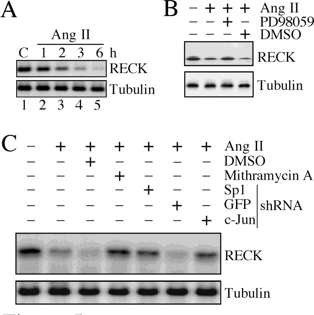
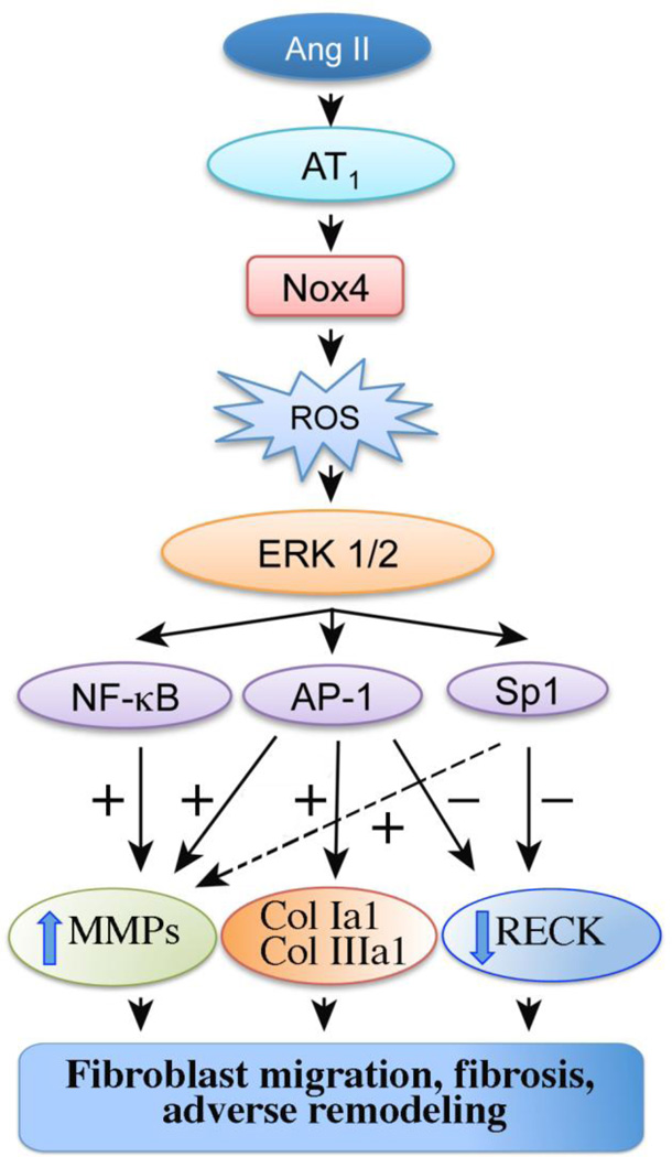
Sustained induction and activation of matrixins (matrix metalloproteinases or MMPs), and the destruction and deposition of extracellular matrix (ECM), are the hallmarks of cardiac fibrosis. The reversion-inducing-cysteine-rich protein with Kazal motifs (RECK) is a unique membrane-anchored endogenous MMP regulator. We hypothesized that elevated angiotensin II (Ang II), which is associated with fibrosis in the heart, differentially regulates MMPs and RECK both in vivo and in vitro. Continuous infusion of Ang II into male C57Bl/6 mice for 2weeks resulted in cardiac fibrosis, with increased expressions of MMPs 2, 7, 9 and 14, and of collagens Ia1 and IIIa1. The expression of RECK, however, was markedly suppressed. These effects were inhibited by co-treatment with the Ang II type 1 receptor (AT1) antagonist losartan. In vitro, Ang II suppressed RECK expression in adult mouse cardiac fibroblasts (CF) via AT1/Nox4-dependent ERK/Sp1 activation, but induced MMPs 2, 14 and 9 via NF-κB, AP-1 and/or Sp1 activation. Further, while forced expression of RECK inhibits, its knockdown potentiates Ang II-induced CF migration. Notably, RECK overexpression reduced Ang II-induced MMPs 2, 9 and 14 activation, but enhanced collagens Ia1 and IIIa1 expression and soluble collagen release. These results demonstrate for the first time that Ang II suppresses RECK, but induces MMPs both in vivo and in vitro, and RECK overexpression blunts Ang II-induced MMP activation and CF migration in vitro. Strategies that upregulate RECK expression in vivo have the potential to attenuate sustained MMP expression, and blunt fibrosis and adverse remodeling in hypertensive heart diseases.

Keywords: 12-O-tetradecanoylphorbol-13-acetate; 3-(4,5-dimethythiazol-2-yl)-2,5-diphenyl tetrazolium bromide; ADAM; AP-1; ARB; AT1; AT2; Adverse remodeling; CF; CMV; CVD; Cardiac fibrosis; DPI; ECM; EGFP; ERK; Elk; Ets-like protein; GAPDH; GFP; GPI; JNK; LV; MAPK; MMP; MOI; MT1-MMP; MTT; NADPH; NADPH oxidase; NF-κB; Nox; Nox4; RECK; ROS; SBP; Sp1; TACE; TIMP; TPA; TPA DNA response element; TRE; UTR; WT; a disintegrin and metalloproteinase domain; activator protein-1; angiotensin II type 1 receptor; angiotensin II type II receptor; angiotensin receptor blockers; c-Jun amino-terminal kinase; cardiac fibroblasts; cardiovascular disease; cytomegalovirus; diphenylene iodonium; enhanced green fluorescent protein; extracellular matrix; extracellular signal-regulated kinase; glyceraldehyde-3-phosphate dehydrogenase; glycophosphatidylinositol; green fluorescent protein; left ventricle; matrix metalloproteinase; membrane type 1-MMP; mitogen-activated protein kinase; multiplicity of infection; nicotinamide adenine dinucleotide phosphate; nuclear factor kappa B; reactive oxygen species; reversion-inducing-cysteine-rich protein with Kazal motifs; shRNA; short hairpin RNA; siMMP; small inhibitory RNA against MMP; specific protein 1; systolic blood pressure; tissue inhibitor of metalloproteinase; tumor necrosis factor, alpha, converting enzyme; untranslated region; wild-type.


Conflict of interest statement

Conflict of interest: None

Figures

Fig. 1
Fig. 1. Ang II/AT1-induced myocardial fibrosis is characterized by increased MMPs and suppressed RECK expression
A, Ang II infusion increases cardiac fibrosis via AT1. Male C57Bl/6 mice were continuously infused with Ang II (1.5 µg/kg body wt/min) for 14 days via miniosmotic pumps. Saline served as a control. One group of mice receiving Ang II was co-treated with the AT1 antagonist losartan in drinking water (0.6 g/L). Collagen deposition was analyzed by Picrosirius Red staining of cryosections (8 µm), and photographed at 100× magnification (n=6). B, Ang II increases ECM protein gene expression. Left ventricular (LV) tissue from mice described in A was analyzed for collagens type Ia1 and IIIa1 by RT-qPCR. Expression of 18S rRNA served as an invariant control. *P < 0.01 vs. saline, †P < 0.05 vs. Ang II (n = 6/group). C, Ang II induces MMPs expression. LV tissue from mice described in A was analyzed for MMP2 and 9 activation by immunoblotting using antibodies that detect both pro and active forms (left hand panel). MMPs 7 and 14 expressions were also analyzed by immunoblotting (right panel) (n=2–4). D, E, Ang II suppresses RECK expression. LV tissue from mice described in A were analyzed for RECK mRNA expression by RT-qPCR (D) and protein levels by immunoblotting (E; n=2–4). D*P < at least 0.01 vs. saline, †P < 0.05 vs. Ang II (n = 6/group).
Fig. 2
Fig. 2. Ang II-induced cardiac fibroblast migration is differentially regulated by RECK and MMPs
A, Ang II stimulates CF migration via AT1. At 70% confluency, CF were made quiescent by incubating in medium supplemented with 0.5% BSA for 48h. The quiescent CF were trypsinized, re-suspended in medium containing 0.5% BSA, layered on Matrigel™ basement membrane matrix-coated filters, incubated with or without losartan (10 µM for 1 h) and then with Ang II (10−7M for 12 h). The lower chamber contained media with 10% serum. Cells migrating to the other side of the membrane were quantified using MTT assay. B, Forced expression of RECK inhibits, but its knockdown potentiates Ang II-induced CF migration. CF transduced with Ad.RECK (moi 40 for 24 h) or lentiviral RECK shRNA (moi 0.5 for 48 h) were made quiescent, layered on Matrigel™ basement membrane matrix-coated filters, incubated with Ang II (10−7M) for 12 h, and analyzed for migration by MTT assay. Knockdown of RECK was confirmed by immunoblotting (inset). Akt served as an off target (n=3). *P < 0.001 vs. untreated; †P < 0.01 vs. Ang II, §P < 0.05 vs. Ang II (n=6). C, Ang II induces CF migration via MMP2 and MMP9. CF transduced with adenoviral MMP2 or MMP9 siRNA (moi 100 for 48 h) were analyzed for migration as in B. Knockdown of MMP2 and MMP9 was confirmed by immunoblotting as shown on the right (n=3). *P < 0.001 vs. untreated; †P < at least 0.05 vs. Ang II (n=6).
Fig. 3
Fig. 3. Ang II induces CF migration via Nox4-dependent ERK activation
A, Ang II induces ERK activation. The quiescent CF incubated with Ang II (10−7M) were analyzed for ERK activation by an in vitro immune complex kinase assay using Elk-1 as a substrate. A representative of three independent experiments is shown. B, Ang II stimulates Nox4-dependent H2O2 generation. CF infected with Ad.siNox4 (moi 100 for 48 h) or treated with DPI (10 µM for 30 min) prior to Ang II addition (10−7M) for 30 min were analyzed for H2O2 production using the Amplex® Red assay. *P < 0.01 vs. untreated; †P < at least 0.05 vs. Ang II ± DMSO or Ad.siGFP (n=6). C, Ang II induces ERK activation via Nox4 and ROS. CF transduced with Ad.siNox4 (moi 100 for 48 h) or pretreated with DPI (10 µM for 30 min) were incubated with Ang II (10−7M) for 30 min. ERK activation was analyzed as in A (n=3). Knockdown of Nox4 was confirmed by immunoblotting (right hand panel). D, PD98059 inhibits Ang II-induced ERK activation. The quiescent CF were treated with the ERK inhibitor PD98059 (10 µM for 1 h), p38MAPK inhibitor SB203580 (1 µM for 30 min) or JNK inhibitor SP600125 (20 µM for 30 min) prior to Ang II addition (10−7M for 30 min). ERK activation was analyzed as in A (n=3). E, Ang II stimulates CF migration via ERK. The quiescent CF were layered on Matrigel™ basement membrane matrix-coated filters, incubated with PD98059 (10 µM for 1 h) followed by Ang II (10−7M for 12 h). Cell migration was analyzed by MTT assay. *P < 0.001 vs. untreated; †P < 0.01 vs. Ang II (n=6). F, Knockdown of Nox4 attenuates Ang II-induced CF migration. CF transduced with Ad.siNox4 (moi 100 for 48 h) were incubated with Ang II (10−7M) for 12 h were analyzed for migration as in A. A–D *P < at least 0.01 vs. untreated; †P < at least 0.05 vs. Ang II (n=6).
Fig. 4
Fig. 4. Ang II induces CF migration and collagen expression via NF-κB, AP-1 and/or Sp1-dependent MMP expression
A, B, Ang II induces MMP2 and MMP9 expression via p65 and c-Jun. CF transduced with lentiviral shRNA (moi 0.5 for 48 h) were incubated with Ang II (10−7M) for 2 h. MMP2 (A) and MMP9 (B) mRNA expression was analyzed by RT-qPCR. Knockdown of p65 and c-Jun was confirmed by immunoblotting as shown on the right (n=3). *P < 0.01 vs. untreated; †P < 0.05 vs. Ang II (n=3). C, Ang II stimulates CF migration via p65 and c-Jun. CF transduced with lentiviral p65 or c-Jun shRNA (moi 0.5 for 48 h) were made quiescent, layered on Matrigel™ basement membrane matrix-coated filters, and incubated with Ang II (10−7M) for 12 h. Cell migration was analyzed by MTT assay. c-Jun and p65 served as respective off targets. *P < 0.001 vs. untreated; †P < 0.01 vs. Ang II (n=6). D, Ang II induces p65 and c-Jun activation via ERK. The quiescent CF were incubated with PD98059 (10 µM for 1 h) prior to Ang II (10−7M for 30 min) addition. Phospho-p65 and phospho-c-Jun levels were analyzed by immunoblotting using nuclear protein extracts. Lamin A/C (nuclear) and GAPDH (cytoplasmic) served as loading and purity controls (n=3). E, F, Ang II activates Sp1 via ERK. The quiescent CF were treated PD98059 (10 µM for 1 h) prior to Ang II addition (10−7M for 3 h). Sp1 activation was analyzed by ELISA using nuclear protein extracts (n=6). Lamin A/C and GAPDH served as loading and purity control (inset). *P < 0.001 vs. untreated; †P < 0.01 vs. Ang II ± DMSO (n=6). F, Ang II induces MMP14 via Sp1. CF transduced with lentiviral Sp1 shRNA (moi 0.5 for 48 h) prior to Ang II addition (10−7M for 3 h) were analyzed for MMP14 expression by immunoblotting using membrane fractions (n=3). Knockdown of Sp1 was confirmed by immunoblotting and is shown on the right. c-Jun served as an off-target. G, H, Ang II induces collagens type Ia1 (G) and IIIa1 (H) expression in part via AP-1. CF treated as A, but for 24 h were analyzed for collagen Ia1 (G) and collagen IIIa1 (H) mRNA expression by RT-qPCR and protein levels by immunoblotting (n=3). G, H*P < 0.01 vs. untreated; †P < 0.01 vs. Ang II ± GFP shRNA (n=6).
Fig. 5
Fig. 5. Ang II suppresses RECK expression via Sp1
A, Time-dependent suppression of RECK expression by Ang II. The quiescent CF treated with Ang II (10−7M) for the indicated time periods were analyzed for RECK expression by immunoblotting using cleared whole cell lysates (n=3). B, Ang II suppresses RECK expression via ERK. The quiescent CF were treated with PD98059 (10 µM for 1 h) prior to Ang II addition (10−7M for 3 h). RECK expression was analyzed as in A (n=3). C, Ang II suppresses RECK via Sp1. CF transduced with lentiviral Sp1, GFP and c-Jun shRNA (moi 0.5 for 48 h) or treated with mithramycin (100 nM in DMSO for 45 min) prior to Ang II addition (10−7M for 3 h) were analyzed for RECK expression by immunoblotting (n=3).
Fig. 6
Fig. 6. RECK overexpression inhibits MMP activation
A–C, Forced expression of wild type RECK (Ad.RECK) inhibits MMP9 (A), MMP2 (B) and MMP14 (C) activation. CF were transduced with Ad.RECK (moi 40 for 24 h) and then treated with Ang II (10−7M) for an additional 24 h. MMP9 (A) and MMP2 (B) enzymatic activity was analyzed by immunoblotting using equal amounts of culture supernatants and antibodies that detect both pro and active forms (left hand panels). MMP7 expression (C) was analyzed by immunoblotting using membrane fractions. Tubulin (whole cell homogenates) or GAPDH (cytoplasmic extracts) served as loading controls. The immunoreactive bands were semiquantified by densitometry and results from three independent experiments are summarized in the middle panels. Enzymatic activities were also analyzed by Biotrak activity assay kits (right hand panels) using concentrated culture supernatants containing 1 µg of total protein (MMPs 9 and 2). MMP7 enzymatic activity was also analyzed by the Biotrak activity assay kit using whole cell lysates (C, right hand panel). A–C, *P < 0.01 vs. respective untreated; †P < at least 0.05 vs. Ang II ± Ad.GFP (n=6).
Fig. 7
Fig. 7. RECK overexpression enhances Ang II-induced collagen expression
A, B, Forced expression of wild type RECK enhances Ang II-induced collagens type Ia1 (A) and type IIIa1 (B) levels. CF transduced with Ad.RECK (moi 40 for 24 h) were treated with Ang II (10−7M) for 12 h, and then analyzed for collagen expression by immunoblotting using cleared whole cell lysates. The immunoreactive bands from three independent experiments were semiquantified by densitometry and are shown in the respective lower panels. A, B*P < 0.05 vs. respective untreated; †P < 0.05 vs. Ang II ± Ad.GFP (n=3). C, RECK overexpression enhances total collagen release. CF were transduced with Ad.RECK as in A, and then treated with Ang II for 48 h. Equal amounts of culture supernatants were analyzed for recently synthesized collagens by Sircol collagen assay and normalized to cell numbers. In addition to cell numbers, Tubulin in whole cell lysates also served as a control (inset). *P < at least 0.01 vs. untreated; †P < 0.05 vs. Ang II ± Ad.GFP (n=6).
Fig. 8
Fig. 8
Schema showing possible signal transduction pathways involved in the differential regulation of MMPs and RECK in Ang II-induced collagen synthesis, fibroblast migration, fibrosis, and adverse remodeling. +: positive regulation, −: negative regulation, broken arrow: though we have not investigated it here, Sp1 has also been shown to transcriptionally upregulate MMP9.

References

    1. de Gasparo M, Catt KJ, Inagami T, Wright JW, Unger T. International union of pharmacology. XXIII. The angiotensin II receptors. Pharmacol Rev. 2000 Sep;52(3):415–472. - PubMed
    1. Fan D, Takawale A, Lee J, Kassiri Z. Cardiac fibroblasts, fibrosis and extracellular matrix remodeling in heart disease. Fibrogenesis Tissue Repair. 2012;5(1):15. - PMC - PubMed
    1. Porter KE, Turner NA. Cardiac fibroblasts: at the heart of myocardial remodeling. Pharmacol Ther. 2009 Aug;123(2):255–278. - PubMed
    1. Patterson NL, Iyer RP, de Castro Bras LE, Li Y, Andrews TG, Aune GJ, et al. Using proteomics to uncover extracellular matrix interactions during cardiac remodeling. Proteomics Clin Appl. 2013 Aug;7(7–8):516–527. - PMC - PubMed
    1. Chen W, Frangogiannis NG. The role of inflammatory and fibrogenic pathways in heart failure associated with aging. Heart Fail Rev. 2010 Sep;15(5):415–422. - PMC - PubMed

Publication types

MeSH terms