

Plasma biomarkers of liver injury and inflammation demonstrate a lack of apoptosis during obstructive cholestasis in mice
- PMID: 24096036
- PMCID: PMC3860754
- DOI: 10.1016/j.taap.2013.09.023
Plasma biomarkers of liver injury and inflammation demonstrate a lack of apoptosis during obstructive cholestasis in mice
Abstract
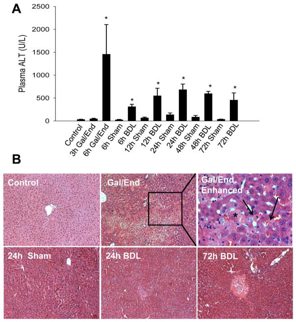
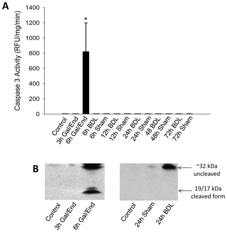
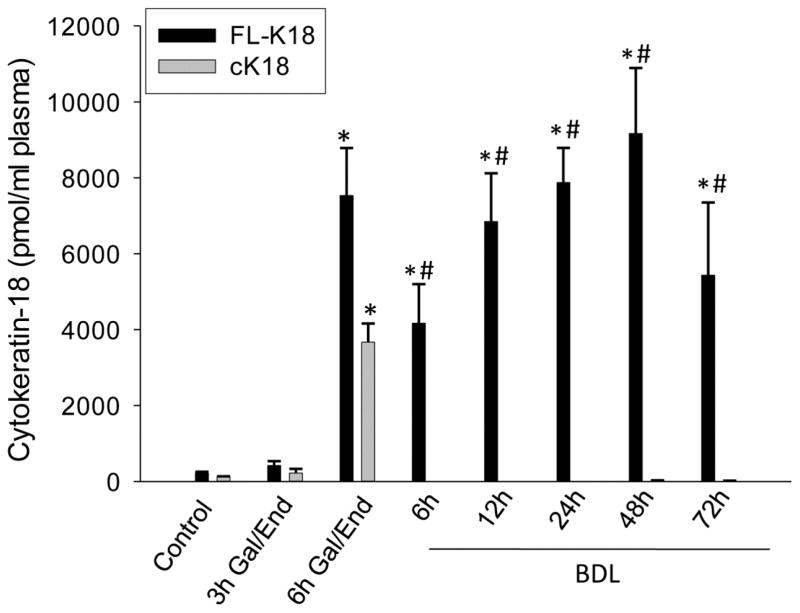
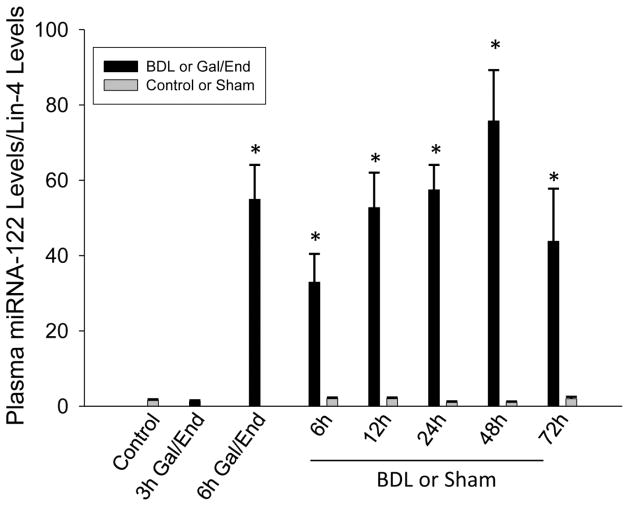
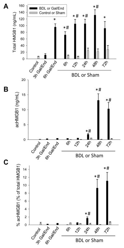
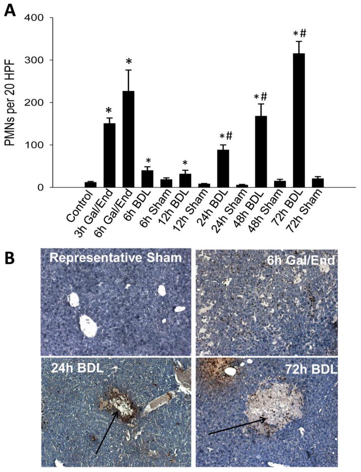
Cholestasis is a pathological common component of numerous liver diseases that results in hepatotoxicity, inflammation, and cirrhosis when untreated. While the predominant hypothesis in cholestatic liver injury remains hepatocyte apoptosis due to direct toxicity of hydrophobic bile acid exposure, recent work suggests that the injury occurs through inflammatory necrosis. In order to resolve this controversy, we used novel plasma biomarkers to assess the mechanisms of cell death during early cholestatic liver injury. C57Bl/6 mice underwent bile duct ligation (BDL) for 6-72 h, or sham operation. Another group of mice were given d-galactosamine and endotoxin as a positive control for apoptosis and inflammatory necrosis. Plasma levels of full length cytokeratin-18 (FL-K18), microRNA-122 (miR-122) and high mobility group box-1 protein (HMGB1) increased progressively after BDL with peak levels observed after 48 h. These results indicate extensive cell necrosis after BDL, which is supported by the time course of plasma alanine aminotransferase activities and histology. In contrast, plasma caspase-3 activity, cleaved caspase-3 protein and caspase-cleaved cytokeratin-18 fragments (cK18) were not elevated at any time during BDL suggesting the absence of apoptosis. In contrast, all plasma biomarkers of necrosis and apoptosis were elevated 6 h after Gal/End treatment. In addition, acetylated HMGB1, a marker for macrophage and monocyte activation, was increased as early as 12 h but mainly at 48-72 h. However, progressive neutrophil accumulation in the area of necrosis started at 6h after BDL. In conclusion, these data indicate that early cholestatic liver injury in mice is an inflammatory event, and occurs through necrosis with little evidence for apoptosis.
Keywords: ALT; Apoptosis; BDL; Bile duct ligation; Biomarkers; Cytokeratin-18; DAMP; FL-K18; GCDC; Gal/End; H&E; HMGB1; High mobility group box-1; ICAM-1; TNF-α; alanine aminotransferase; bile duct ligation; cK18; caspase-cleaved cytokeratin-18 fragment; damage associated molecular pattern; full length cytokeratin-18; galactosamine/endotoxin; glycochenodeoxycholate; hematoxylin and eosin; high mobility group box 1; intercellular adhesion molecule-1; miR-122; microRNA-122; tumor necrosis factor-alpha.
© 2013.
Conflict of interest statement
The authors declare no competing financial interest.
Figures
References
-
- Antoine DJ, Dear JW, Lewis PS, Platt V, Coyle J, Masson M, Thanacoody RH, Gray AJ, Webb DJ, Moggs JG, Bateman DN, Goldring CE, Park BK. Mechanistic biomarkers provide early and sensitive detection of acetaminophen-induced acute liver injury at first presentation to hospital. Hepatology. 2013;58:777–787. - PMC - PubMed
-
- Antoine DJ, Jenkins RE, Dear JW, Williams DP, McGill MR, Sharpe MR, Craig DG, Simpson KJ, Jaeschke H, Park BK. Molecular forms of HMGB1 and keratin-18 as mechanistic biomarkers for mode of cell death and prognosis during clinical acetaminophen hepatotoxicity. J Hepatol. 2012;56:1070–1079. - PMC - PubMed
-
- Antoine DJ, Williams DP, Kipar A, Jenkins RE, Regan SL, Sathish JG, Kitteringham NR, Park BK. High-mobility group box-1 protein and keratin-18, circulating serum proteins informative of acetaminophen-induced necrosis and apoptosis in vivo. Toxicol Sci. 2009;112:521–531. - PubMed
Publication types
MeSH terms
Substances
Grants and funding
- R01 DK070195/DK/NIDDK NIH HHS/United States
- R01 AA12916/AA/NIAAA NIH HHS/United States
- 5P20RR021940-07/RR/NCRR NIH HHS/United States
- P20 RR021940/RR/NCRR NIH HHS/United States
- G0700654/MRC_/Medical Research Council/United Kingdom
- 8 P20 GM103549-07/GM/NIGMS NIH HHS/United States
- T32 ES007079-26A2/ES/NIEHS NIH HHS/United States
- P20 GM12345/GM/NIGMS NIH HHS/United States
- Wellcome Trust/United Kingdom
- P20 GM103549/GM/NIGMS NIH HHS/United States
- T32 ES007079/ES/NIEHS NIH HHS/United States
- R01 AA012916/AA/NIAAA NIH HHS/United States
LinkOut - more resources
Full Text Sources
Other Literature Sources
Medical
Research Materials
