. 2013 Nov;20(11):1298-303.
doi: 10.1038/nsmb.2683. Epub 2013 Oct 6.

Structure of human mitochondrial RNA polymerase elongation complex

Affiliations

Structure of human mitochondrial RNA polymerase elongation complex

Kathrin Schwinghammer et al. Nat Struct Mol Biol. 2013 Nov.

Abstract

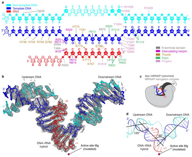
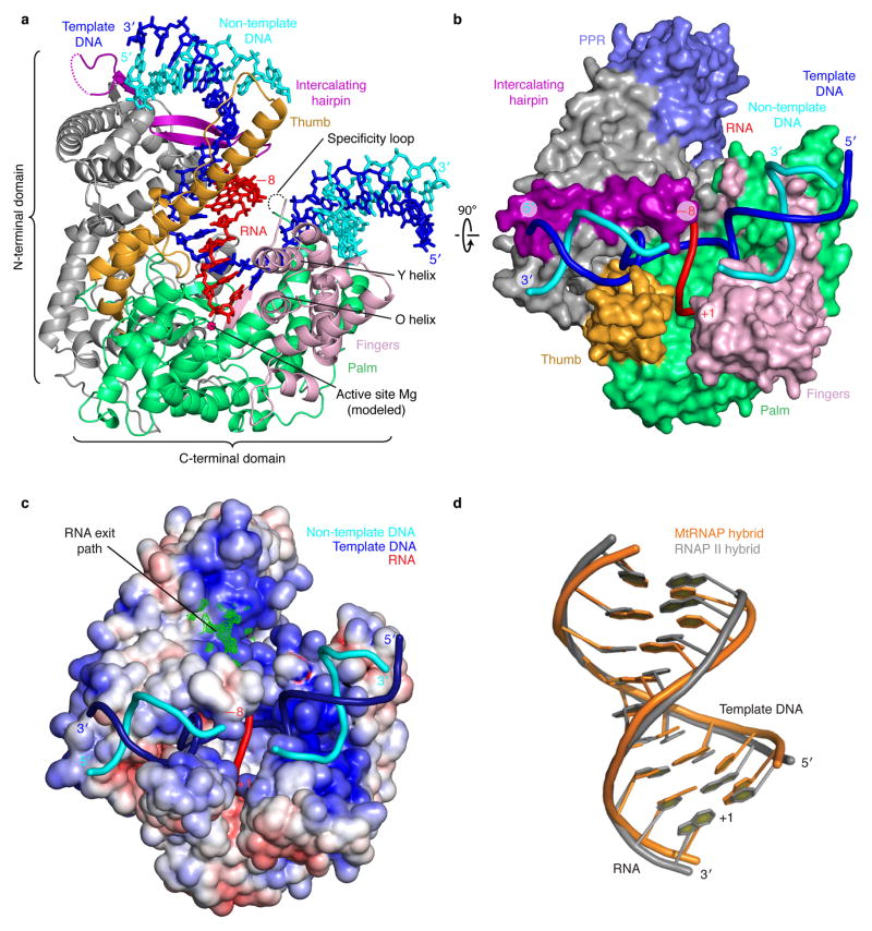
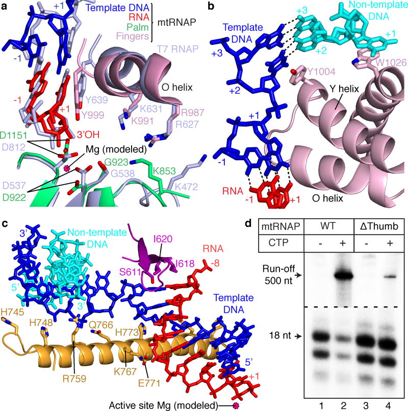
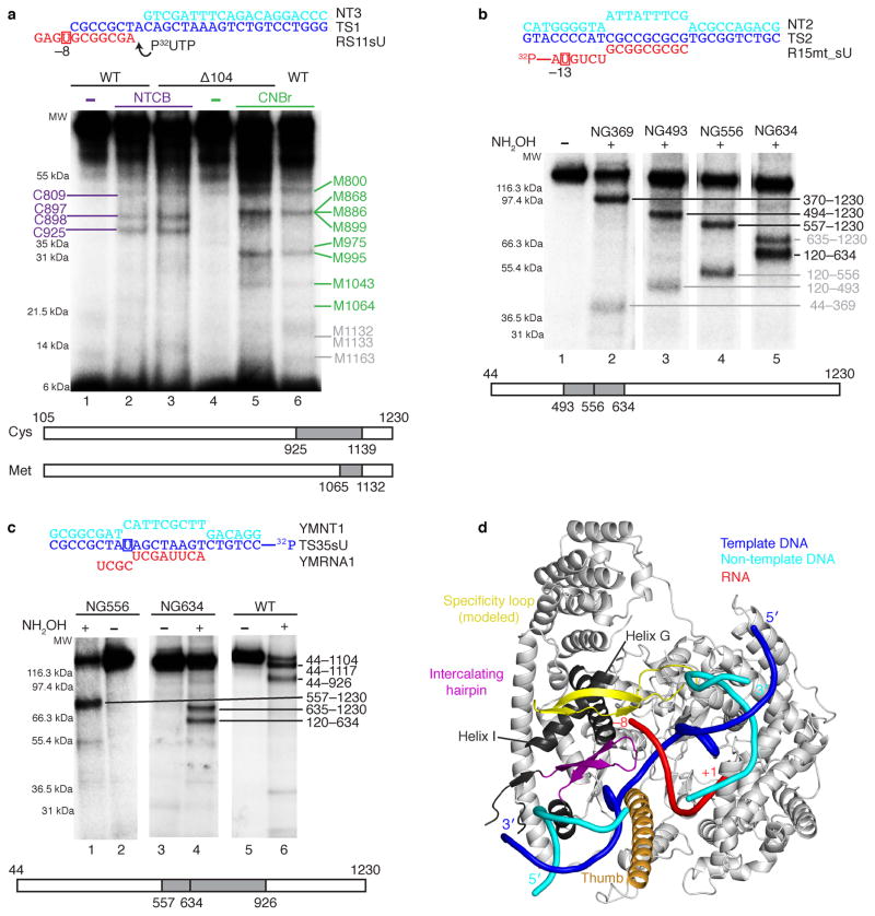
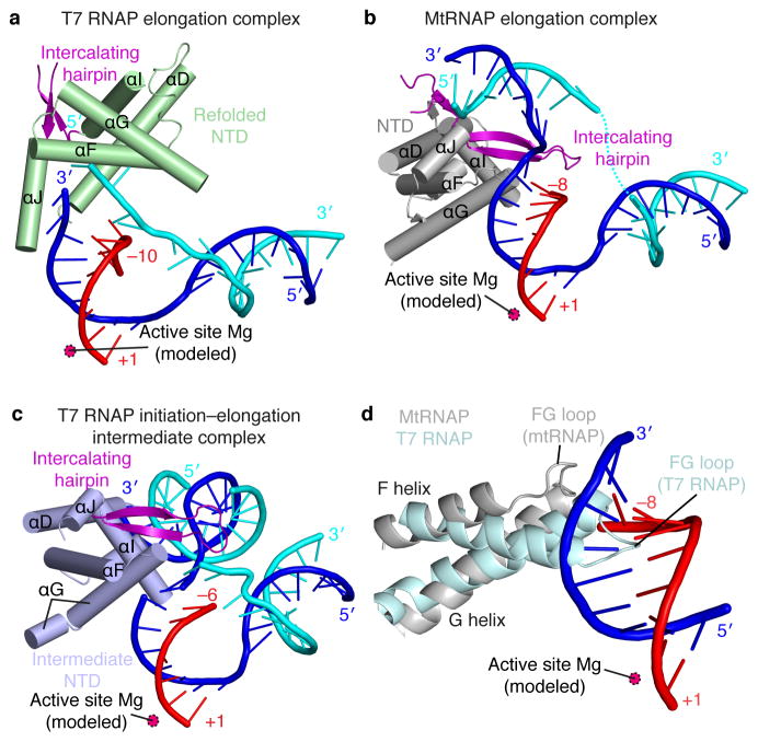
Here we report the crystal structure of the human mitochondrial RNA polymerase (mtRNAP) transcription elongation complex, determined at 2.65-Å resolution. The structure reveals a 9-bp hybrid formed between the DNA template and the RNA transcript and one turn of DNA both upstream and downstream of the hybrid. Comparisons with the distantly related RNA polymerase (RNAP) from bacteriophage T7 indicates conserved mechanisms for substrate binding and nucleotide incorporation but also strong mechanistic differences. Whereas T7 RNAP refolds during the transition from initiation to elongation, mtRNAP adopts an intermediary conformation that is capable of elongation without refolding. The intercalating hairpin that melts DNA during T7 RNAP initiation separates RNA from DNA during mtRNAP elongation. Newly synthesized RNA exits toward the pentatricopeptide repeat (PPR) domain, a unique feature of mtRNAP with conserved RNA-recognition motifs.


Conflict of interest statement

COMPETING FINANCIAL INTERESTS

The authors declare no competing financial interests.

Figures

Figure 1
Figure 1. Nucleic acid structure and mtRNAP interactions observed in the crystal structure
(a) Schematic overview of interactions between mtRNAP and nucleic acids. The nucleic acid scaffold contains template DNA (blue), non-template DNA (cyan) and RNA (red). Unfilled elements were not visible in the electron density map. Interactions with mtRNAP residues are indicated as lines (hydrogen bonds, ≤3.6 Å), dashed lines (electrostatic contacts, 3.6–4.2 Å), or arrows (stacking interactions). (b) Refined nucleic acid structure with 2Fo-Fc electron density omit map contoured at 1.5σ. (c) Polymerase opening from the clenched conformation of free mtRNAP (PDB 3SPA, dark grey) to the elongation complex (light grey). Structures were superimposed based on their NTDs. (d) Angles between duplex axes of upstream DNA, DNA–RNA hybrid, and downstream DNA.
Figure 2
Figure 2. Structure of mtRNAP elongation complex determined by x-ray crystallography
(a) Overview with mtRNAP depicted as a ribbon (thumb, orange; palm, green; fingers, pink; intercalating hairpin, purple), and nucleic acids as sticks (color code as in Fig. 1). A Mg2+ ion (magenta) was placed according to a T7 RNAP structure. PPR domain was omitted for clarity. (b) View of the structure rotated by 90° around a horizontal axis. The polymerase is depicted as a surface model and includes the PPR domain (slate). Nucleic acids are depicted as ribbons. (c) Electrostatic surface representation of the mtRNAP elongation complex with template DNA (blue), non-template DNA (cyan) and RNA (red). The Fo-Fc electron density of the mobile 5′-RNA tail is shown as a green mesh (contoured at 2.5 σ). (d) Superimposition of RNA–DNA hybrids in elongation complexes of mtRNAP (orange) and RNAP II (PDB 1I6H, grey).
Figure 3
Figure 3. Active center and nucleic acid strand separation observed in the crystal structure
(a) Conservation of active centers in mtRNAP (color code as in Figs. 1 and 2) and T7 RNAP (PDB 3E2E, light blue). Structures were superimposed based on their palm subdomains and selected residues were depicted as stick models. (b) Downstream DNA strand separation. (c) RNA separation from DNA at the upstream end of the hybrid and thumb–hybrid interactions. (d) Primer extension assays showed that a thumb subdomain plays a key role in elongation complex stability. Elongation complexes of WT (lanes 1 and 2) and Δthumb (lanes 3 and 4) mtRNAP variants were halted 18 nucleotides (nts) downstream of the light-strand promoter (LSP) by omitting cytidine triphosphate (CTP) (see also Supplementary Fig. 5a online).
Figure 4
Figure 4. Analysis of mtRNAP–nucleic acid contacts by cross-linking experiments (see also Supplementary Fig. 5b and c online)
(a) RNA nucleotide –9 cross-links to the specificity loop of mtRNAP. The cross-linked complexes were treated with 2-nitro-5-thiocyano-benzoic acid (NTCB, lanes 2 and 3) or cyanogen bromide (CNBr, lanes 5 and 6). Positions of the cysteine (cys) and methionine (met) residues that produced labeled peptides are indicated in purple and green, correspondingly. Grey numbers indicate methionine residues that did not produce labeled peptides and the expected migration of these peptides. (b) Mapping of the RNA–mtRNAP cross-link at RNA nucleotide –13 with different mtRNAP variants having a single hydroxylamin clevage site (NG) at a defined position. The cross-links were treated with hydroxylamine (NH2OH). The major cross-linked peptides are highlighted in black, minor (less than 10%) cross-linking sites in grey. (c) Mapping of the template strand DNA–mtRNAP cross-link at nucleotide –8. The cross-links were treated with NH2OH as described above. (d) Location of the cross-linked regions in mtRNAP elongation complex. The T7 RNAP specificity loop was built into the mRNAP structure by homology modeling. The structural elements that belong to the identified cross-linked regions and lie within 3–5 Å from the photo cross-linking probe include the modeled specificity loop (yellow, residues 1080–1108), part of the thumb (orange, residues 752–791) and part of the intercalating hairpin (purple, residues 605–623). Cross-linked regions that are not part of a defined structural element are shown in dark grey (e.g. helix G residues 587–571 and helix I residues 570–586).
Figure 5
Figure 5. Lack of NTD refolding upon mtRNAP elongation observed in the crystal structure
(a–c) Structures of the NTDs of T7 RNAP and mtRNAP. The NTD of T7 RNAP (a) is refolded in the elongation complex (PDB 1MSW), whereas the NTD of mtRNAP (b) is not, and resembles the NTD in the T7 intermediate (PDB 3E2E) (c). Helices are depicted as cylinders and nucleic acids as ribbons with sticks for protruding bases. (d) The FG loop of T7 RNAP (PDB 1QLN, pale cyan) protrudes into the hybrid-binding site but is shorter and positioned differently in mtRNAP (silver).

References

    1. Masters BS, Stohl LL, Clayton DA. Yeast mitochondrial RNA polymerase is homologous to those encoded by bacteriophages T3 and T7. Cell. 1987;51:89–99. - PubMed
    1. Mercer TR, et al. The human mitochondrial transcriptome. Cell. 2011;146:645–658. - PMC - PubMed
    1. Gaspari M, Larsson NG, Gustafsson CM. The transcription machinery in mammalian mitochondria. Biochim Biophys Acta. 2004;1659:148–152. - PubMed
    1. Cheetham GM, Steitz TA. Structure of a transcribing T7 RNA polymerase initiation complex. Science. 1999;286:2305–2309. - PubMed
    1. Ringel R, et al. Structure of human mitochondrial RNA polymerase. Nature. 2011;478:269–273. - PubMed

Publication types

Associated data

LinkOut - more resources