

Estradiol increases ER-negative breast cancer metastasis in an experimental model
- PMID: 24096710
- PMCID: PMC4151354
- DOI: 10.1007/s10585-012-9559-0
Estradiol increases ER-negative breast cancer metastasis in an experimental model
Abstract
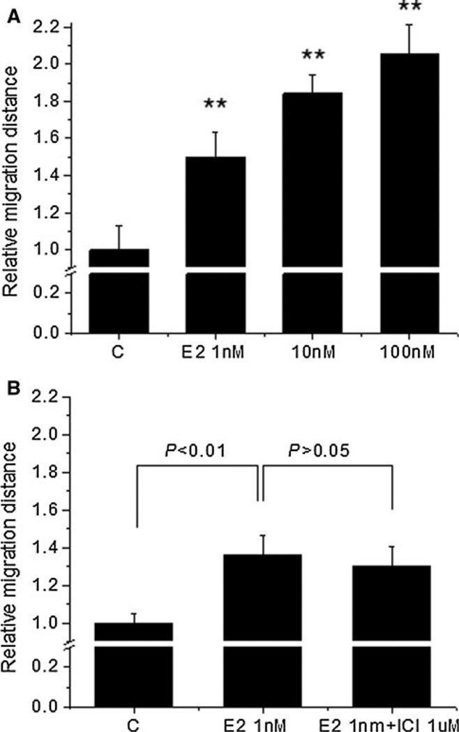
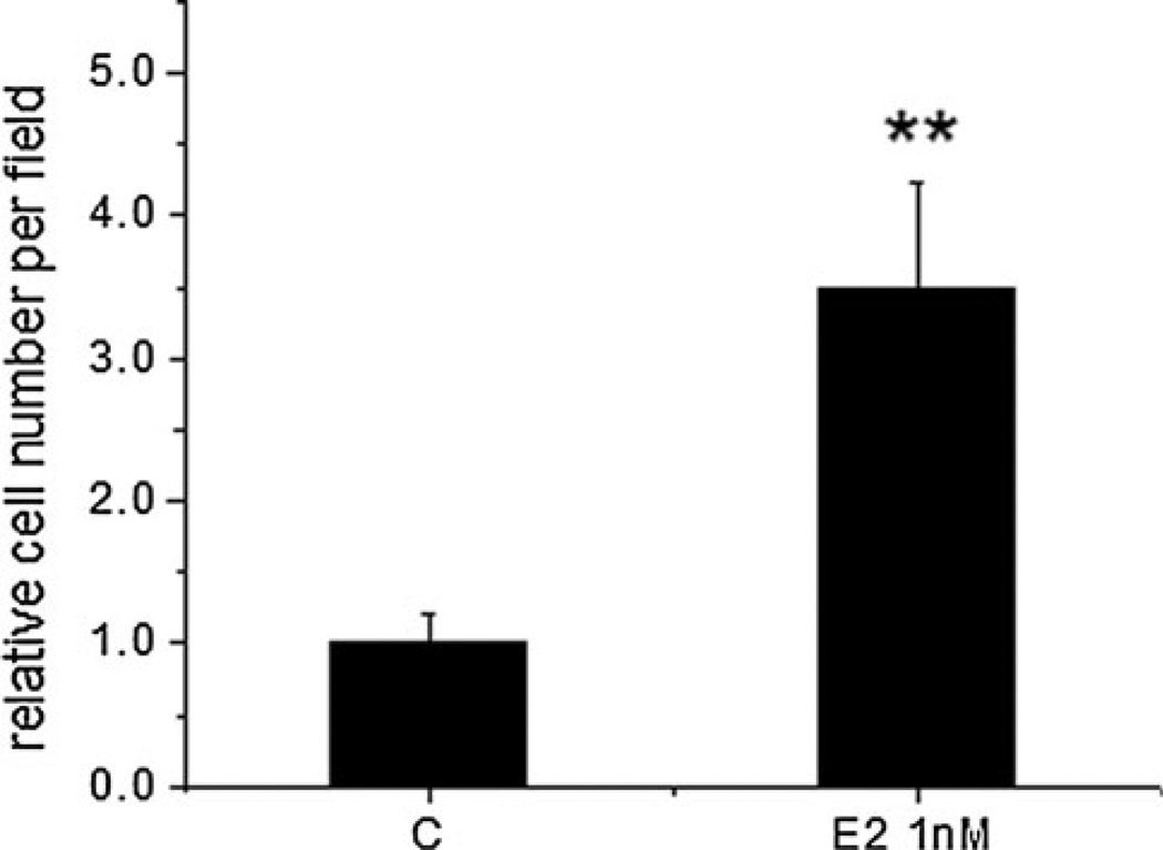
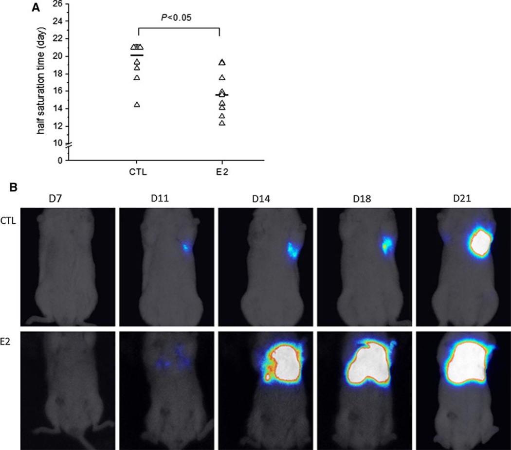
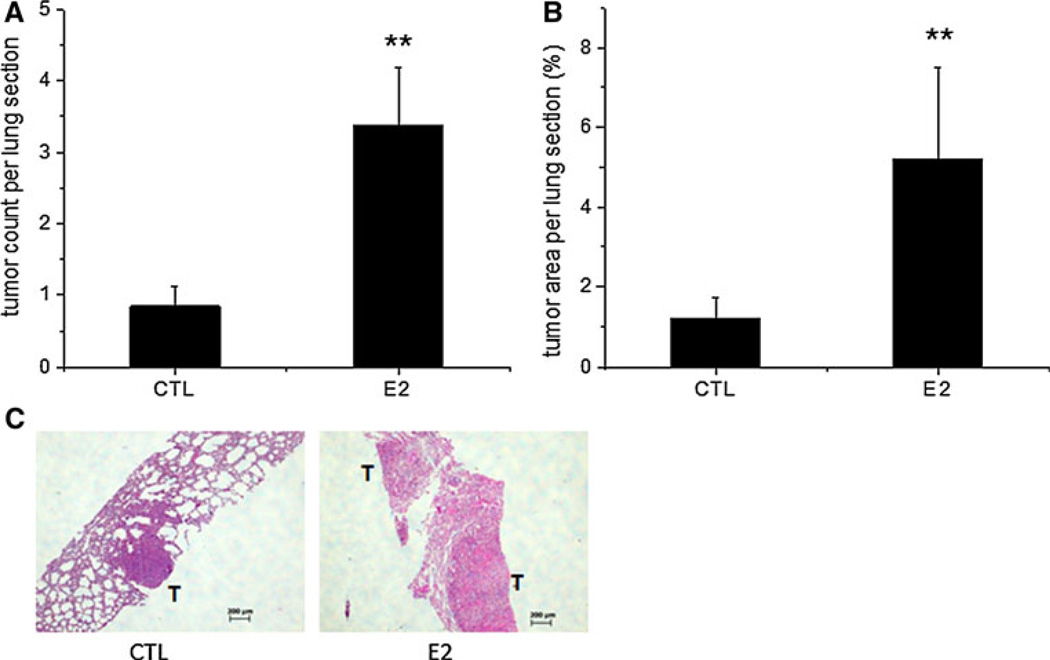
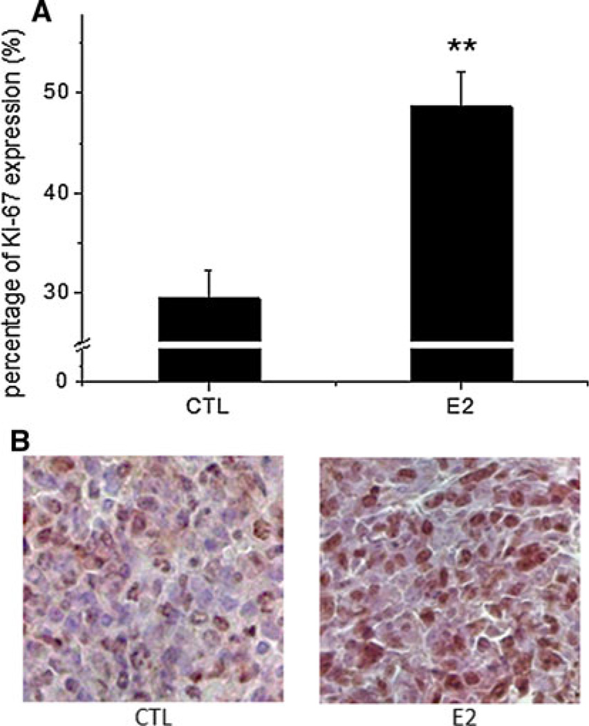
Breast cancer (BC) is the most common cancer affecting women in the United States and metastatic breast cancer is the leading cause of death. The role estradiol plays in ER-positive BC is well-documented, but the way it contributes to ER-negative BC remains unclear. In the present study, we utilized an experimental model of BC metastasis into lung by injecting ER-negative murine 4T1 cells into mice via the lateral tail vein. A 56 % metastasis occurrence rate following the injection of 5 × 10(3) cells was observed, thus this cell number was selected to study the potential stimulatory effect of estradiol on ER-negative BC metastasis. Female ovariectomized mice were randomized into estradiol and control groups with 16 mice per group, and estradiol pellets were implanted subcutaneously in the estradiol group. Results demonstrated that estradiol accelerated BC metastasis as indicated by bioluminescent imaging. In addition, estradiol enhanced metastatic tumor colony formation and increased the size of tumor nodules in the lungs, which were due, in part, to the increase in proliferative cells in the metastatic tumors. In vitro, estradiol increased the motility and invasion of 4T1 cells, and the stimulatory effect on cell motility was not blocked by ICI 182, 780, confirming that ER was not involved in the process. Results from the present study suggest that estradiol plays a role in ER-negative BC metastasis in whole animals.
Conflict of interest statement
Figures
Similar articles
-
Effects of letrozole on breast cancer micro-metastatic tumor growth in bone and lung in mice inoculated with murine 4T1 cells.Clin Exp Metastasis. 2016 Jun;33(5):475-85. doi: 10.1007/s10585-016-9792-z. Epub 2016 May 21. Clin Exp Metastasis. 2016. PMID: 27209469
-
Dietary soy isoflavones increase metastasis to lungs in an experimental model of breast cancer with bone micro-tumors.Clin Exp Metastasis. 2015 Apr;32(4):323-33. doi: 10.1007/s10585-015-9709-2. Epub 2015 Mar 8. Clin Exp Metastasis. 2015. PMID: 25749878 Free PMC article.
-
Dietary fat increases solid tumor growth and metastasis of 4T1 murine mammary carcinoma cells and mortality in obesity-resistant BALB/c mice.Breast Cancer Res. 2011 Aug 11;13(4):R78. doi: 10.1186/bcr2927. Breast Cancer Res. 2011. PMID: 21834963 Free PMC article.
-
Both estradiol and tamoxifen decrease proliferation and invasiveness of cancer cells transfected with a mutated estrogen receptor.J Steroid Biochem Mol Biol. 1997 Apr;61(1-2):11-7. doi: 10.1016/s0960-0760(96)00255-5. J Steroid Biochem Mol Biol. 1997. PMID: 9328205
-
[Estrogens, cathepsin D and metastasis in cancers of the breast and ovary: invasion or proliferation?].C R Seances Soc Biol Fil. 1998;192(2):241-51. C R Seances Soc Biol Fil. 1998. PMID: 9841098 Review. French.
Cited by
-
L-Arginine supplementation inhibits the growth of breast cancer by enhancing innate and adaptive immune responses mediated by suppression of MDSCs in vivo.BMC Cancer. 2016 Jun 1;16:343. doi: 10.1186/s12885-016-2376-0. BMC Cancer. 2016. PMID: 27246354 Free PMC article.
-
The complex nature of oestrogen signalling in breast cancer: enemy or ally?Biosci Rep. 2016 Jun 30;36(3):e00352. doi: 10.1042/BSR20160017. Print 2016 Jul. Biosci Rep. 2016. PMID: 27160081 Free PMC article. Review.
-
Time-dependent interactions of blood platelets and cancer cells, accompanied by extramedullary hematopoiesis, lead to increased platelet activation and reactivity in a mouse orthotopic model of breast cancer - implications for pulmonary and liver metastasis.Aging (Albany NY). 2020 Mar 19;12(6):5091-5120. doi: 10.18632/aging.102933. Epub 2020 Mar 19. Aging (Albany NY). 2020. PMID: 32191918 Free PMC article.
-
Estrogen-induced SDF-1α production promotes the progression of ER-negative breast cancer via the accumulation of MDSCs in the tumor microenvironment.Sci Rep. 2016 Dec 20;6:39541. doi: 10.1038/srep39541. Sci Rep. 2016. PMID: 27996037 Free PMC article.
-
Unfavorable effect of calcitriol and its low-calcemic analogs on metastasis of 4T1 mouse mammary gland cancer.Int J Oncol. 2018 Jan;52(1):103-126. doi: 10.3892/ijo.2017.4185. Epub 2017 Nov 2. Int J Oncol. 2018. PMID: 29115583 Free PMC article.
References
-
- Hagemeister FB, Jr, et al. Causes of death in breast cancer: a clinicopathologic study. Cancer. 1980;46(1):162–167. - PubMed
-
- Jemal A, et al. Cancer statistics, 2003. CA Cancer J Clin. 2003;53(1):5–26. - PubMed
-
- Rosen PR, et al. A long-term follow-up study of survival in stage I (T1N0M0) and stage II (T1N1M0) breast carcinoma. J Clin Oncol. 1989;7(3):355–366. - PubMed
Publication types
MeSH terms
Substances
Grants and funding
LinkOut - more resources
Full Text Sources
Other Literature Sources
