Review
. 2013 Nov;47(17):1105-11.
doi: 10.1136/bjsports-2013-092835. Epub 2013 Oct 4.

Sports-related extensor carpi ulnaris pathology: a review of functional anatomy, sports injury and management

Affiliations
Free PMC article
Review

Sports-related extensor carpi ulnaris pathology: a review of functional anatomy, sports injury and management

Doug Campbell et al. Br J Sports Med. 2013 Nov.
Free PMC article

Abstract

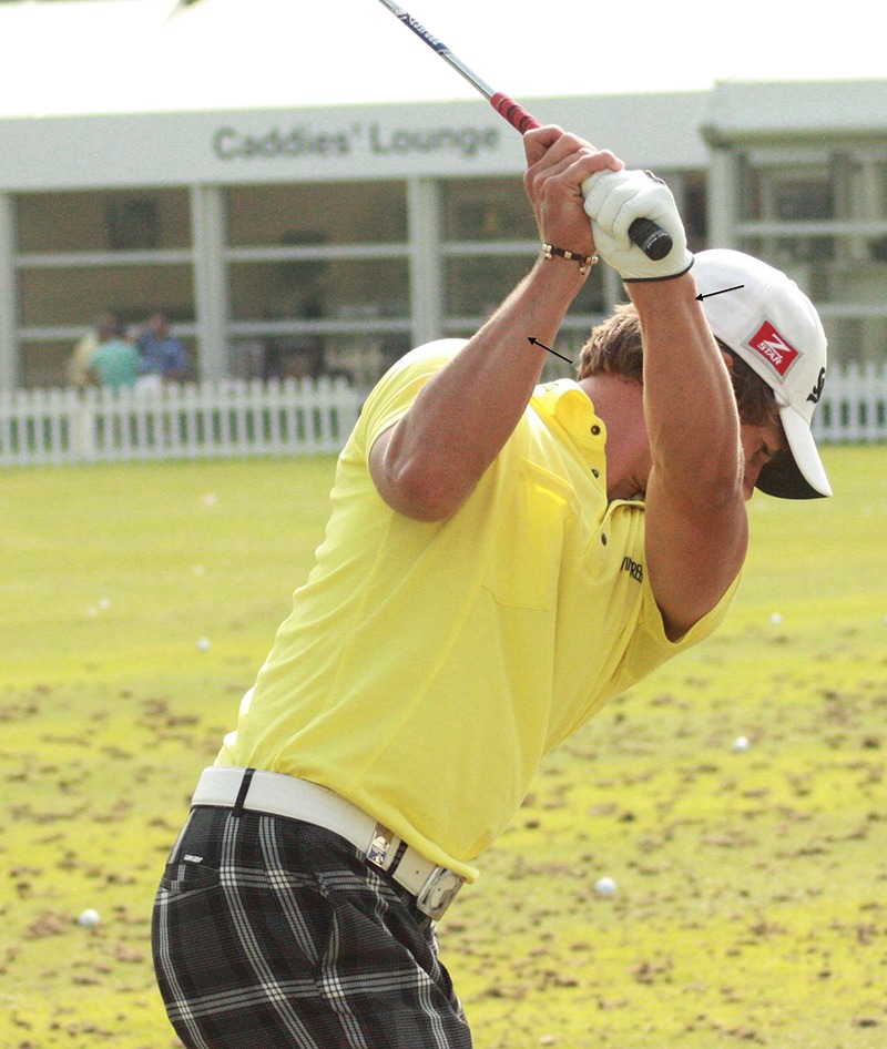
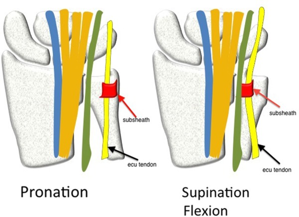
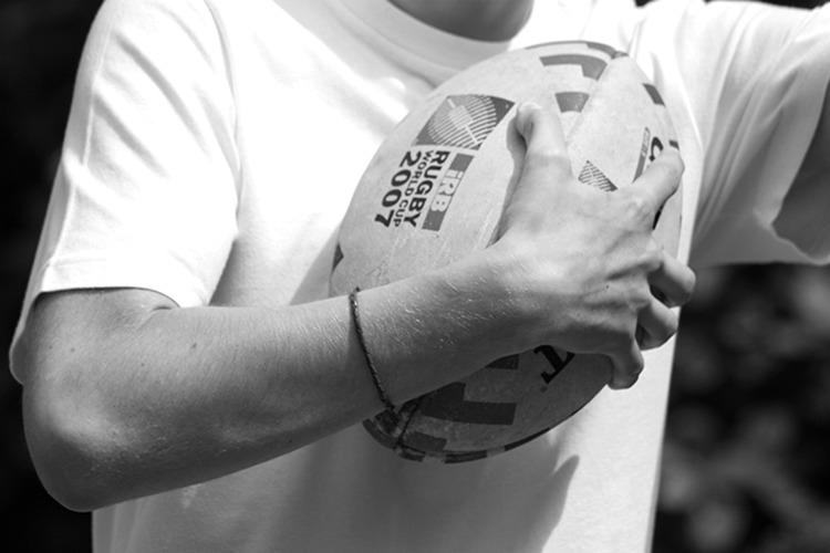
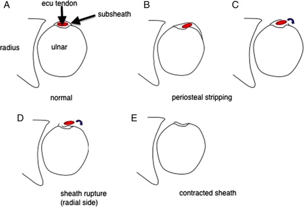
The extensor carpi ulnaris (ECU) muscle plays a key role not only in the active movements of wrist extension and ulnar deviation but also in providing stability to the ulnar side of the wrist. Its position relative to the other structures in the wrist changes with forearm pronation and supination. As such, it must be mobile yet stable. The ECU tendon relies on specific stabilising structures to hold it in the correct positions to perform its different functions. These structures can be injured in a variety of different athletic activities such as tennis, golf and rugby league, yet their injury and disruption is predictable when the mechanics of the ECU and the techniques of the sport are understood. The ECU tendon is also vulnerable to tendon pathologies other than instability. It lies subcutaneously and is easily palpated and visualised with diagnostic ultrasound, allowing early diagnosis and management of its specific conditions. Treatment includes rest, splintage and surgery with each modality having specific indications and recognised outcomes. This review described the functional anatomy in relevant sporting situations and explained how problems occur as well as when and how to intervene.

Keywords: Sporting Injuries; Tendons; Ultrasound; Wrist Injuries.


Figures

Figure 1
Figure 1
Extensor carpi ulnaris muscle seen on the ulnar side of both of this golfer's wrists.
Figure 2
Figure 2
Diagrammatic representation of the change of position of the extensor carpi ulnaris tendon between pronation and supination. There is an angulation of the tendon as it exits the subsheath in supination and flexion.
Figure 3
Figure 3
The vulnerable extensor carpi ulnaris tendon in supination, flexion and ulnar deviation in a rugby player.
Figure 4
Figure 4
(Subsheath disruption) The types of subsheath rupture comprise (A) normal, (B) periosteal stripping resulting in a false sheath, (C) fibro-osseous sheath rupture at the ulnar side, (D) fibro-osseous sheath rupture at the radial side and (E) a contracted but intact fibro-osseous sheath.
Figure 5
Figure 5
Axial ultrasound images of the normal extensor carpi ulnaris (ECU) tendon. In wrist pronation (A) the tendon lies within the ulnar groove (white arrows). The subsheath (black arrows) is immediately superficial to the tendon and attaches to the ulna. Distal to the ulna (B) the tendon (curved white arrow) lies superficial to the meniscal homologue of the triangular fibrocartilage complex (asterix) and the triquetrum. The extensor retinaculum (broken black arrows) displays hyper-reflective and hyporeflective properties due to the effects of anisotropy. In wrist supination (C) the tendon (curved white arrow) moves to the ulnar aspect of the groove (white arrows), and a small area of echo bright fatty tissue lies in the radial aspect of the groove (black asterisk). The tendons of the fourth and fifth compartments (broken white arrows) now lie in closer relation to the ECU tendon.
Figure 6
Figure 6
Longitudinal ultrasound image (A) of extensor carpi ulnaris tenosynovitis with prominent areas of anechoic fluid in the tendon sheath (white arrows). The tendon is normal. There is only minor inflammatory change in the tendon sheath on axial power Doppler imaging (B). (Reproduced from Imaging of Pain, Waldman SD & Campbell RSD, Ch 129 Extensor Carpi Ulnaris, p330, Copyright 2011, with permission from Elsevier.)
Figure 7
Figure 7
Axial fat saturated T2-weighted image of the wrist. There is a linear area of high signal intensity within the extensor carpi ulnaris tendon (white arrow) representing a longitudinal partial cleft tear.
Figure 8
Figure 8
Axial T2 fat saturated MRI of the wrist in a rugby league player following an acute extensor carpi ulnaris subsheath injury. The tendon (white arrow) is subluxed in an ulnar direction and the subsheath is torn at its radial insertion on the ulna (black arrow). There is associated marrow oedema in the head of the ulna (curved white arrow). There were associated injuries including an acute triangular fibrocartilage tear, and there is an effusion in the distal radioulnar joint.
Figure 9
Figure 9
Axial fat saturated T2-weighted images of the wrist following a subsheath injury. In supination (A), the ECU tendon is subluxed and lies in the ulnar portion of the bony groove. This position is fixed and the tendon does not return to it normal position in pronation (B). There is also marrow oedema within the head of the ulna indicating the acute nature of the subsheath injury (white arrows).

Comment in

References

    1. Brigstocke G, Hearnden A, Holt CA, et al. The functional range of movement of the human wrist. J Hand Surg Eur Vol 2013;38:554–6 - PubMed
    1. Brigstocke GH, Hearnden A, Holt C, et al. In-vivo confirmation of the use of the dart thrower's motion during activities of daily living. J Hand Surg Eur Vol 2012. [Epub ahead of print] - PubMed
    1. Horii E, An KN, Linscheid RL. Excursion of prime wrist tendons. J Hand Surg [Am] 1993;18:83–90 - PubMed
    1. Hajj AA, Wood MB. Stenosing tenosynovitis of the extensor carpi ulnaris. J Hand Surg [Am] 1986;11:519–20 - PubMed
    1. Cook JL, Purdam CR. Is tendon pathology a continuum? A pathology model to explain the clinical presentation of load-induced tendinopathy. Br J Sports Med 2009;43:409–16 - PubMed

Publication types

LinkOut - more resources