Review
. 2013 Dec;12(12):1578-87.
doi: 10.1128/EC.00213-13. Epub 2013 Oct 4.

Strategies to discover the structural components of cyst and oocyst walls

Affiliations
Review

Strategies to discover the structural components of cyst and oocyst walls

John Samuelson et al. Eukaryot Cell. 2013 Dec.

Abstract

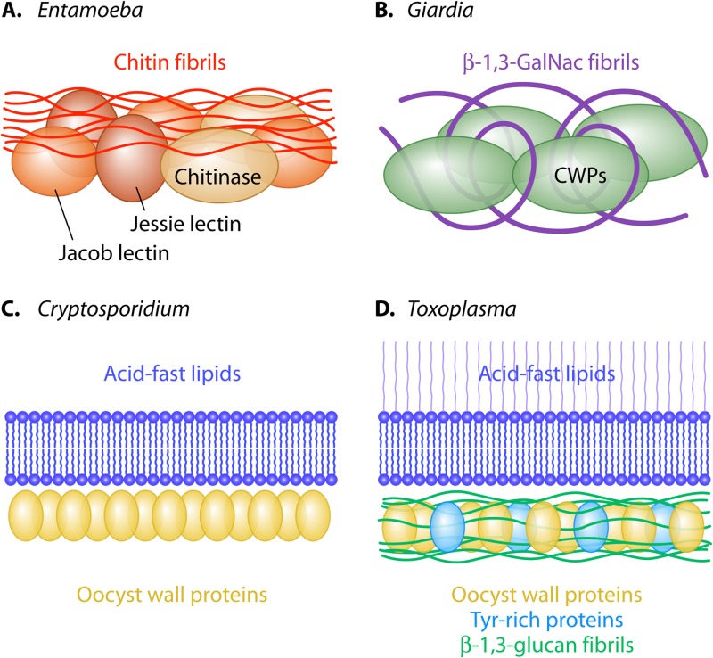
Cysts of Giardia lamblia and Entamoeba histolytica and oocysts of Toxoplasma gondii and Cryptosporidium parvum are the infectious and sometimes diagnostic forms of these parasites. To discover the structural components of cyst and oocyst walls, we have developed strategies based upon a few simple assumptions. Briefly, the most abundant wall proteins are identified by monoclonal antibodies or mass spectrometry. Structural components include a sugar polysaccharide (chitin for Entamoeba, β-1,3-linked glucose for Toxoplasma, and β-1,3-linked GalNAc for Giardia) and/or acid-fast lipids (Toxoplasma and Cryptosporidium). Because Entamoeba cysts and Toxoplasma oocysts are difficult to obtain, studies of walls of nonhuman pathogens (E. invadens and Eimeria, respectively) accelerate discovery. Biochemical methods to dissect fungal walls work well for cyst and oocyst walls, although the results are often unexpected. For example, echinocandins, which inhibit glucan synthases and kill fungi, arrest the development of oocyst walls and block their release into the intestinal lumen. Candida walls are coated with mannans, while Entamoeba cysts are coated in a dextran-like glucose polymer. Models for cyst and oocyst walls derive from their structural components and organization within the wall. Cyst walls are composed of chitin fibrils and lectins that bind chitin (Entamoeba) or fibrils of the β-1,3-GalNAc polymer and lectins that bind the polymer (Giardia). Oocyst walls of Toxoplasma have two distinct layers that resemble those of fungi (β-1,3-glucan in the inner layer) or mycobacteria (acid-fast lipids in the outer layer). Oocyst walls of Cryptosporidium have a rigid bilayer of acid-fast lipids and inner layer of oocyst wall proteins.


Figures

Fig 1
Fig 1
Cyst and oocyst walls of human pathogens contain a structural sugar polymer (chitin, β-1,3-GalNAc, β-1,3-glucan, or cellulose) and/or acid-fast lipids. Saccharomyces walls contain chitin and β-1,3-glucan. Toxoplasma has three walled forms (oocyst, sporocyst, and tissue cyst). The structural component(s) of tissue cysts of Toxoplasma has not been identified. Bradyzoites are not shown within Toxoplasma tissue cysts. No attempt was made to draw structures to scale.
Fig 2
Fig 2
Fluorescence and transmission electron micrographs (TEMs) show important structural features of cyst walls of Entamoeba and Giardia and oocyst walls of Cryptosporidium and Toxoplasma. (A) Antibodies (red) show Jessie lectins bound to chitin fibrils of the Entamoeba cyst wall. (B) Antibodies (green) show CWP1 in a shard of the cyst wall of Giardia broken by sonication. (C) Carbol fuchsin (red) stains acid-fast lipids in Cryptosporidium oocyst walls. (D) Dityrosines (blue) in oocyst wall (white arrow) and sporocyst walls (blue arrows) of Toxoplasma are autofluorescent in the UV channel. (E) Three Entamoeba cyst walls isolated by centrifugation have uniform thickness and appearance. (F) Recombinant CWP1 binds to fibrils of β-1,3-GalNAc in cyst walls of Giardia deproteinated with NaOH. (G) The Cryptosporidium oocyst wall has a fibrillar glycocalyx (G), a bilayer (Bil), an inner layer containing oocyst wall proteins (OWPs), and tethers (T) that appear globular after they have broken. (H) Recombinant dectin-1 (red) binds to the oocyst wall (white arrow) but not to sporocyst walls of Toxoplasma (same oocyst as shown in panel D). (I) Recombinant Jessie lectin of Entamoeba self-aggregates and forms a biofilm composed of branched fibrils. (J) Deproteinated cyst walls of Giardia form a hollow sphere of curled fibrils of β-1,3-GalNAc. (K) A sonicated and pronase-treated wall of Cryptosporidium contains a rigid bilayer (Bil) and nothing else. (L) A sonicated Eimeria oocyst wall has an inner layer, which is a porous scaffold of fibrils of β-1,3-glucan. Micrographs of Entamoeba are reprinted from references (A and I) and (E), of Giardia from reference (F), of Cryptosporidium from references (G and K) and (C), and of Toxoplasma from reference (D, H, and L). Panels B and J are original here.
Fig 3
Fig 3
Venn diagrams show strategies to identify proteins that are common to oocyst walls of all coccidia (Cryptosporidium, Toxoplasma, and Eimeria) (purple) (oocyst wall proteins [OWPs], polyketide synthases [PKSs], O-GalNAc transferases [GalNAcTs], and N-glycans with Man5GlcNac2) but absent in hemoparasites (green). There are also proteins specific to Cryptosoporidium (blue) (possible oocyst wall proteins [POWPs]) or to Toxoplasma and Eimeria (red), which make similar oocysts with two walled forms.
Fig 4
Fig 4
Lectins present in cyst and oocyst walls (8). (A) Entamoeba and Saccharomyces chitinases have catalytic domains that share common ancestry (purple), but they have chitin-binding domains (ChBD) that do not share common ancestry and are at different ends of the lectins (24, 53). The Entamoeba chitin-binding domain (orange) is also present at the N terminus of the Jessie lectin, which has a self-aggregating or daub domain (khaki) (6, 50). (B) Entamoeba Jacob lectins have multiple chitin-binding domains (gray), which are distinct from those of the amebic chitinase and Jessie lectin (24, 52). The spacer (pink) has Ser- and Thr-rich regions, which contain O-phosphodiester-linked glycans (51). Acanthamoeba proteins with three cellulose-binding domains (CeBD) have a structure similar to that of the Entamoeba Jacob lectins (36). (C) Giardia cyst wall proteins (CWP1 to CWP3) have a Leu-rich repeat (blue-green) that binds fibrils of the β-1,3-GalNAc polymer (7). The Cys-rich region (CRR in pink) is necessary for binding to the wall (70). (D) Toxoplasma and Schizosaccharomyces glucan hydrolases have catalytic domains that share common ancestry (dark blue), but they have glucan-binding domains (GBD) that do not share common ancestry and are at different ends of the lectins (9).
Fig 5
Fig 5
Models for cyst walls of Entamoeba and Giardia and for oocyst walls of Cryptosporidium and Toxoplasma. (A) The Entamoeba cyst wall contains chitin fibrils and three chitin-binding lectins (chitinase, Jessie, and Jacob) (, , , , , –54). (B) The Giardia cyst wall contains cyst wall proteins (CWPs), which are lectins that bind to curled fibrils of the β-1,3-GalNAc polymer (7, 19, 25, 29). (C) The Cryptosporidium oocyst wall contains a layer of acid-fast lipids and a layer of oocyst wall proteins (OWPs) (10, 11). (D) The Toxoplasma oocyst wall contains a layer of acid-fast lipids and a layer that contains fibrils of β-1,3-glucan, oocyst wall proteins, and Tyr-rich proteins that form dityrosines (9, 11, 16, 21).

References

    1. Baldursson S, Karanis P. 2011. Waterborne transmission of protozoan parasites: review of worldwide outbreaks—an update 2004–2010. Water Res. 45:6603–6614 - PubMed
    1. Esch KJ, Petersen CA. 2013. Transmission and epidemiology of zoonotic protozoal diseases of companion animals. Clin. Microbiol. Rev. 26:58–85 - PMC - PubMed
    1. Ximénez C, Morán P, Rojas L, Valadez A, Gómez A. 2009. Reassessment of the epidemiology of amebiasis: state of the art. Infect. Genet. Evol. 9:1023–1032 - PubMed
    1. Elmore SA, Jones JL, Conrad PA, Patton S, Lindsay DS, Dubey JP. 2010. Toxoplasma gondii: epidemiology, feline clinical aspects, and prevention. Trends Parasitol. 26:190–196 - PubMed
    1. Kotloff KL, Nataro JP, Blackwelder WC, Nasrin D, Farag TH, Panchalingam S, Wu Y, Sow SO, Sur D, Breiman RF, Faruque AS, Zaidi AK, Saha D, Alonso PL, Tamboura B, Sanogo D, Onwuchekwa U, Manna B, Ramamurthy T, Kanungo S, Ochieng JB, Omore R, Oundo JO, Hossain A, Das SK, Ahmed S, Qureshi S, Quadri F, Adegbola RA, Antonio M, Hossain MJ, Akinsola A, Mandomando I, Nhampossa T, Acacio S, Biswas K, O'Reilly CE, Mintz ED, Berkeley LY, Muhsen K, Sommerfelt H, Robins-Browne RM, Levine MM. 2013. Burden and aetiology of diarrhoeal disease in infants and young children in developing countries (the Global Enteric Multicenter Study, GEMS): a prospective, case-control study. Lancet 382:209–222 - PubMed

Publication types

LinkOut - more resources