. 2013 Oct 7:3:2880.
doi: 10.1038/srep02880.

Mechanism of amyloid β-protein dimerization determined using single-molecule AFM force spectroscopy

Affiliations

Mechanism of amyloid β-protein dimerization determined using single-molecule AFM force spectroscopy

Zhengjian Lv et al. Sci Rep. .

Abstract

Aβ42 and Aβ40 are the two primary alloforms of human amyloid β-protein (Aβ). The two additional C-terminal residues of Aβ42 result in elevated neurotoxicity compared with Aβ40, but the molecular mechanism underlying this effect remains unclear. Here, we used single-molecule force microscopy to characterize interpeptide interactions for Aβ42 and Aβ40 and corresponding mutants. We discovered a dramatic difference in the interaction patterns of Aβ42 and Aβ40 monomers within dimers. Although the sequence difference between the two peptides is at the C-termini, the N-terminal segment plays a key role in the peptide interaction in the dimers. This is an unexpected finding as N-terminal was considered as disordered segment with no effect on the Aβ peptide aggregation. These novel properties of Aβ proteins suggests that the stabilization of N-terminal interactions is a switch in redirecting of amyloids form the neurotoxic aggregation pathway, opening a novel avenue for the disease preventions and treatments.


Figures

Figure 1
Figure 1. Schematic of the experimental setup.
(a) One Aβ molecule immobilized on the mica substrate is picked up by another Aβ molecule functionalized on the AFM tip. Putative interaction segments within a dimer are highlighted in pink bands. A rupture event occurs when a misfolded dimer is torn apart. For clarity, only one linker molecule is drawn on AFM tip and substrate, and the sizes of objects in the scheme are not to scale. N and C denote N−terminus and C−terminus, respectively. (b) Typical force−distance curve that shows either no interactions (black) or specific interactions (red). In the case of specific interactions, breaking a misfolded Aβ dimer resulted in a rupture distance at ~ 48 nm and a rupture force at ~ 73 pN. A vertical black arrow indicates the rupture position. For clarity, only the retraction curves are shown. Y offset was performed with the red line but not with the black line. A typical histogram of rupture force distribution at a loading rate of 6215 pN/s for Aβ40 (c). The most probable rupture force for Aβ40 was 57.0 pN. A typical histogram of rupture force distribution at a loading rate of 6068 pN/s for [VPV]Aβ40 (d). The most probable rupture force for [VPV]Aβ40 was 83.6 pN. The solid lines represent the fit of probability density function.
Figure 2
Figure 2. Representative dynamic force spectra of Aβ40.
The solid red line represents the results fit with the Bell−Evans model. The obtained energy profile parameters are summarized in Table S1. The inset shows the reconstructed energy landscapes of misfolded Aβ dimers. Error bars represent ± SEM.
Figure 3
Figure 3. The distributions of contour length (a) and rupture force (b) at loading rates of 5000−7000 pN/s for Aβ40.
The contour length distribution showed a major data cluster at ~ 33 nm. The most probable rupture force was 63.4 ± 3.2 pN. The scatter distribution of rupture forces with varying contour lengths is shown in (c). The total number of data was 170. Error is shown by ± SEM.
Figure 4
Figure 4. The distributions of contour lengths (a) and rupture forces (b) at loading rates of 5000−7000 pN/s for [VPV]Aβ40.
The contour length distribution showed a major data cluster at ~ 42 and a comparable cluster at ~ 57 nm. The most probable rupture force was 79.3 ± 1.5 pN. The scatter distribution of rupture forces with varying contour lengths is shown in (c). The total number of data was 280. Error is shown by ± SEM.
Figure 5
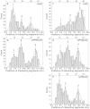
Figure 5. The contour length distributions for Aβ40, [VPV]Aβ40, Aβ42, [VPV]Aβ42, and [pP]Aβ42 dimers are shown in (a), (b), (c), (d) and (e), respectively.
The interaction segments are aligned according to the contour length distribution. The dotted lines denote the distribution profiles and peaks are approximated according to the profiles. Notably, there are some uncertainties in length estimation. The arrows indicate interaction segments.

Similar articles

Cited by

References

    1. Chiti F. & Dobson C. M. Protein misfolding, functional amyloid, and human disease. Annu Rev Biochem 75, 333–366 (2006). - PubMed
    1. Roychaudhuri R., Yang M., Hoshi M. M. & Teplow D. B. Amyloid beta-protein assembly and Alzheimer disease. J Biol Chem 284, 4749–4753 (2009). - PMC - PubMed
    1. Benilova I., Karran E. & De Strooper B. The toxic Abeta oligomer and Alzheimer's disease: an emperor in need of clothes. Nat Neurosci 15, 349–357 (2012). - PubMed
    1. De Strooper B., Vassar R. & Golde T. The secretases: enzymes with therapeutic potential in Alzheimer disease. Nat Rev Neurol 6, 99–107 (2010). - PMC - PubMed
    1. Karran E., Mercken M. & De Strooper B. The amyloid cascade hypothesis for Alzheimer's disease: an appraisal for the development of therapeutics. Nat Rev Drug Discov 10, 698–712 (2011). - PubMed

Publication types

Substances