. 2013 Nov;16(11):1576-87.
doi: 10.1038/nn.3541. Epub 2013 Oct 6.

Non-epithelial stem cells and cortical interneuron production in the human ganglionic eminences

Affiliations

Non-epithelial stem cells and cortical interneuron production in the human ganglionic eminences

David V Hansen et al. Nat Neurosci. 2013 Nov.

Abstract

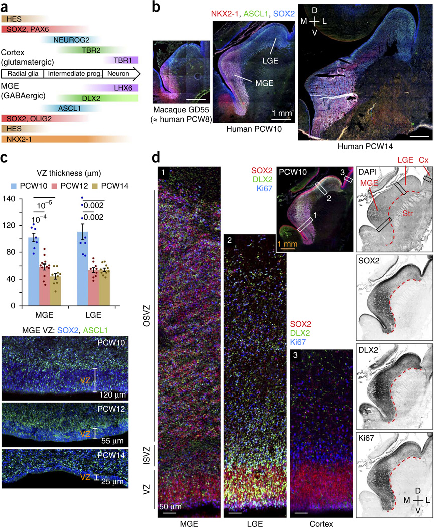
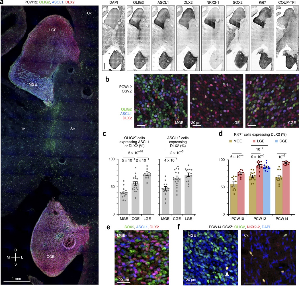
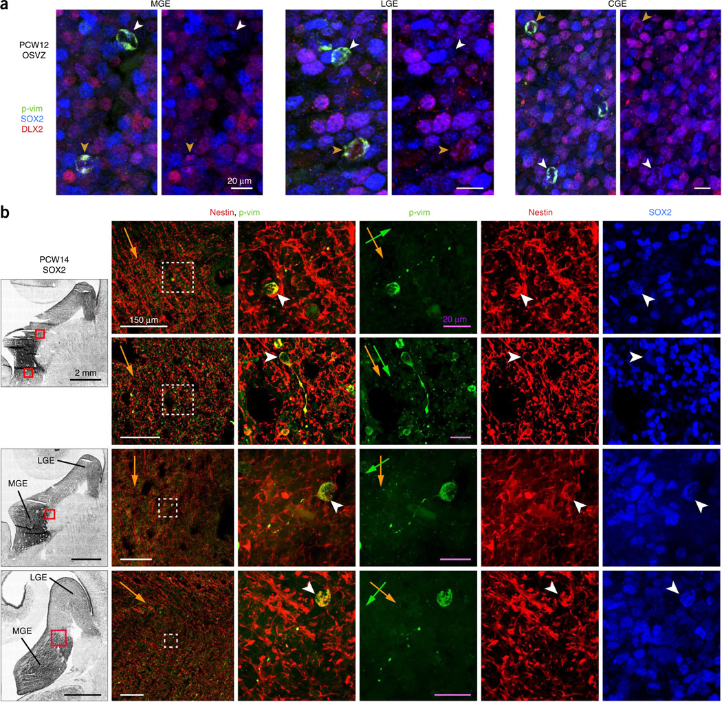
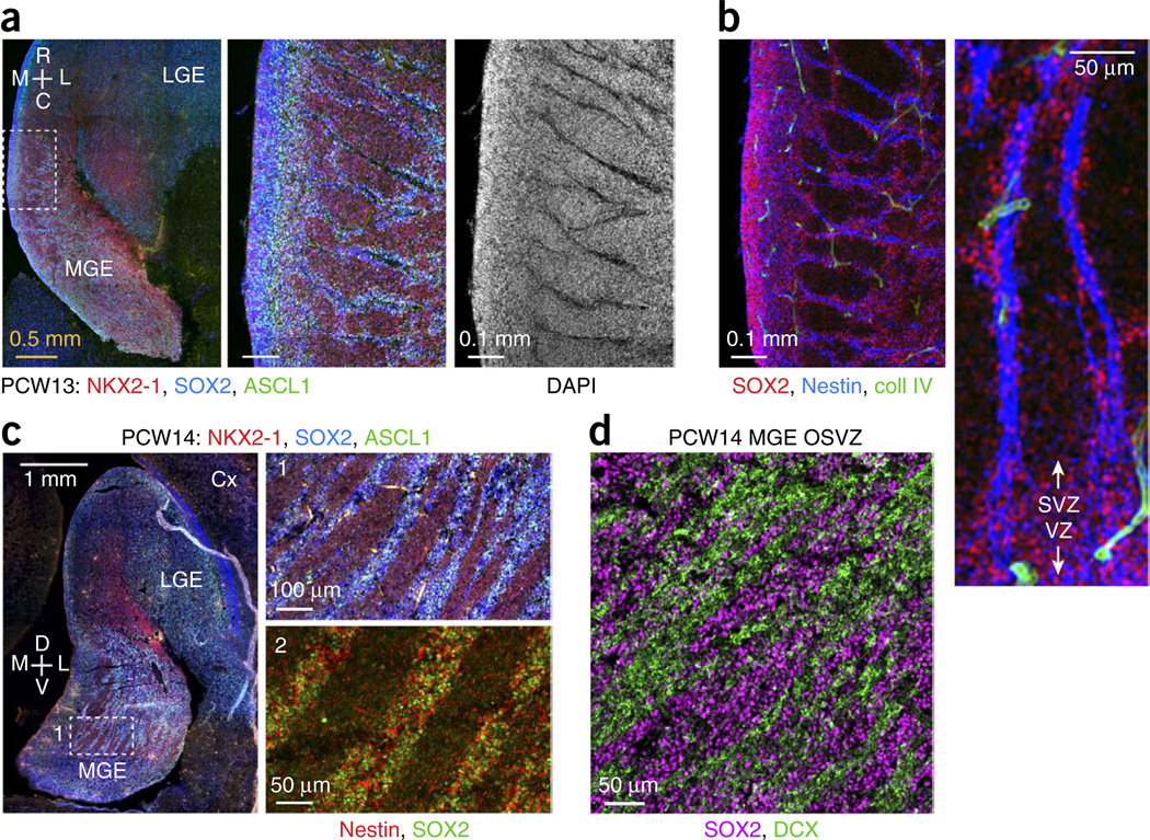
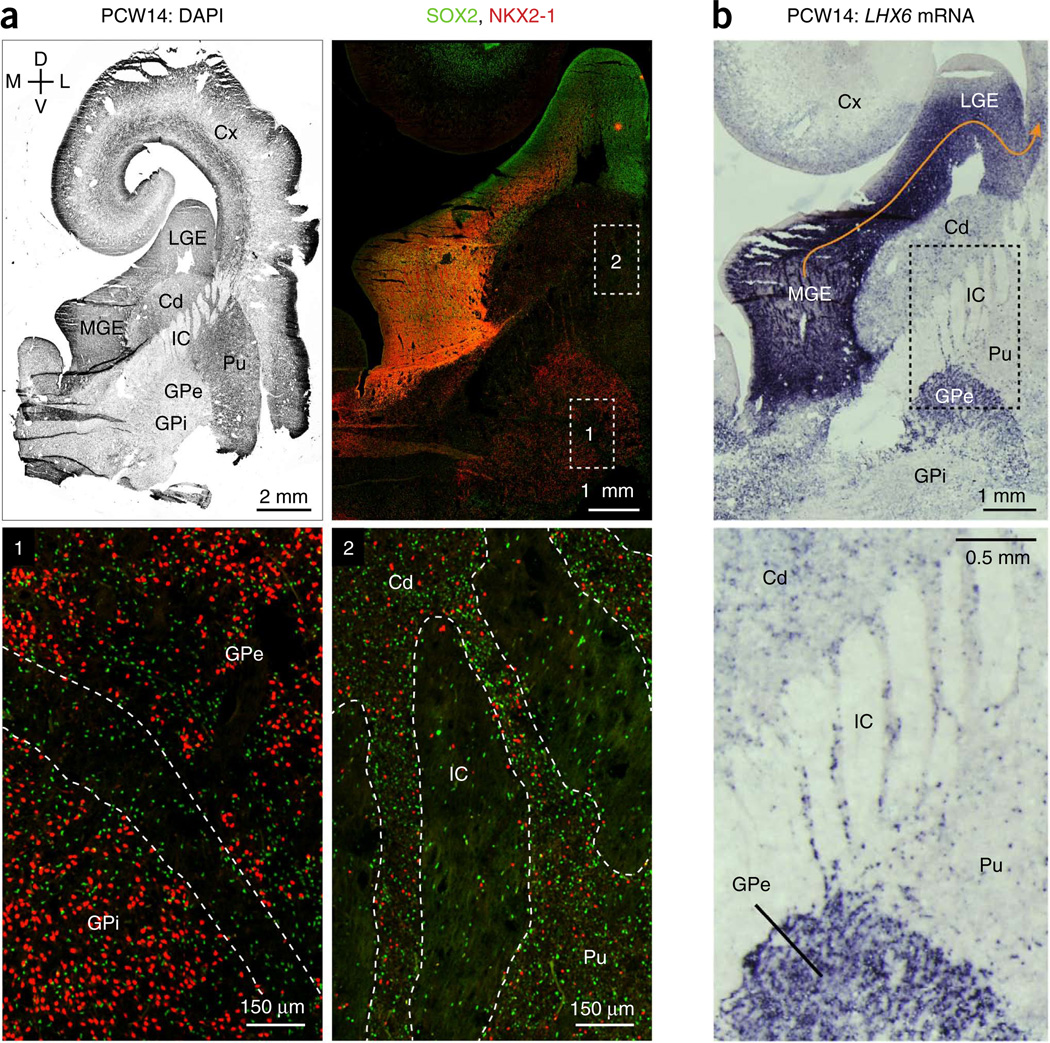
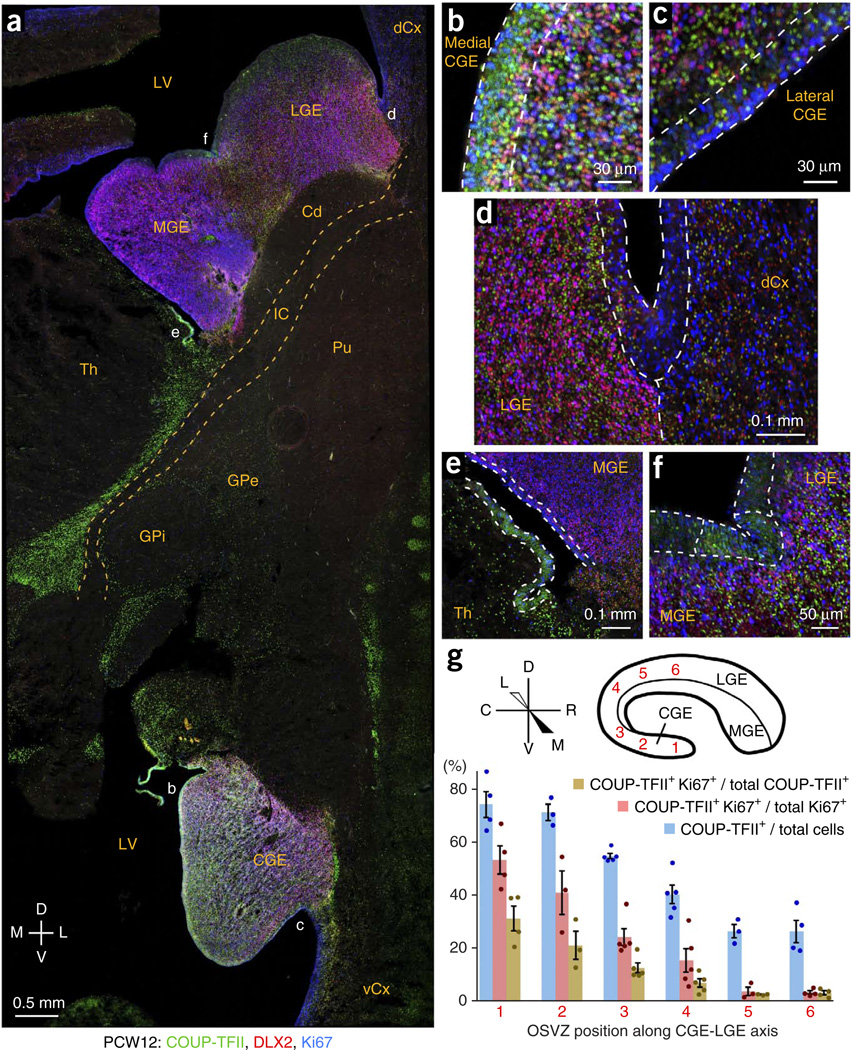
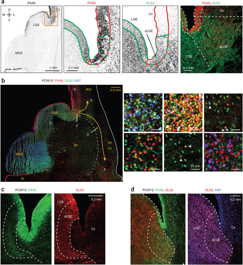
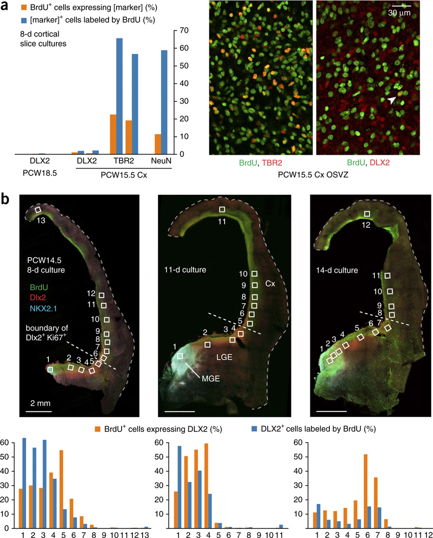
GABAergic cortical interneurons underlie the complexity of neural circuits and are particularly numerous and diverse in humans. In rodents, cortical interneurons originate in the subpallial ganglionic eminences, but their developmental origins in humans are controversial. We characterized the developing human ganglionic eminences and found that the subventricular zone (SVZ) expanded massively during the early second trimester, becoming densely populated with neural stem cells and intermediate progenitor cells. In contrast with the cortex, most stem cells in the ganglionic eminence SVZ did not maintain radial fibers or orientation. The medial ganglionic eminence exhibited unique patterns of progenitor cell organization and clustering, and markers revealed that the caudal ganglionic eminence generated a greater proportion of cortical interneurons in humans than in rodents. On the basis of labeling of newborn neurons in slice culture and mapping of proliferating interneuron progenitors, we conclude that the vast majority of human cortical interneurons are produced in the ganglionic eminences, including an enormous contribution from non-epithelial SVZ stem cells.


Figures

Figure 1
Figure 1
Developmental expansion of the OSVZ in the human ganglionic eminences. (a) Regional transcription factors that specify neuronal subtypes also distinguish progenitor cell types. Neural stem cells in the MGE express NKX2-1 and OLIG2. In intermediate progenitor cells, ASCL1 and DLX2 repress HES and OLIG2 expression and specify differentiation into GABAergic neurons. LHX6 expression and downregulation of NKX2-1 are important for migration to the cortex. (b) The subventricular region of ganglionic eminence progenitor cells, marked by SOX2 and ASCL1, expanded during the early second trimester, reaching a thickness of ~2.5 mm by PCW14 in the MGE. Macaque brain at gestational day 55 was developmentally similar to PCW8 human brain (Supplementary Fig. 1). D, dorsal; L, lateral; M, medial; V, ventral (frontal sections). (c) The ganglionic eminence ventricular zone (VZ) thickness diminished during the early second trimester, to as little as 25 µm in the ventromedial MGE by PCW14. Data are presented as mean ± s.e.m.; t test P values are indicated. (d) Comparison of germinal regions in human MGE, LGE and cortex (Cx) at PCW10 (frontal section). Outlined areas are magnified. The OSVZ in the MGE had a greater density of progenitor cells than in the LGE, and both regions exceeded the cortical OSVZ in thickness and progenitor cell density. Many DLX2+ cells in the ganglionic eminences expressed Ki67, whereas cortical DLX2+ cells were non-proliferative. In the medial aspect of the MGE, the weaker staining was a result of a microhemorrhage in the tissue that masked the signal. Str, striatum.
Figure 2
Figure 2
Neural stem cells are abundant in the MGE OSVZ. (a) Progenitor markers of the ventral forebrain (SOX2, OLIG2, ASCL1, Ki67) labeled the human MGE (marked by NKX2-1) and CGE (marked by strong COUP-TFII) more strongly than the LGE. PCW12 frontal sections are from an intermediate location on the rostral-caudal axis. Th, thalamus. Scale bars represent 1 mm. (b) The MGE OSVZ had an abundance of cells expressing the same markers (OLIG2+, ASCL1, DLX2) as undifferentiated radial glia in the ventricular zone. Shown are representative fields of images quantified in c. (c,d) OSVZ progenitor cells in the MGE were less differentiated than those in the CGE and LGE. OLIG2+ cells were less likely to express markers of neuronal commitment, and ASCL1+ and Ki67+ cells were less likely to express DLX2 in the MGE. The difference in the degree of differentiation among OLIG2+ cells also reached significance when comparing CGE and LGE (t29 = 4.37, P = 0.0002). The CGE was only measured at PCW12, as it is not well developed at PCW10 and our sample of PCW14 brain tissue lacked the CGE and other caudoventral structures. Error bars represent s.e.m.; t test P values are indicated. (e) SOX5, expressed in radial glia of the ganglionic eminence ventricular zone, was abundantly expressed in OSVZ progenitors in the MGE. (f) OLIG2+ cells in the human MGE OSVZ generally did not express NKX2-2, an oligodendroglial lineage marker. The arrowhead marks a rare exception. In the cortex, most or all of the sparse OLIG2+ cells throughout the OSVZ and intermediate zone expressed NKX2-2.
Figure 3
Figure 3
Most OSVZ stem cells in the ganglionic eminences lack M phase fibers and are randomly oriented. (a) In the human ganglionic eminences, nearly all OSVZ progenitors in M phase (labeled by 4A4 antibody for p-vim) displayed a simple, rounded morphology, indicating that any glial fibers were retracted during M phase. This was true for both neural stem cells (SOX2+ DLX2, white arrowheads) and intermediate progenitor cells (DLX2+, orange arrowheads). (b) The few progenitors in the MGE OSVZ that maintained short glial fibers during M phase were unipolar, expressed SOX2 and nestin, and had random orientation. Left, SOX2 stains of ganglionic eminences in frontal PCW14 sections. Second column, magnified images of areas boxed in red, with the overall contour of nestin fibers depicted by orange arrows. Right, magnification of the areas boxed in white showing features of 4A4+ cells that retained fibers during M phase, with cell bodies marked by white arrowheads and orientation of unipolar fibers marked by green arrows. Only one example (second row) was aligned with the overall glial fiber scaffold.
Figure 4
Figure 4
Segregation of progenitor cells and neurons in the MGE OSVZ. (a) Pronounced type I clustering in PCW13 horizontal section. Magnified images of boxed area show streaks of progenitor cells that extended from ISVZ into OSVZ and were devoid of nuclei at their core (DAPI). C, caudal; R, rostral. (b) Dense bundles of radial glial fibers formed the core of type I MGE progenitor cell clusters. SOX2+ progenitors clustered around apparent fascicles of nestin+ fibers. The structure of the neurovasculature (marked by collagen IV) was unrelated, although points of intersection were observed. (c) Type II clustering of progenitor cells in PCW14 frontal sections. Left, superior ganglionic eminences from a relatively caudal location; boxed OSVZ area is magnified in panel 1. Progenitors (SOX2+, ASCL1+) and non-progenitor cells (NKX2-1 only) segregated into broad, elongated streaks in the MGE OSVZ, oriented toward the LGE. Nestin signal was diffusely distributed throughout type II progenitor clusters (panel 2 from an adjacent section). (d) Type II clusters may be formed as neurons coalesce into migratory streams out of the MGE. Cells in the SOX2 clusters expressed DCX, a marker of migrating neurons.
Figure 5
Figure 5
Destinations of human MGE-derived neurons. (a) Distribution of NKX2-1+ cells throughout the developing basal ganglia. Top left, PCW14 frontal section counterstained for nuclei. Ventral structures, including inferior ganglionic eminences and some cortex, were not recovered with the tissue sample. Top right, a broad view of the subpallium stained for SOX2 and NKX2-1. Note the stream of NKX2-1+ cells with downregulated SOX2 migrating tangentially from the MGE into the LGE, toward the cortex. Boxed regions of the globus pallidus (1) and striatum (2) are magnified and shown below. The cellular bridges that traverse the internal capsule and connect the striatal compartments are outlined (2). Cd, caudate; IC, internal capsule; Pu, putamen. (b) In situ hybridization for LHX6 mRNA revealed sparse MGE-derived cells in the caudate and putamen, a more dense population in the GPe, and intense labeling throughout the whole LGE, the corridor for tangential migration to the cortex (orange arrow).
Figure 6
Figure 6
Patterns of COUP-TFII expression in the CGE and LGE. (a) COUP-TFII expression was widespread in the CGE, moderate in the LGE and mostly absent in the MGE. COUP-TFII was also observed in thalamus, GPe and other brain regions. dCx, dorsal cortex; vCx, ventral cortex; LV, lateral ventricle. (b,c) COUP-TFII was highly expressed in the ventricular zone of medial, but not lateral, CGE. OSVZ patterning was similar, with many COUP-TFII+ Ki67+ cells in the medial, but not lateral, CGE OSVZ. (d) Coexpression of DLX2 and Ki67 in the OSVZ of LGE, but not cortex, marked the LGE-cortical boundary. Many non-proliferating (Ki67) DLX2+ cells in the LGE and cortex expressed COUP-TFII, implicating the LGE as a migratory corridor for CGE-derived cortical interneurons. (e) COUP-TFII was mostly absent from the MGE (except at its caudal end; Supplementary Fig. 10), but was highly expressed in the immediately ventral ventricular zone, possibly the source of a ventral stream along the thalamus’ lateral edge (a). (f) COUP-TFII was expressed in a restricted region of ventricular zone (outlined) immediately surrounding the interganglionic sulcus. (g) COUP-TFII expression patterns indicate whether the OSVZ is more characteristic of CGE or LGE. In CGE, ~70% of OSVZ cells expressed COUP-TFII, ~30% of which were proliferating. In the LGE OSVZ, only ~25% of cells expressed COUP-TFII, almost none of which were proliferating. The transition between these extremes was gradual at intermediate points of measurement along the circumferential CGE-LGE axis. Error bars represent s.e.m.; t test P values are listed in the Source Data.
Figure 7
Figure 7
Production of cortical interneurons in the dorsal LGE. (a) The LGE expressed PAX6 at low levels, except in the dLGE, where PAX6 was coexpressed at high levels with DLX2. Shown are magnified images of the boxed area of PCW12 tissue, with ventricular zone regions outlined, PAX6high domain in red and DLX2+ domain in green. Right, dashed lines outlining distinct ventricular zone segments were extended into the corresponding OSVZ regions. (b) The dLGE supplied both the dorsal and lateral cortices with interneurons. Shown is a frontal section stained for PAX6, DLX2 and Ki67. Magnified images of the LGE OSVZ (1), dLGE OSVZ (2), lateral cortical stream (3), lateral cortex (4), dorsal cortex intermediate zone near dLGE (5) and more distal dorsal cortex OSVZ (6) are shown. The strong PAX6 and DLX2 coexpression shown in 4 and 5 is suggestive of interneuron migration from dLGE to lateral and dorsal cortices. Arrowheads in 3 indicate proliferating migratory DLX2+ cells. Arrowheads in panel 5 indicate non-proliferative DLX2+ PAX6+ (dLGE-derived) cortical interneurons. (c,d) Coexpression of DLX2 with high levels of PAX6 defined the dLGE as a progenitor cell compartment distinct from LGE or cortex. In addition, high levels of DLX2 expression indicated the dLGE-Cx boundary in ganglionic eminence progenitor cells, frequently with Ki67, whereas interneurons in the cortex expressed DLX2 at much lower levels and virtually never with Ki67. The precise location of the border between dLGE and cortex with respect to the corticostriatal sulcus varied among tissue samples.
Figure 8
Figure 8
Lack of DLX2+ cell production in cortical slice cultures. (a) Slices of PCW18.5 and PCW15.5 cortical tissues were cultured with BrdU for 8 d and stained for BrdU and DLX2. Thousands of cells were counted and the percentage of BrdU+ cells that expressed DLX2+ cells was consistently less than 1%. Staining for other markers of cortical neurogenic activity revealed large numbers of BrdU+ TBR2+ or NeuN+ cells. For DLX2 and TBR2 in slices from PCW15.5, colabeling with BrdU was measured in two different slices, with both values shown. Images show the regular colocalization of TBR2 and BrdU and an infrequent instance of DLX2+ cell production (arrowhead). (b) Hemispheric slice cultures including LGE/MGE from PCW14.5 were cultured with BrdU for 8–14 d. OSVZ images were captured and analyzed for DLX2 and BrdU colabeling from the numbered locations. Less than 1% of the BrdU+ cells in the cortex expressed DLX2+ at any time point, except in fields bordering the LGE, where we observed limited evidence of tangential migration. Additional images were taken from other locations in the cortical wall to assure that diverse regions were examined for evidence of interneuron production or migration.

Comment in

References

    1. Wonders CP, Anderson SA. The origin and specification of cortical interneurons. Nat. Rev. Neurosci. 2006;7:687–696. - PubMed
    1. Brown KN, et al. Clonal production and organization of inhibitory interneurons in the neocortex. Science. 2011;334:480–486. - PMC - PubMed
    1. Lui JH, Hansen DV, Kriegstein AR. Development and evolution of the human neocortex. Cell. 2011;146:18–36. - PMC - PubMed
    1. Kageyama R, Ohtsuka T, Hatakeyama J, Ohsawa R. Roles of bHLH genes in neural stem cell differentiation. Exp. Cell Res. 2005;306:343–348. - PubMed
    1. Alifragis P, Liapi A, Parnavelas JG. Lhx6 regulates the migration of cortical interneurons from the ventral telencephalon but does not specify their GABA phenotype. J. Neurosci. 2004;24:5643–5648. - PMC - PubMed

Publication types