. 2014 Oct;49(10):1378-91.
doi: 10.1007/s00535-013-0885-z. Epub 2013 Oct 7.

Functions and regulation of MUC13 mucin in colon cancer cells

Affiliations

Functions and regulation of MUC13 mucin in colon cancer cells

Brij K Gupta et al. J Gastroenterol. 2014 Oct.

Abstract

Background: MUC13 is overexpressed and aberrantly localized in colon cancer tissue; however, the specific functions and regulation of MUC13 expression are unknown.

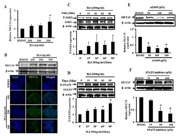
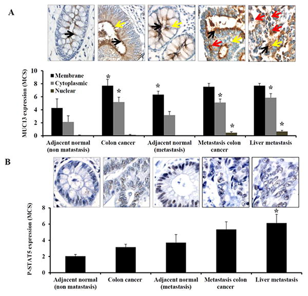
Methods: Stable cell lines with either overexpressed or suppressed MUC13 levels were analyzed to determine cell growth, colony formation, cell migration, and cell invasion assays. The molecular mechanisms involved in MUC13 regulation were elucidated via chromatin immunoprecipitation (ChIP) and analysis of interleukin 6 (IL6) treatments. Colon cancer tissues were analyzed by immunohistochemistry (IHC) for the protein levels of MUC13 and P-STAT5 in colon cancer cells.

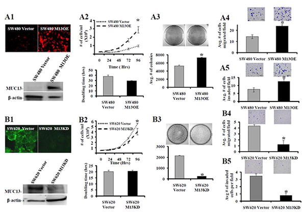
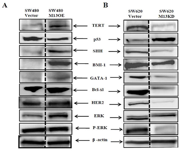
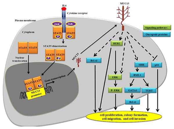
Results: Overexpression of MUC13 increased cell growth, colony formation, cell migration, and invasion. In concordance, MUC13 silencing decreased these tumorigenic features. Overexpression of MUC13 also modulated various cancer-associated proteins, including telomerase reverse transcriptase, sonic hedgehog, B cell lymphoma murine like site 1, and GATA like transcription factor 1. Additionally, MUC13-overexpressing cells showed increased HER2 and P-ERK expression. ChIP analysis revealed binding of STAT5 to the predicted MUC13 promoter. IL6 treatment of colon cancer cells increased the expression of MUC13 via activation of the JAK2/STAT5 signaling pathway. Suppression of JAK2 and STAT5 signaling by chemical inhibitors abolished IL6-induced MUC13 expression. IHC analysis showed increased expression of both P-STAT5 and MUC13 in colon cancer as compared to adjacent normal tissue.

Conclusions: The results of this study, for the first time, suggest functional roles of MUC13 in colon cancer progression and provide information regarding the regulation of MUC13 expression via JAK2/STAT5 which may reveal promising therapeutic approaches for colon cancer treatment.


Conflict of interest statement

Conflict of interest: Authors declare no conflict of interest.

Figures

Fig. 1
Fig. 1. MUC13 expression enhances the tumorigenic features of colon cancer cells
(A) SW480 cells were transfected with GFP tagged full length MUC13 to obtain MUC13 over-expressing (SW480 M13OE) cells. Vector control (SW480 Vector) cells were also selected. (B) SW620 cells were transduced with MUC13 specific shRNA lentiviral particles to obtain MUC13 knock-down (SW620 M13KD) cells. Vector control (SW620 Vector) cells were also obtained. The selected cell populations were screened for MUC13 by immunofluorescence (A1 and B1, top) and confirmed by Western blot (A1 and B1, bottom). Cell growth (A2, B2, top), cell doubling time (A2 and B2, bottom), colony formation (A3 and B3, top and bottom), cell migration (A4 and B4), and cell invasion (A5 and B5) assays were performed with MUC13 over-expressing and MUC13 knock-down cells, respectively, as described in experimental procedures. Representative images are shown above bar graph for the corresponding assays. Bar indicates the mean, error bar indicates the SEM, N=3, * P<0.05.
Fig. 2
Fig. 2. MUC13 modulates the expression of multiple cancer-associated pathways
Total cell lysates were collected from MUC13 over-expressing cells (SW480 M13OE) (A), MUC13 knock-down cells (SW620 M13KD) (B) and from corresponding vector controls SW480 Vector and SW620 Vector cells, respectively. The lysates were immunoblotted for multiple proteins, including: TERT, p53, SHH, BMI-1, GATA1, Bcl-xl, HER2, ERK and P-ERK. β-actin was used as the internal loading control for all immunoblot analysis. A representative blot from at least 2 independent lysates is shown.
Fig. 3
Fig. 3. IL6 induces MUC13 expression via binding of transcription factor STAT5 to the promoter of MUC13
(A) MUC13 expression levels vary among cell lines: RNA was isolated from colon cancer (SW48, SW480, SW620, T84), ovarian cancer (SKOV-3, CaOV-3) and pancreatic cancer (HPAFII, MiaPaca) cell lines and analyzed by Q-RT-PCR for MUC13 mRNA. β2-microglobulin was used as the housekeeping gene control. For quantitative analysis, the relative MUC13 RNA expression is normalized to SW48 cell line. (B) Expression profile of transcription factors predicted to bind to the MUC13 promoter: RNA isolated from T84 (top) and HPAFII (bottom) cell lines was analyzed by Q-RT-PCR to determine the expression level of the panel of potential transcription factors identified by in silico analysis. Relative mRNA expression of transcription factors were calculated using ΔΔCt method. (C) Schematic representation of STAT5 DNA binding sequence in the predicted promoter of MUC13: Location of the STAT5 binding sequence is boxed and the STAT5 binding sequence is in red and underlined, illustrating the STAT5 binding site is in a 1 kbp upstream region from the translational start site (TSS). (D) STAT5 binds to the predicted promoter of MUC13: ChIP analysis was performed as described in experimental procedures and MUC13 DNA that was pulled down by anti- STAT5 was amplified using semi quantitative PCR in T84 (D, top left) and HPAFII (D, top right) cells. Densitometry analysis of PCR product is shown in T84 (D, bottom left) and HPAFII (D, bottom right). (E) The MUC13 amplification was confirmed by real time PCR in T84 (right) and HPAFII (left).
Fig. 4
Fig. 4. IL6 treatment enhances MUC13 expression via the JAK2/STAT5 pathway
(A and B) IL6 increases MUC13 expression HT-29 cells were serum starved and treated with IL6 (100-300 ng/ml) and vehicle control (DMSO) for 48-72 hrs. A) At 48 hrs RNA was isolated and analyzed by Q-RT-PCR for MUC13 expression. B) At 72 hrs cell lysates were collected and analyzed by Western blot (top) or cells were fixed and processed for immunofluorescence analysis (bottom). (C and D) IL6 treatment increases P-JAK2 and P-STAT5 expression: Cells were treated with IL6 (200 ng/ml) or DMSO for 15-90 mins after an 8 hr serum starvation. SDS lysates were collected and subjected for Western blot analysis to detect total JAK2, P-JAK2 (4C), total STAT5 and P-STAT5 (4D) expression. Quantification of P-JAK2 and P-STAT5 expression is shown below corresponding Western blots. (E and F) Treatment with JAK2 and STAT5 inhibitors attenuated MUC13 expression: Cells were serum starved for 8 hrs and then treated with JAK2 (AG490) and STAT5 inhibitors. Cells were further treated with JAK2 and STAT5 inhibitors in the presence of IL6 for 48-72 hrs. Expression of MUC13 was detected by Western blot analysis following treatment with JAK2 inhibitor (4E) and STAT5 inhibitor (4F). Quantitative analysis is shown below corresponding Western blots. All experiments were repeated at least two times and representative blot is shown. Bar indicates the mean, error bar indicates the SEM, * P<0.05.
Fig. 5
Fig. 5. P-STAT5 expression is associated with MUC13 expression in colon cancer tissues
Tissue microarrays containing tissues from patients with non-metastatic disease (adjacent normal and colon cancer), and from patients with metastatic disease (adjacent normal, metastatic colon cancer, and liver metastasis tissues) were processed for immunostaining using MUC13 and P-STAT5 antibodies. Representative images of (A) MUC13 and (B) P-STAT5 immunostaining are shown and quantification was done as described in experimental methods. Black, yellow and red arrow indicates membranous, cytoplasmic and nuclear MUC13 expression respectively. Bar indicates the mean, error bar indicates the SEM, * P<0.05.
Fig. 6
Fig. 6. Schematic representation of MUC13 regulation and associated cancer pathways influencing MUC13 induced tumorigenesis
IL6 treatment induces phosphorylation of P-JAK2 and P-STAT5. Once phosphorylated, P-STAT5 translocates from cytoplasm to nucleus where it binds to the predicted promoter of MUC13 and increases MUC13 production at RNA and protein levels. MUC13 modulates the expression of various oncogenic proteins, resulting in increased tumorigenic features such as cell growth, cell doubling time, cell migration and cell invasion.

References

    1. Siegel R, Naishadham D, Jemal A. Cancer statistics, 2012. CA: a cancer journal for clinicians. 2012;62(1):10–29. Epub 2012/01/13. - PubMed
    1. Siegel R, Ward E, Brawley O, Jemal A. Cancer statistics, 2011: the impact of eliminating socioeconomic and racial disparities on premature cancer deaths. CA Cancer J Clin. 2011;61(4):212–36. Epub 2011/06/21. - PubMed
    1. Jemal A, Siegel R, Xu J, Ward E. Cancer statistics, 2010. CA Cancer J Clin. 2010;60(5):277–300. Epub 2010/07/09. - PubMed
    1. Hollingsworth MA, Strawhecker JM, Caffrey TC, Mack DR. Expression of MUC1, MUC2, MUC3 and MUC4 mucin mRNAs in human pancreatic and intestinal tumor cell lines. International journal of cancer Journal international du cancer. 1994;57(2):198–203. Epub 1994/04/15. - PubMed
    1. Ogata S, Uehara H, Chen A, Itzkowitz SH. Mucin gene expression in colonic tissues and cell lines. Cancer research. 1992;52(21):5971–8. Epub 1992/11/01. - PubMed

Publication types

MeSH terms