. 2013 Oct 7:3:2884.
doi: 10.1038/srep02884.

Macrophages lift off surface-bound bacteria using a filopodium-lamellipodium hook-and-shovel mechanism

Affiliations

Macrophages lift off surface-bound bacteria using a filopodium-lamellipodium hook-and-shovel mechanism

Jens Möller et al. Sci Rep. .

Abstract

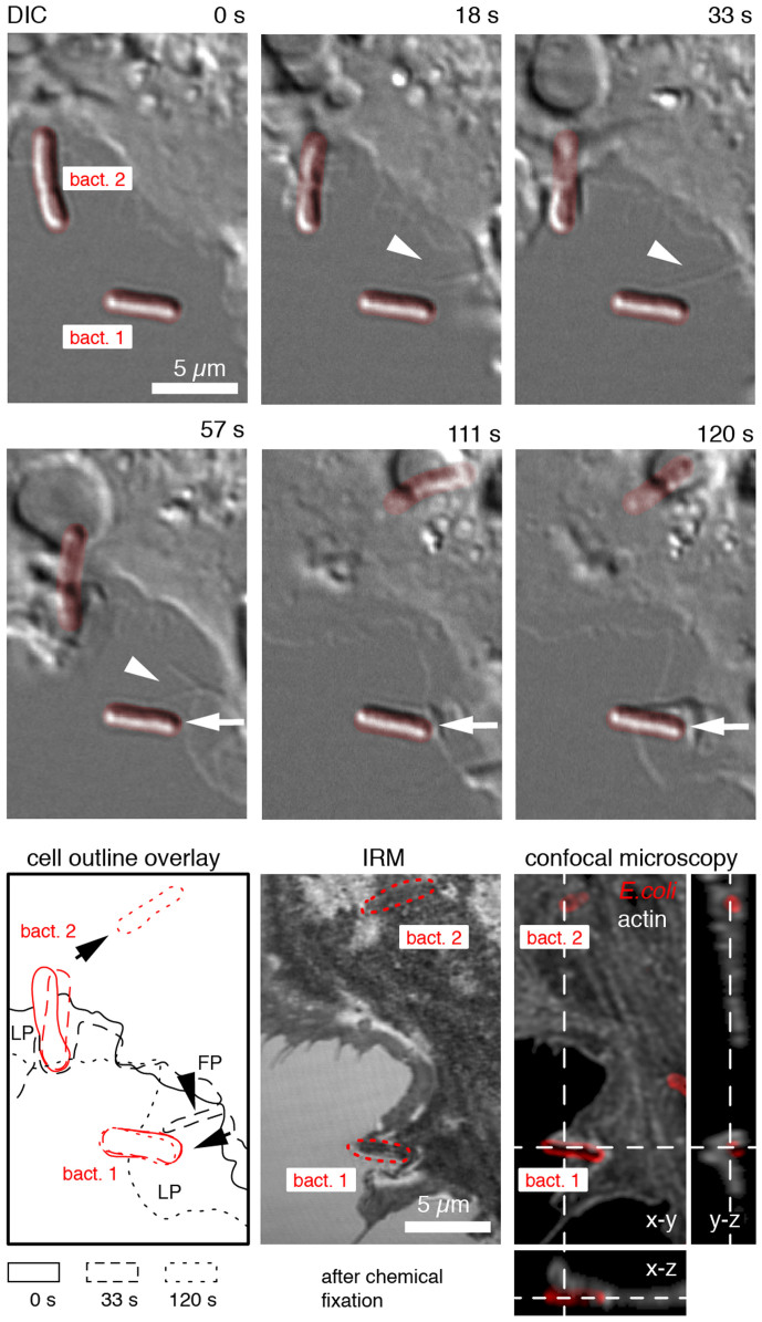
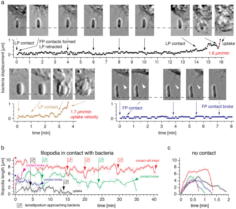
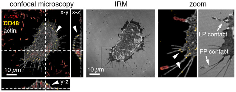
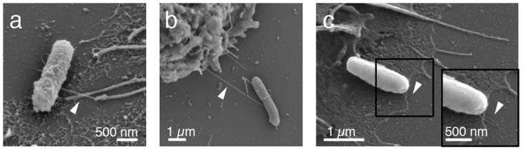
To clear pathogens from host tissues or biomaterial surfaces, phagocytes have to break the adhesive bacteria-substrate interactions. Here we analysed the mechanobiological process that enables macrophages to lift-off and phagocytose surface-bound Escherichia coli (E. coli). In this opsonin-independent process, macrophage filopodia hold on to the E. coli fimbriae long enough to induce a local protrusion of a lamellipodium. Specific contacts between the macrophage and E. coli are formed via the glycoprotein CD48 on filopodia and the adhesin FimH on type 1 fimbriae (hook). We show that bacterial detachment from surfaces occurrs after a lamellipodium has protruded underneath the bacterium (shovel), thereby breaking the multiple bacterium-surface interactions. After lift-off, the bacterium is engulfed by a phagocytic cup. Force activated catch bonds enable the long-term survival of the filopodium-fimbrium interactions while soluble mannose inhibitors and CD48 antibodies suppress the contact formation and thereby inhibit subsequent E. coli phagocytosis.


Figures

Figure 1
Figure 1. Multistep macrophage uptake of surface-bound E. coli.
Macrophage encountered surface-bound E. coli (bact.1, bact.2, false coloured red) by filopodia (FP, arrowhead)) and lamellipodia (LP, arrow) as visualized by live cell DIC microscopy (Supplementary Movie 1). For bacterium 1, the initial FP contact remained intact sufficiently long for the macrophage to locally protrude a LP towards the bacterium (arrow, 33–57 s). Upon LP contact, the membrane locally ruffled in front of the bacterium (111 s) followed by LP protrusion under the bacterium (120 s). LP protrusion underneath the bacterium was confirmed by interference reflection microscopy (IRM) and 3D confocal fluorescence microscopy of the same cell after chemical fixation at 120 s. Bacteria were labelled with primary anti-E. coli and secondary Dylight 649 antibody (red). The macrophage F-actin cytoskeleton was stained with Alexa-488 phalloidin (white). Confocal stacks were deconvolved using Huygens software. For bacterium 2, the LP was already in contact at the start of the time series. The uptake of bact. 2, as indicated by the rapid displacement of the bacterium (57–120 s, see outline overlay) was confirmed by 3D confocal microscopy (y-z cross section, 120 s). Due to the fast macrophage dynamics, the last frame of the DIC sequence (120 s) does not overlay exactly the IRM as well as with the confocal micrograph of the fixed sample.
Figure 2
Figure 2. Formation of long-lived interactions between filopodia and surface-bound E. coli.
(a) Kinetic analysis of bacterial displacements during filopodia (FP) contact and lamellipodia (LP) uptake. Upper row: With intact filopodia-bacterium contacts, no substantial bacterial displacement was observed for 14 min (Supplementary Movie 4, see Methods for displacement analysis). Only after the lamellipodium contacted the bacterium, it was displaced from its original position and the uptake proceeded with 1.6 μm/min. Lower row left: Bacterial displacement initiated by a direct lamellipodium contact without detection of a previous filopodium contact (Supplementary Movie 5). Uptake proceeded with 1.7 μm/min. Lower row right: A filopodium contact alone was not sufficient to displace and pick up a bacterium that firmly adhered to the surface (Supplementary Movie 6). (b) Kinetic analysis of individual filopodium-bacterium contact lengths. 5 representative traces are plotted, including the filopodium-bacterium contacts analysed in (a) (black and blue traces, Supplementary Movies 2 and 3). Long-lived interactions of up to 40 min were observed. The onset of a lamellipodium protrusion towards the captured bacterium is indicated (LP). Note that some contacts were already present at the start of the time series (asterisk). Lines represent adjacent 3-point averages. (c) Extension and withdrawal traces of individual filopodia that were not in contact with surface-bound E. coli (Supplementary Movie 2). If no contact was formed, filopodia retracted within 2 minutes.
Figure 3
Figure 3. Immunostaining of the GPI-anchored mannosylated FimH receptor CD48 on the J774A.1 macrophage cell membrane.
Deconvolved confocal fluorescence and interference reflection micrographs (IRM). Samples were stained for macrophage actin (white), CD48 (yellow) and E. coli (red). CD48 exclusively localized within the macrophage membrane including filopodia (see arrowheads and x-z and y-z cross sections). Filopodia (FP) and lamellipodia (LP) – bacteria contacts were suggested by IRM.
Figure 4
Figure 4. Macrophage - E. coli contacts as seen in SEM micrographs.
(a) Macrophage filopodium bound to an individual E. coli type 1 fimbrium. (b) Long-distance macrophage – bacterium contact (c) Lamellipodium localized underneath a surface-bound bacterium. The arrowhead points towards the last two fimbriae that are still in contact with the mannosylated surface.
Figure 5
Figure 5. Bacterial uptake was blocked by mannose inhibitor, was substantially reduced by CD48 antibodies and was independent of the direction of fluid flow.
(a) Addition of 2% soluble α−D-mannopyranoside inhibitor (αMM) and CD48 antibodies substantially reduced the rate of E. coli phagocytosis. Uptake rates were analysed for 10 independent macrophages during 10 min live cell experiments and normalized by the time-averaged number of bacteria available in the zone explored by filopodia. Mean values are given as horizontal line. (b) Bacterial uptake was observed at all sides of the macrophages and was independent of the direction of fluid flow (0.1 ml/min flow rate/0.06 pN/μm2 shear stress). (c) Filopodia length distribution as analysed from fixed macrophages. 95% of the filopodia (n = 400) have a length less than 8 μm, which we defined here as the filopodia sensing zone (yellow area).
Figure 6
Figure 6. Shear stress dependent FimH-specific E. coli adhesion to mannosylated CD48 surfaces.
(a) Accumulation of type I fimbriated E. coli FimH-j96 bacteria on flow chamber bottom glass surfaces coated with CD48, mono-mannose bovine serum albumin (1 M) and tri-mannose RNaseB (3 M) under varying shear stresses τ (1 pN μm−2 = 1 Pascal or 10 dynes cm−2). E. coli accumulation was analysed after 5 min. As negative controls, either 2% of a mono-mannose inhibitor (αMM) was added to the media (formula image), or the non-fimbriated E. coli parent strain AAEC191A was used (Δfim, formula image). (b) Fraction of E. coli FimH-j96 that adhered firmly on CD48, 1 M and 3 M. Bacteria were defined firmly adhering if they moved less than one-half of a bacterial diameter over >30 s. (c) Effect of different FimH variants on bacterial accumulation to 1 M and CD48. While the low binding FimH-f18 strain only adhered to CD48 and 1 M above a critical shear stress, FimH-j96 E. coli accumulated on CD48 without any shear threshold.
Figure 7
Figure 7. Proposed “Hook-and-Shovel” mechanism for macrophage pick up of surface-bound type 1 fimbriated E. coli.
(a) The mannosylated membrane anchored surface receptor CD48 of macrophages specifically binds to the bacterial fimbrial tip adhesin FimH, which contains a single mannose-binding pocket in the lectin domain (Hook). (b) Due to filopodia retractions, the bacterial fimbriae are elongated. As bacteria adhere tightly to substrate surfaces via multiple bonds, the macrophages fail to pull bacteria off the surface via the hook alone. (c) To facilitate uptake, macrophages protrude lamellipodia towards the bacterium to sequentially break the bonds that anchor E. coli to the surface (Shovel). (d) Once the bacterium is completely lifted off the substrate and lies on the lamellipodium, a phagocytic cup is formed to internalize the bacterium.

References

    1. Aderem A. Phagocytosis and the inflammatory response. J. Infect. Dis. 187 Suppl 2, S340–345 (2003). - PubMed
    1. Mahdavian Delavary B., van der Veer W. M., van Egmond M., Niessen F. B. & Beelen R. H. J. Macrophages in skin injury and repair. Immunobiology 216, 753–762 (2011). - PubMed
    1. Anderson J. M. Biological responses to materials. Annu. Rev. Mater Res. 31, 81–110 (2001).
    1. Nielubowicz G. R. & Mobley H. L. T. Host-pathogen interactions in urinary tract infection. Nat. Rev. Urol. 7, 430–441 (2010). - PubMed
    1. Murray P. J. & Wynn T. A. Protective and pathogenic functions of macrophage subsets. Nat. Rev. Immunol. 11, 723–737 (2011). - PMC - PubMed

Publication types

MeSH terms