Review
. 2014 Jan;99(1):35-51.
doi: 10.1113/expphysiol.2013.071969. Epub 2013 Oct 4.

Structure and function of voltage-gated sodium channels at atomic resolution

Affiliations
Review

Structure and function of voltage-gated sodium channels at atomic resolution

William A Catterall. Exp Physiol. 2014 Jan.

Abstract

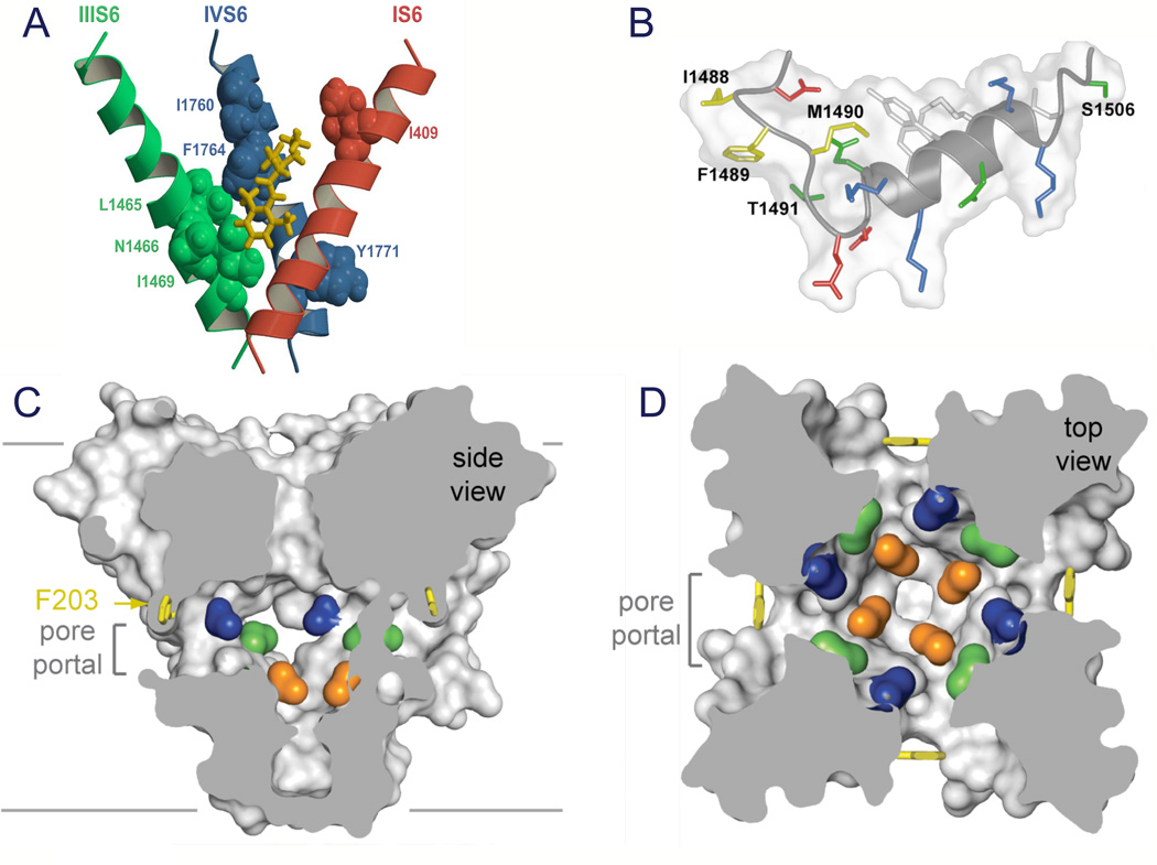
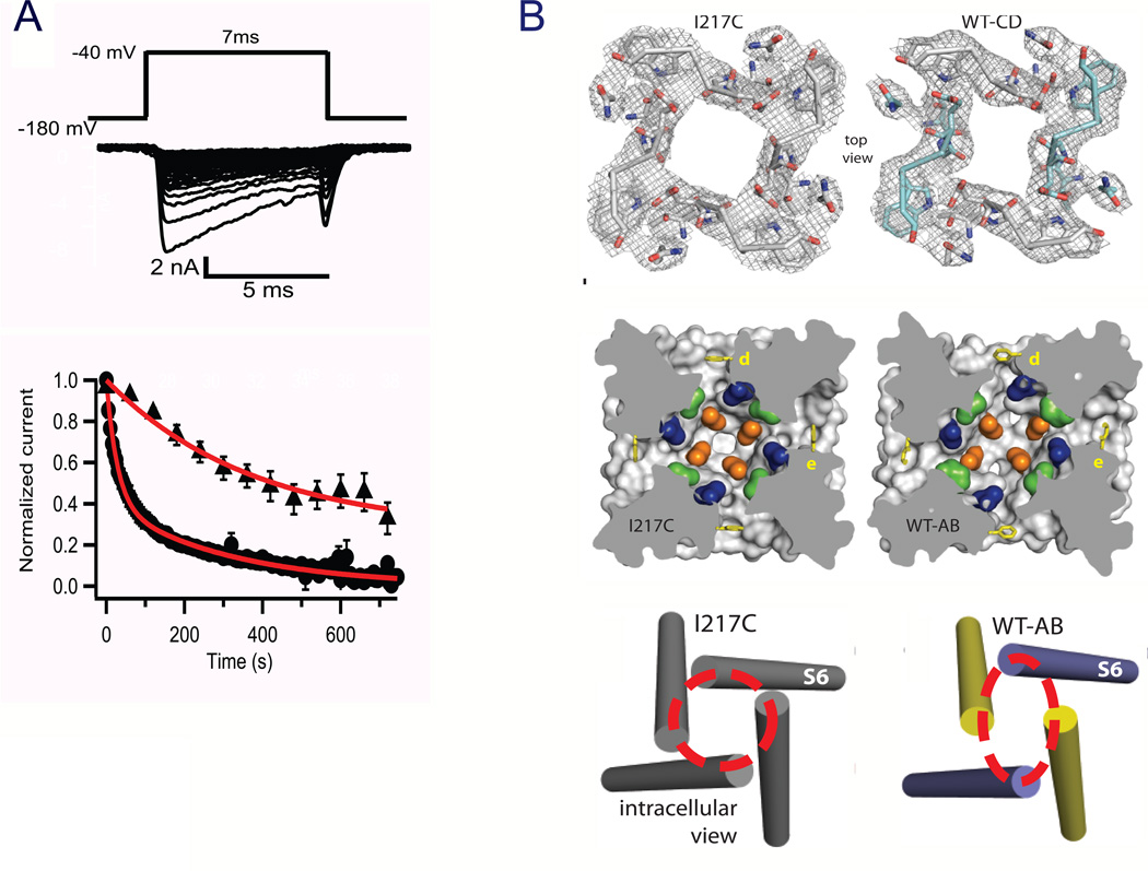
Voltage-gated sodium channels initiate action potentials in nerve, muscle and other excitable cells. Early physiological studies described sodium selectivity, voltage-dependent activation and fast inactivation, and developed conceptual models for sodium channel function. This review article follows the topics of my 2013 Sharpey-Schafer Prize Lecture and gives an overview of research using a combination of biochemical, molecular biological, physiological and structural biological approaches that have elucidated the structure and function of sodium channels at the atomic level. Structural models for voltage-dependent activation, sodium selectivity and conductance, drug block and both fast and slow inactivation are discussed. A perspective for the future envisions new advances in understanding the structural basis for sodium channel function and the opportunity for structure-based discovery of novel therapeutics.


Figures

Figure 1
Figure 1. Subunit structure of voltage-gated sodium channels
A. SDS polyacrylamide gel electrophoresis patterns illustrating the α and (β subunits of the brain sodium channels. Left. α and (βi subunits covalently labeled with [ 125 I]-labeled scorpion toxin (Beneski and Catterall, 1980). Lane 1, specific labeling; lane 2, nonspecific labeling. Right. Sodium channel purified from rat brain showing the α, β1, and β2 subunits and their molecular weights (Hartshorne et al., 1982). As illustrated, the α and β2 subunits are linked by a disulfide bond. Tetrodotoxin and scorpion toxins bind to the α subunits of sodium channels as indicated and were used as molecular tags to identify and purify the sodium channel protein (Beneski & Catterall, 1980; Hartshorne et al., 1982; Hartshorne & Catterall, 1984). Inset. Single channel currents conducted by a single purified sodium channel incorporated into a planar bilayer (Hartshorne et al., 1985).
Figure 2
Figure 2. The primary structures of the subunits of the voltage-gated sodium channels
Cylinders represent alpha helical segments. Bold lines represent the polypeptide chains of each subunit with length approximately proportional to the number of amino acid residues in the brain sodium channel subtypes. The extracellular domains of the β1 and β2 subunits are shown as immunoglobulin-like folds. □, sites of probable N-linked glycosylation; P in red circles, sites of demonstrated protein phosphorylation by PKA (circles) and PKC (diamonds); green, pore-lining segments; white circles, the outer (EEEE) and inner (DEKA) rings of amino residues that form the ion selectivity filter and the tetrodotoxin binding site; yellow, S4 voltage sensors; h in blue circle, inactivation particle in the inactivation gate loop; blue circles, sites implicated in forming the inactivation gate receptor (Catterall, 2000). Sites of binding of α- and β-scorpion toxins and a site of interaction between α and β1 subunits are also shown. Tetrodotoxin is a specific blocker of the pore of sodium channels (Hille, 1975b), whereas the α- and β-scorpion toxins block fast inactivation and enhance activation, respectively, and thereby generate persistent sodium current that causes depolarization block of nerve conduction (Catterall et al., 2007). Tetrodotoxin has been used as a tool to probe the pore of the sodium channel, whereas the scorpion toxins have been valuable as probes of voltage sensor function.
Figure 3
Figure 3. Structure of NaVAb
A. Top view of NaVAb channels colored according to crystallographic temperature factors of the main-chain (blue < 50 Å2 to red > 150 Å2). B. Architecture of the NaVAb pore. Glu177 side-chains, purple; pore volume, grey. The S5 and S6 segments and the P loop from two lateral subunits are shown (Payandeh et al., 2011).
Figure 4
Figure 4. Structure of the voltage sensor
A. Structure of the voltage sensor in an activated state. Side views of the structures of NaVAb(yellow (Payandeh et al., 2011)) and KV1.2 (purple (Long et al., 2005a)) are superimposed. Extracellular negative cluster (ENC), red; hydrophobic constriction site (HCS), green; intracellular negative cluster (INC), red. B. Model of the resting state of the NaChBac voltage sensor. Gating charges R1–R4, blue; T0, Thr in the position of the R0 gating charge in some KV channels. L112C, Cys substituted for Leu adjacent to R1 in S4 segment forming a disulfide bond with Cys substituted for Asp60 (D60C) in S2 segment in the resting state as observed in disulfide locking experiments. E43, Glu 43 in S1 segment, a component of the extracellular negative cluster. E70, Glu70 in S2 segment, a component of the intracellular negative cluster.
Figure 5
Figure 5. Disulfide locking the voltage sensor in resting and activated states
NaChBac wild-type (WT), single Cys mutants D60C and R3CA, and double Cys mutant D60C:R3C were expressed in tsA-201 cells and recorded using the whole-cell voltage clamp mode. A. Reversible disulfide locking. Mean normalized peak currents elicited by a 0.1 Hz train of 500-ms depolarizations to 0 mV from a holding potential of −140 mV in tsA cells transfected with D60C, R3C, or D60C:R3C channels. After 2 min in control saline conditions, cells were exposed to 1 mM β-mercaptoethanol (DeCaen et al., 2008). B. Time course of voltage-sensor locking. D60C:R3C channels were first unlocked by a 5-s prepulse to −160 mV. Cells were then depolarized for the indicated times to approximately V1/2 + 20 mV (WT, −20 mV; D60C & R3C, 0 mV; D60C:R3C, −30 mV), returned to −120 mV for 5 s and depolarized for 100 ms test pulse to 0 mV. Peak test pulse current at 0 mV was normalized to the control pulse current in the absence of a prepulse and mean (± SEM) was plotted versus prepulse duration (red circles with error bars). Sodium current recorded during a −30 mV prepulse (black); time course of activation in the absence of inactivation (blue trace) estimated by fitting an exponential to the current decay and adding the inactivated component back to the total current (DeCaen et al., 2008). C. Disulfide locking of R1 and Glu43 in the resting state. D. Disulfide locking of R2 and Glu43 in resting and activated states. E. Disulfide locking of R3 and Glu43 in the activated state. Disulfide locking was induced by depolarization in the presence of 1 mM H2O2 (DeCaen et al., 2011).
Figure 6
Figure 6. Model for pore opening transition
Superposition of NaVAb (yellow (Payandeh et al., 2011)) and KV1.2/2.1 (green (Long et al., 2007)) viewed from the membrane.
Figure 7
Figure 7. The ion selectivity filter of NaVAb
A. Top view of the ion selectivity filter. Symmetry-related molecules are colored white and yellow; P-helix residues are colored green. Hydrogen bonds between Thr175 and Trp179 are indicated by grey dashes. Electron-densities from Fo-Fc omit maps are contoured at 4.0 σ (blue and grey) and subtle differences can be appreciated (small arrows) (Payandeh et al., 2011). B. Side view of the selectivity filter. Glu177 (purple) interactions with Gln172, Ser178 and the backbone of Ser180 are shown in the far subunit. Fo-Fc omit map, 4.75 σ (blue); putative cations or water molecules (red spheres, IonEX). Electron-density around Leu176 (grey; Fo-Fc omit map at 1.75 σ) and a putative water molecule is shown (grey sphere). Na+-coordination sites: SiteHFS, SiteCEN and SiteIN. (Payandeh et al., 2011)
Figure 8
Figure 8. Molecular dynamics of sodium conductance
Entry and exit of sodium ions in the pore of NaVAb were analyzed without constraints for 23 µs at 150 mM Na+ and V=0. A. Axial distribution of Na+ in the selectivity filter (SF) and central cavity (CC), distinguishing between states in which Na+ is directly bound to Glu177 (“E”, green), to both Glu177 and Leu176 (“EL”, yellow), or to neither (brown). The selectivity filter is defined by two spatially resolved Na+ binding sites, E and EL. The small peaks at z = −0.65 and z = 0.40 nm in the brown distribution correspond to direct Na+ coordination by the hydroxyl O atom of Ser178 and water-mediated coordination to the carbonyl O atom of Thr175, respectively. B. Mechanism and kinetics of Na+ translocation through the selectivity filter. The black box represents the selectivity filter, with the central cavity to the right below and the extracellular mouth to the left. The populations of all four states 1’, 2, 2’, and 3, which differ in the occupancy of the channel and of the selectivity filter, are shown in %, and the rate constants computed from the molecular dynamics trajectories are shown above or below each arrow in units of µs−1. At this ionic concentration (150 mM), states 2 and 2’ correspond to the resting state of the system. The exchange between states 2 and 2’, which corresponds to a unitary ionic translocation through the selectivity filter, involves either one-ion or three-ion intermediate states (Chakrabarti et al., 2013).
Figure 9
Figure 9. Drug receptor site and fast inactivation gate
A. Model of the local anesthetic receptor site in mammalian NaV1.2 channels (Yarov-Yarovoy et al., 2002). B. Structure of the inactivation gate of mammalian NaV1.2 channels in solution determined by NMR (Rohl et al., 1999). C. Side-view through the pore module illustrating fenestrations (portals) and hydrophobic access to central cavity. Phe203 side-chains, yellow sticks. Surface representations of NaVAb residues aligning with those implicated in drug binding and block, Thr206, blue; Met209, green; Val213, orange. Membrane boundaries, grey lines. Electrondensity from an Fo-Fc omit map is contoured at 2.0 σ. D. Top-view sectioned below the selectivity filter, colored as in C (Payandeh et al., 2011).
Figure 10
Figure 10. Structure of the slow-inactivated state in NaVAb
A. Use-dependent development of slow inactivation. Depolarizations from a holding potential of −180 mV to −40 mV, 7 ms in duration, were applied at 0.2 Hz (circles) or once per min (triangles), and the peak current elicited by each pulse was measured. Currents were normalized to the peak inward current during the first pulse. BTop, selectivity filter. Stick representation of the selectivity filter with a 2Fo-Fc map calculated at 3.2 Å resolution (grey mesh) contoured at 1.5 σ for NaVAb -I217C and 1.0 σ for NaVAb -CD. Symmetry-related subunits in WT-CD are colored white (Chains A and D) and cyan (Chains B and C), respectively. Middle, central cavity. A view through the pore module sectioned below the selectivity filter illustrates the lateral pore fenestrations, hydrophobic access to the central cavity, and structural asymmetry in the NaVAb-AB pore domain. Phe203 side-chains are yellow sticks. NaVAb residues implicated in drug binding in vertebrate NaV channels are colored: Thr206 (blue), Met209 (green), and Val213 (orange). Bottom, activation gate. Red dashed lines indicate the Cα location of D219 (the last S6 residue modeled in WT-AB), where the S6 helices are shown as cylinders. WT-chain A, purple; WT-chain B, yellow (Payandeh et al., 2012).

References

    1. Adelman WJ, Jr, Palti Y. The effects of external potassium and long duration voltage conditioning on the amplitude of sodium currents in the giant axon of the squid, Loligo pealei. J Gen Physiol. 1969;54:589–606. - PMC - PubMed
    1. Armstrong CM. Interaction of tetraethylammonium ion derivatives with the potassium channels of giant axon. J Gen Physiol. 1971;58:413–437. - PMC - PubMed
    1. Armstrong CM. Sodium channels and gating currents. Physiol Rev. 1981;61:644–682. - PubMed
    1. Armstrong CM, Benzanilla F. Charge movement associated with the opening and closing of the activation gates of the Na channels. J Gen Physiol. 1974;63:533–552. - PMC - PubMed
    1. Armstrong CM, Bezanilla F. Currents related to movement of the gating particles of the sodium channels. Nature. 1973;242:459–461. - PubMed

Publication types

MeSH terms

LinkOut - more resources