Review
. 2013 Dec 21;42(24):9207-18.
doi: 10.1039/c3cs60198f. Epub 2013 Oct 7.

Cellular nanotechnology: making biological interfaces smarter

Affiliations
Review

Cellular nanotechnology: making biological interfaces smarter

Paula M Mendes. Chem Soc Rev. .

Abstract

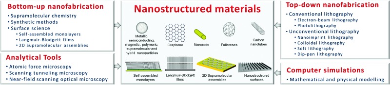
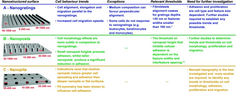
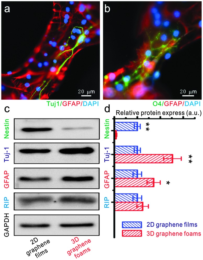
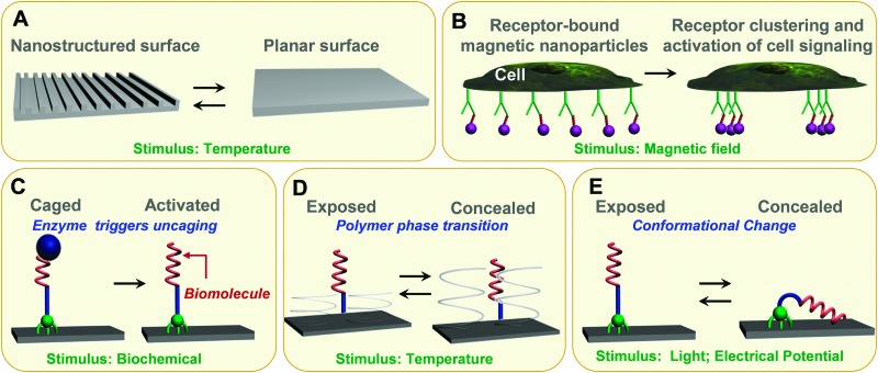
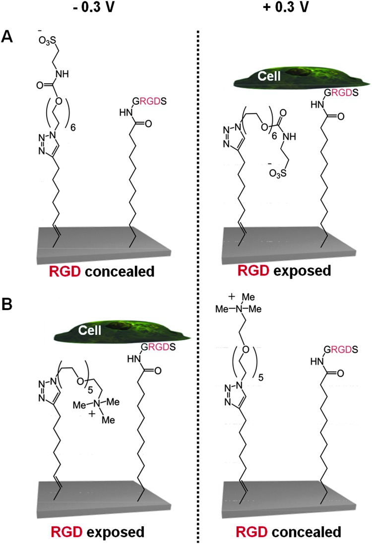
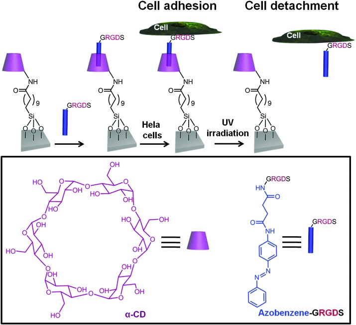
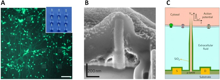
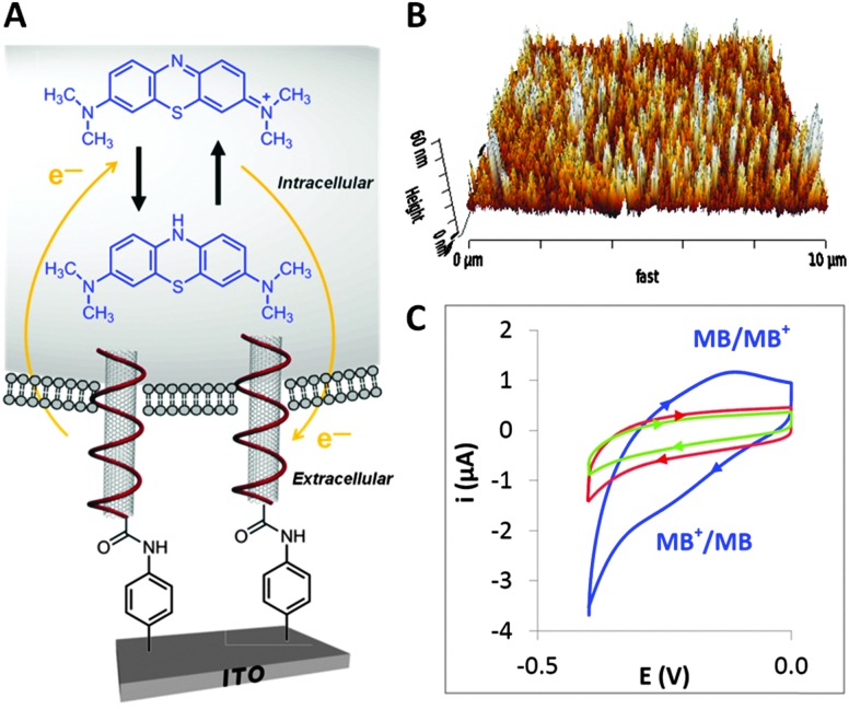
Recently, there has been an outburst of research on engineered cell-material interfaces driven by nanotechnology and its tools and techniques. This tutorial review begins by providing a brief introduction to nanostructured materials, followed by an overview of the wealth of nanoscale fabrication and analysis tools available for their development. This background serves as the basis for a discussion of early breakthroughs and recent key developments in the endeavour to develop nanostructured materials as smart interfaces for fundamental cellular studies, tissue engineering and regenerative medicine. The review covers three major aspects of nanostructured interfaces - nanotopographical control, dynamic behaviour and intracellular manipulation and sensing - where efforts are continuously being made to further understand cell function and provide new ways to control cell behaviour. A critical reflection of the current status and future challenges are discussed as a conclusion to the review.


Figures

Fig. 1
Fig. 1. Schematic representation of examples of nanostructured materials, which can be fabricated and controlled by a powerful range of nanotechnology tools and techniques – bottom-up and top-down nanofabrication technologies, new or improved analytical techniques and high-performance computer simulations.
Fig. 2
Fig. 2. Summary of the main effects of the interaction of mammalian cells with nanoscale topography in the form of (A) nanogratings, (B) nanoposts and (C) nanopits with different feature widths, depths and interfeature spacings. Progress has been made, but yet there is still a great deal of work to be done in fully understanding the influence of nanotopography on cell behaviour. * The threshold is associated with a particular spacing (ridge-grating ratio) and the cell type investigated. Different threshold numbers are expected depending on the cell type and ridge-grating ratio used. ** The threshold is based on several studies with different cell types and features with different aspect ratios and interfeature spacing.
Fig. 3
Fig. 3. The differentiation of NSCs on a 3D graphene foam scaffold. (a and b) Fluorescence images of differentiated NSCs that were immunostained for Tuj-1 (marker for neurons – green, a), GFAP (marker for astrocytes – red, a and b), O4 (marker for oligodendrocytes – green, b) and DAPI (marker for nuclei – blue, a and b). Western blot analysis of nestin (marker for NSCs), Tuj-1, GFAP and RIP (marker for oligodendrocyte) protein expression of differentiated NSCs on 2D graphene films and 3D graphene foam scaffold. (d) Relative optical densities of nestin, Tuj-1, GFAP and RIP bands shown in (c). Reproduced with permission from ref. 25 (copyright 2012, Nature Publishing Group).
Fig. 4
Fig. 4. Stimuli-responsive interfaces as in vitro model systems. (A) Shape-memory polymer surfaces, which transitions are induced by temperature, allows for dynamic topographical control of cell behaviour. (B) Superparamagnetic nanoparticles (purple), each linked with a single ligand, bind evenly across the plasma membrane to cell surface receptors. By applying a magnetic field, the nanoparticles aggregate together, driving the receptors into a clustered arrangement and activating cell signalling. (C) Enzyme triggers activation of a surface-tethered RGD peptide, thereby promoting cell adhesion. (D) Specific interactions between cell integrins and immobilised RGD moieties can be non-invasively thermally regulated for cell attachment/detachment using surface-grafted thermoresponsive polymers. (E) Surface-tethered electrical and photoresponsive molecules are able to expose or conceal biomolecules on demand.
Fig. 5
Fig. 5. Schematic representation of two electro-switchable surfaces that are able to control cell adhesion under an electrical potential. In one case (A), the surface is constituted by a RGD and EG6-sulfonate mixed SAM that promotes cell adhesion under a positive potential of +0.3 V, while in the other case (B) the surface comprises a RGD and EG5-ammonium mixed SAM that prevents cell adhesion at the same positive potential of +0.3 V.
Fig. 6
Fig. 6. Schematic representation of a α-CD terminated-silane SAM which can form an inclusion complex via host–guest recognition with an azobenzene-GRGDS peptide. Hela cells were cultured on the substrate when the azobenzene was in its trans conformation. Upon UV irradiation at 365 nm, the trans-azobenzene underwent isomerization to the cis isomer and both the azo-GRGDS and the cells were detached from the substrate.
Fig. 7
Fig. 7. (A) Rat cortical neurons cultured on top of a device consisting of 16 stimulation/recording pads for parallel multi-site interrogation of neuronal circuits. Scale bar, 120 μm. Inset: SEM micrograph of the nine silicon nanowires that constitute each pad. Scale bar, 1 μm. Reproduced with permission from ref. 55 (copyright 2012, Nature Publishing Group). (B) SEM micrograph illustrating that a Pt nanopillar electrode is engulfed tightly by the cell following electroporation. Focused ion beam (FIB) milling was employed to expose the interface cross-section. Reproduced with permission from ref. 56 (copyright 2012, Nature Publishing Group). (C) Schematic diagram showing a branched intracellular nanotube FET device, in which a hollow silica nanotube (green) penetrates the cell membrane, bringing the cytosol (pink) into contact with a p-type silicon nanowire (p-SiNW). Reproduced with permission from ref. 57 (copyright 2011, Nature Publishing Group).
Fig. 8
Fig. 8. (A) DNA-wrapped CNTs vertically anchored to an ITO surface that are naturally taken up by a mouse macrophage cell and used for electrochemical probing the intracellular environment. (B) AFM image of the CNT modified ITO surface. (C) Cyclic voltammograms of methylene blue (MB) stained cells on DNA-wrapped CNTs vertically anchored to an ITO surface (blue curve) and MB stained cells on CNTs vertically anchored to an ITO surface (green curve). Cyclic voltammogram of cells not stained on DNA-wrapped CNTs vertically anchored to an ITO surface (red curve). The control experiments (green and red curves) did not display any redox peaks, providing strong evidences that the voltammetric peaks observed on the DNA-wrapped CNTs vertically anchored to an ITO surface (blue curve) are due to the reduction and oxidation of MB within the cells. Reproduced with permission from ref. 58 (copyright 2013, American Chemical Society).
Fig. 9
Fig. 9. Schematic representation of the vision for 3D nanostructured surface platforms containing nanopads with vertically oriented nanomaterials that can be addressed individually to stimulate and record processes inside cells, across membranes and between neighbouring cells.
None
Paula M. Mendes

References

    1. Collins F. S., Morgan M., Patrinos A. Science. 2003;300:286–290. - PubMed
    1. Kholodenko B. N. Nat. Rev. Mol. Cell Biol. 2006;7:165–176. - PMC - PubMed
    1. The Chemistry of Nanomaterials: Synthesis, Properties and Applications, ed. C. N. R. Rao, A. Müller and A. K. Cheetham, Wiley-VCH Verlag GmbH, Germany, 2004
    1. Iqbal P., Preece J. A. and Mendes P. M., in Supramolecular Chemistry: From Molecules to Nanomaterials, ed. J. W. Steed and P. A. Gale, John Wiley & Sons Ltd, Chichester, UK, 2012, vol. 8, pp. 3589–3602
    1. Northrop B. H., Braunschweig A. B., Mendes P. M., Dichtel W. R. and Stoddart J. F., Handbook of Nanoscience, Engineering, and Technology, CRC Press, 2007

Publication types

MeSH terms

Substances