. 2013 Oct 24;502(7472):513-8.
doi: 10.1038/nature12602. Epub 2013 Oct 6.

Spatial organization within a niche as a determinant of stem-cell fate

Affiliations

Spatial organization within a niche as a determinant of stem-cell fate

Panteleimon Rompolas et al. Nature. .

Abstract

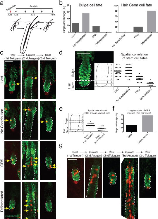
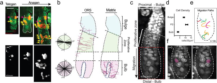
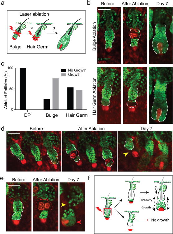
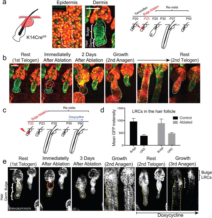
Stem-cell niches in mammalian tissues are often heterogeneous and compartmentalized; however, whether distinct niche locations determine different stem-cell fates remains unclear. To test this hypothesis, here we use the mouse hair follicle niche and combine intravital microscopy with genetic lineage tracing to re-visit the same stem-cell lineages, from their exact place of origin, throughout regeneration in live mice. Using this method, we show directly that the position of a stem cell within the hair follicle niche can predict whether it is likely to remain uncommitted, generate precursors or commit to a differentiated fate. Furthermore, using laser ablation we demonstrate that hair follicle stem cells are dispensable for regeneration, and that epithelial cells, which do not normally participate in hair growth, re-populate the lost stem-cell compartment and sustain hair regeneration. This study provides a general model for niche-induced fate determination in adult tissues.


Figures

Figure 1
Figure 1. Niche location can predict the fate of hair follicle stem cells
a) Scheme of single stem cell lineage tracing in live mice. b) Statistical analysis of the fate of stem cells originating from the bulge or hair germ (n = 108 and 20 lineages, respectively in 8 mice). c) Representative examples of single stem cell lineages (arrows) traced during a full hair cycle. Each sequence represents a different fate that correlates with a specific niche location. d) Graphical correlation between the original location of a single stem cell and its fate after a full hair cycle (n = 128; error bars represent S.E.M.). e) Spatial relocation of ORS lineages after a full hair cycle (n = 23). f) Quantification of the fate of ORS lineages in the 2nd hair cycle (n = 9). g) In vivo lineage tracing of bulge stem cells over two consecutive hair cycles. Scale bars: 50μm.
Figure 2
Figure 2. Mode of ORS growth
a) In vivo lineage tracing sequence (top) and corresponding 3D renderings of the Cre reporter (bottom) showing ORS expansion during hair growth. Arrows denote different lineages. b) Graphical representation of the location and axis of cell divisions in the ORS and Matrix (Bulb) in advanced hair follicle growth (Anagen III-IV). c) ORS cell distribution during active hair growth. d) Quantification of cell density in different regions of the outer hair follicle epithelial layer (n = 9 per region; error bars represent S.E.M.). e) Individual traces of migrating ORS nuclei (see also Video S1). f) Traces of ORS nuclei depicting their relative positions at two time-points eight hours apart (see also Video S3). Scale bars: 50μm.
Figure 3
Figure 3. Bulge and hair germ are mutually dispensable for hair regeneration
a) Scheme of the laser ablation experiment. b) Sequential snapshots of hair follicles immediately or a week after bulge and hair germ ablation. The red dotted line represents the ablated region. c) Quantification of the regenerative capacity of follicles with ablated dermal papilla, bulge and hair germ, respectively (n = 30, 32, 28 follicles, respectively in 9 mice). d) Example of hair growth impairment due to the physical separation of the epithelium from the mesenchymal dermal papilla after ablation. e) Example of hair follicle growth initiation before full niche recovery. f) Scheme of hair follicle niche responses after laser ablation. Scale bars: 50μm.
Figure 4
Figure 4. Functional reconstitution of the stem cell niche from non-hair epithelial populations
a) Scheme of in vivo lineage tracing of non-hair epithelial populations following bulge laser ablation. b) Example of bulge-ablated hair follicle showing the influx of labeled non-hair epithelial cell populations into the niche and their contribution to hair growth. c) Scheme of label retention experiment following bulge ablation. d) Quantification of label retention in control and bulge-ablated hair follicles (n = 15 and 12 follicles; error bars represent S.E.M.). e) Example of label retention in the niche following bulge ablation and recovery. Scale bars: 50μm.

References

    1. Scadden DT. The stem-cell niche as an entity of action. Nature. 2006;441:1075–1079. - PubMed
    1. Spradling AC, et al. Stem cells and their niches: integrated units that maintain Drosophila tissues. Cold Spring Harb Symp Quant Biol. 2008;73:49–57. - PubMed
    1. Fuchs E. The tortoise and the hair: slow-cycling cells in the stem cell race. Cell. 2009;137:811–819. - PMC - PubMed
    1. Li L, Clevers H. Coexistence of quiescent and active adult stem cells in mammals. Science. 2010;327:542–545. - PMC - PubMed
    1. Greco V, Guo S. Compartmentalized organization: a common and required feature of stem cell niches? Development. 2010;137:1586–1594. - PMC - PubMed

Publication types