Neurovascular decoupling is associated with severity of cerebral amyloid angiopathy
- PMID: 24097810
- PMCID: PMC3812103
- DOI: 10.1212/01.wnl.0000435291.49598.54
Neurovascular decoupling is associated with severity of cerebral amyloid angiopathy
Abstract
Objectives: We used functional MRI (fMRI), transcranial Doppler ultrasound, and visual evoked potentials (VEPs) to determine the nature of blood flow responses to functional brain activity and carbon dioxide (CO2) inhalation in patients with cerebral amyloid angiopathy (CAA), and their association with markers of CAA severity.
Methods: In a cross-sectional prospective cohort study, fMRI, transcranial Doppler ultrasound CO2 reactivity, and VEP data were compared between 18 patients with probable CAA (by Boston criteria) and 18 healthy controls, matched by sex and age. Functional MRI consisted of a visual task (viewing an alternating checkerboard pattern) and a motor task (tapping the fingers of the dominant hand).
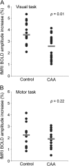
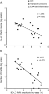
Results: Patients with CAA had lower amplitude of the fMRI response in visual cortex compared with controls (p = 0.01), but not in motor cortex (p = 0.22). In patients with CAA, lower visual cortex fMRI amplitude correlated with higher white matter lesion volume (r = -0.66, p = 0.003) and more microbleeds (r = -0.78, p < 0.001). VEP P100 amplitudes, however, did not differ between CAA and controls (p = 0.45). There were trends toward reduced CO2 reactivity in the middle cerebral artery (p = 0.10) and posterior cerebral artery (p = 0.08).
Conclusions: Impaired blood flow responses in CAA are more evident using a task to activate the occipital lobe than the frontal lobe, consistent with the gradient of increasing vascular amyloid severity from frontal to occipital lobe seen in pathologic studies. Reduced fMRI responses in CAA are caused, at least partly, by impaired vascular reactivity, and are strongly correlated with other neuroimaging markers of CAA severity.
Figures


Comment in
-
A physiologic biomarker for cerebral amyloid angiopathy.Neurology. 2013 Nov 5;81(19):1650-1. doi: 10.1212/01.wnl.0000435303.10519.15. Epub 2013 Oct 4. Neurology. 2013. PMID: 24097811
References
-
- Knudsen KA, Rosand J, Karluk D, Greenberg SM. Clinical diagnosis of cerebral amyloid angiopathy: validation of the Boston criteria. Neurology 2001;56:537–539 - PubMed
-
- Shin HK, Jones PB, Garcia-Alloza M, et al. Age-dependent cerebrovascular dysfunction in a transgenic mouse model of cerebral amyloid angiopathy. Brain 2007;130:2310–2319 - PubMed
Publication types
MeSH terms
Substances
Grants and funding
LinkOut - more resources
Full Text Sources
Other Literature Sources
