. 2013 Dec;195(24):5526-39.
doi: 10.1128/JB.01026-13. Epub 2013 Oct 4.

Identification and characterization of novel Helicobacter pylori apo-fur-regulated target genes

Affiliations

Identification and characterization of novel Helicobacter pylori apo-fur-regulated target genes

Beth M Carpenter et al. J Bacteriol. 2013 Dec.

Abstract

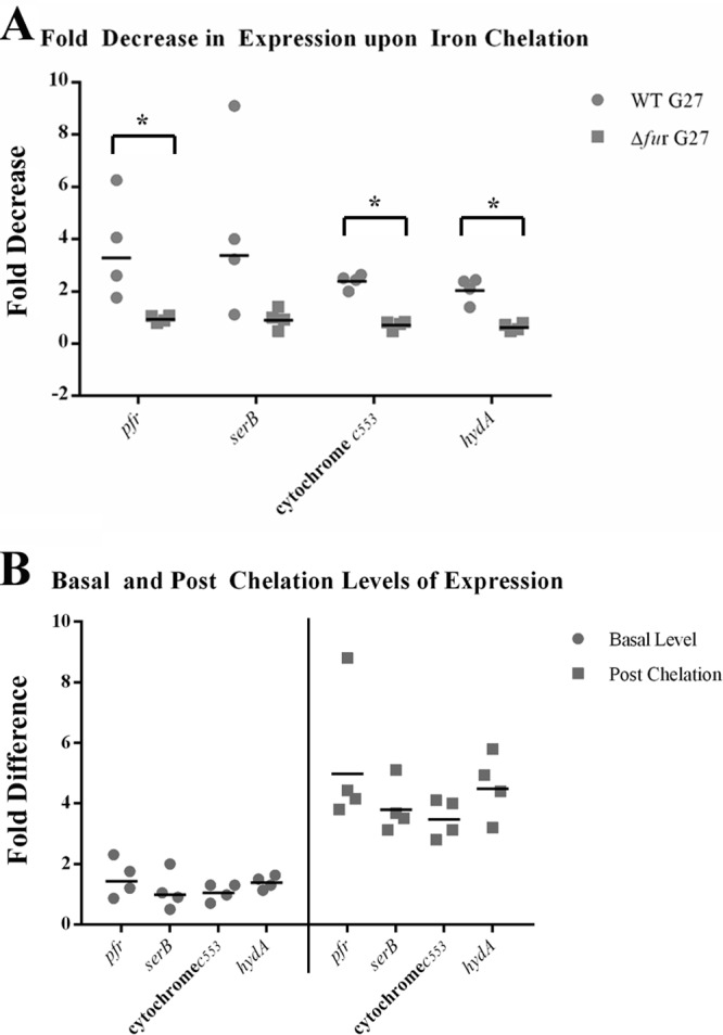
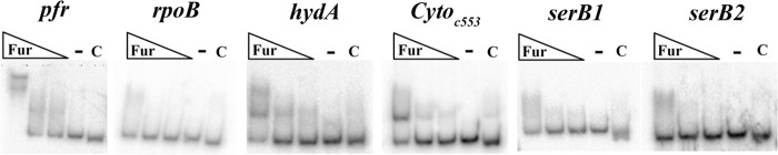
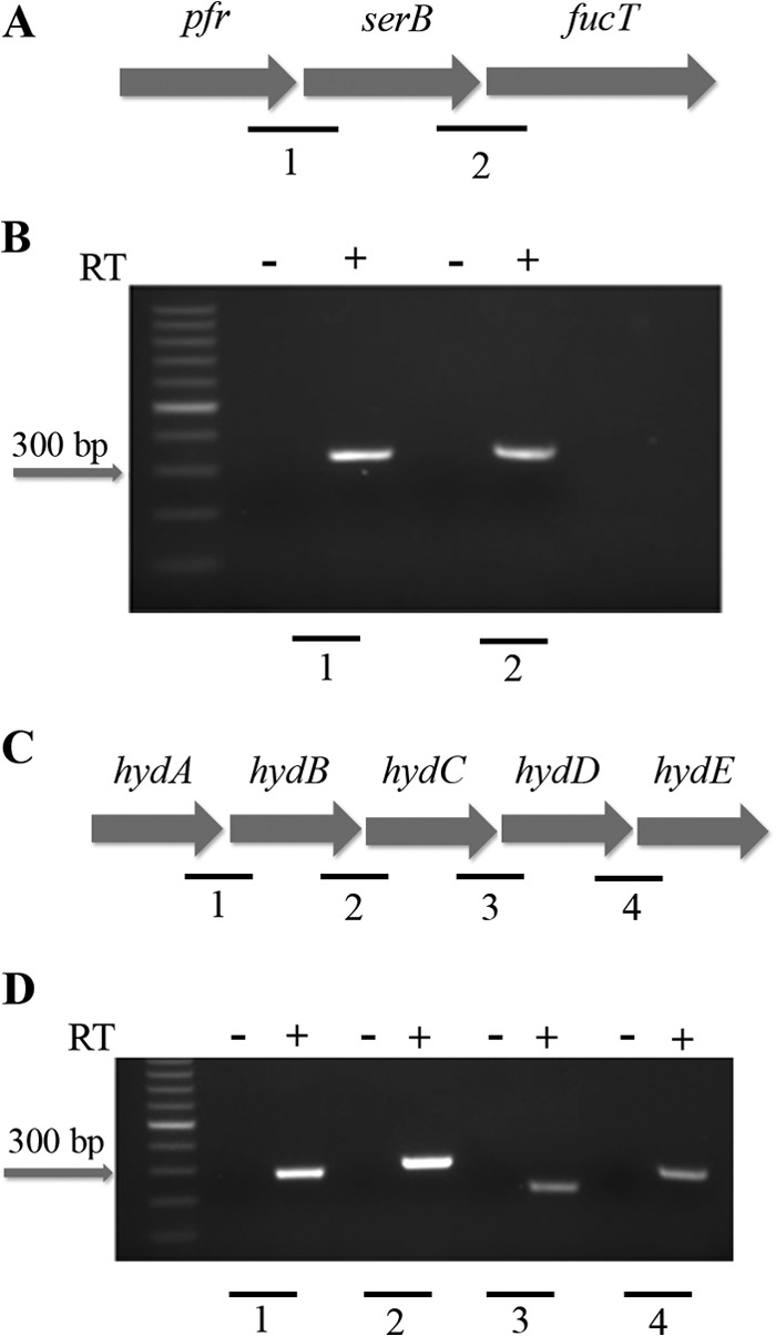
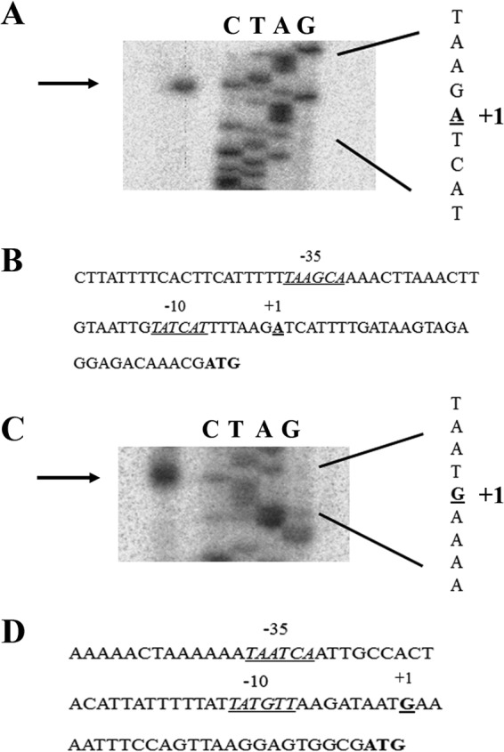
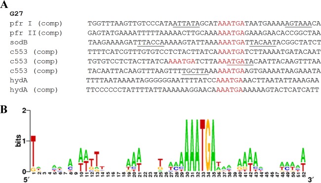
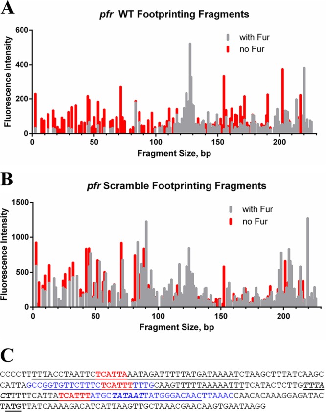
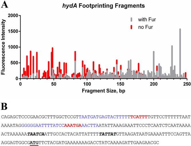
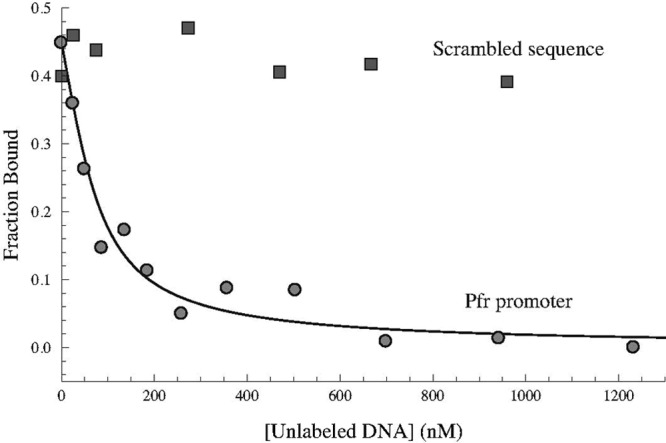
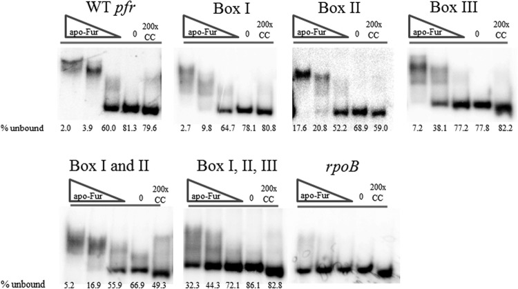
In Helicobacter pylori, the ferric uptake regulator (Fur) has evolved additional regulatory functions not seen in other bacteria; it can repress and activate different groups of genes in both its iron-bound and apo forms. Because little is understood about the process of apo-Fur repression and because only two apo-Fur-repressed genes (pfr and sodB) have previously been identified, we sought to expand our understanding of this type of regulation. Utilizing published genomic studies, we selected three potential new apo-Fur-regulated gene targets: serB, hydA, and the cytochrome c553 gene. Transcriptional analyses confirmed Fur-dependent repression of these genes in the absence of iron, as well as derepression in the absence of Fur. Binding studies showed that apo-Fur directly interacted with the suspected hydA and cytochrome c553 promoters but not that of serB, which was subsequently shown to be cotranscribed with pfr; apo-Fur-dependent regulation occurred at the pfr promoter. Alignments of apo-regulated promoter regions revealed a conserved, 6-bp consensus sequence (AAATGA). DNase I footprinting showed that this sequence lies within the protected regions of the pfr and hydA promoters. Moreover, mutation of the sequence in the pfr promoter abrogated Fur binding and DNase protection. Likewise, fluorescence anisotropy studies and binding studies with mutated consensus sequences showed that the sequence was important for apo-Fur binding to the pfr promoter. Together these studies expand the known apo-Fur regulon in H. pylori and characterize the first reported apo-Fur box sequence.


Figures

Fig 1
Fig 1
apo-Fur regulation of the cytochrome c553 gene, hydA, and serB. RNA was isolated from exponential-phase cultures of WT and Δfur strains of H. pylori under iron-replete and iron chelation conditions as detailed in Materials and Methods. Riboprobes for the cytochrome c553 gene, hydA, serB, and the control promoter, pfr, were used to evaluate changes in expression of these genes. The fold decrease in expression upon iron limitation is shown in panel A. The fold difference in the basal levels of gene expression between the Δfur and WT strains is shown on the left side of panel B. The fold difference in the postchelation levels of gene expression between the Δfur and WT strains is shown on the right side of panel B. The geometric means of the fold decrease and fold difference in the relative levels of expression from the four biological replicates are shown as black lines. An asterisk above a bracket indicates that there was a statistically significant difference in the fold decrease between WT and Δfur strains upon iron chelation (P ≤ 0.01).
Fig 2
Fig 2
Determination of apo-Fur binding to the cytochrome c553 gene, hydA, and serB promoters. EMSAs were performed using purified rFur protein and end-labeled PCR promoter fragments for the cytochrome c553 gene, hydA, and serB. Labeled pfr and rpoB promoter fragments were used as the positive and negative controls, respectively. Increasing concentrations of protein are indicated by the triangles, the no-protein control reactions are indicated by a minus signs, and the cold (unlabeled) competition reactions are indicated by the letter C. EMSAs were performed with apo-Fur (0.093 μg/ml, 0.186 μg/ml, and 0.93 μg/ml of protein), and data are representative of 2 or 3 experimental replicates.
Fig 3
Fig 3
Organization of the pfr and hydA operons. The genes encoding pfr, serB, and fucT are organized into an operon as shown in panel A. Panel B shows the PCR amplification of the gene junctions between pfr and serB and serB and fucT using cDNA (+) and no-RT (−) control reactions as templates. The organization of the hydACBDE operon is shown in panel C. Panel D shows the PCR amplification of the hydAB, hydBC, hydCD, and hydDE gene junctions using cDNA (+) and no-RT (−) control reactions as templates. The individual junctions are indicated by numbers (A and C), which correspond to the numbers shown below the gel images (B and D). Each junctional PCR was conducted twice using biologically independent cDNA templates, with similar results.
Fig 4
Fig 4
Mapping of the cytochrome c553 gene and hydA TSSs. The TSS of the cytochrome c553 gene was mapped using WT H. pylori RNA. Using the cyto_c553_PE primer, which lies within the cytochrome c553 coding region, first-strand cDNA was synthesized. Only a single band was detected, indicating a single TSS for this gene as shown in panel A. Panel B shows the cytochrome c553 gene promoter region, with the core promoter elements indicated. The TSS of the hydA promoter was mapped in a similar manner using the hydA_PE primer. A single band was detected for this promoter, which indicated a single TSS for hydA as shown in panel C. The hydA promoter region with the core promoter elements indicated is shown in panel D. The TSS is indicated by “+1”; the −10 and −35 promoter elements are underlined and labeled accordingly. The translational start codon is given in bold. Data shown are representative of a minimum of two experimental replicates.
Fig 5
Fig 5
Identification of the apo-Fur box consensus sequence. The promoter regions of the four characterized H. pylori apo-Fur repressed genes, pfr, sodB, the cytochrome c553 gene (c553), and hydA, were aligned (A), and a sequence logo (28) was generated (B). The logo is composed of stacks of letters, one for each position of the sequence. Sequence conservation at each position is indicated by the height of the letter stack (measured in bits). The apo-Fur box sequence(s) found in each promoter is colored in red. Other nearby promoter elements (−10 and −35 conserved sequences) are underlined. “comp” indicates that the complementary strand of DNA was used in the alignment. Designations of pfr I and pfr II are from the previously described protected regions (5).
Fig 6
Fig 6
DNase I footprinting of the pfr promoter. A fragment of the pfr promoter fluorescently labeled at the 3′ end was subjected to DNase I digestion in the absence and presence of apo-Fur (A). A fragment of the pfr promoter fluorescently labeled at the 3′ end and containing ACACAC scrambled sequence in place of AAATGA in the first apo-Fur box was subjected to DNase I digestion in the absence and presence of apo-Fur (B). Protected regions are those with reduced peak height and/or entirely missing peaks in the presence of apo-Fur (gray lines) compared to digestion fragments in the absence of Fur (red lines). (C) Sequence of the pfr promoter fragment utilized in the footprinting experiments. The conserved apo-Fur box sequences are in red, and the protected regions as identified through DNase I footprinting are in blue. The −10 and −35 promoter elements are shown in bold italics; the ATG start codon is in bold and underlined. The previously reported protected regions for the pfr promoter (5) are underlined.
Fig 7
Fig 7
DNase I footprinting of the hydA promoter. A fragment of the hydA promoter fluorescently labeled at the 5′ end was subjected to DNase I digestion in the absence and presence of apo-Fur (A). Protected regions are those with reduced peak height and/or entirely missing peaks in the presence of apo-Fur (gray lines) compared to digestion fragments in the absence of Fur (red lines). (B) Sequence of the hydA promoter fragment utilized in the footprinting experiments. The conserved apo-Fur box sequences are in red, and the protected regions as identified through DNase I footprinting are in blue. The −10 and −35 promoter elements are shown in bold italics; the ATG start codon is in bold and underlined.
Fig 8
Fig 8
Specific interaction between apo-Fur and the apo-Fur box consensus sequence. Unlabeled WT and scrambled pfr promoter sequences were used to titrate labeled WT pfr promoter bound to apo-Fur in fluorescence anisotropy studies. As the concentration of unlabeled WT pfr promoter was increased (circles), it competed with the labeled promoter for binding to apo-Fur and successfully titrated the labeled promoter fragment. The Kd was determined to be 14 ± 5 nM, and the R-squared value for the best fit line was 0.981. Increasing concentrations of unlabeled scrambled pfr promoter fragment (squares) were unable to titrate the labeled WT pfr fragments from apo-Fur.
Fig 9
Fig 9
Determination of the role of the AAATGA sequences in apo-Fur binding to the pfr promoter. EMSAs were performed using purified rFur protein and end-labeled promoter fragments for pfr. Box I, box II, and box III indicate fragments where the AAATGA sequence has been scrambled alone or in various combinations. Labeled WT pfr and rpoB promoter fragments were used as the positive and negative controls, respectively. Increasing concentrations of protein are indicated by the triangles, the no-protein control reactions are indicated by minus signs, and the cold (unlabeled) competition reactions are indicated by the letter C. EMSAs were performed with apo-Fur (0.406 μg/ml, 0.703 μg/ml, and 0.141 μg/ml of protein), and data are representative of 2 or 3 experimental replicates. The average percentage of unbound labeled promoter from all of the experimental replicates is given below each reaction lane.

References

    1. Dunn BE, Cohen H, Blaser MJ. 1997. Helicobacter pylori. Clin. Microbiol. Rev. 10:720–741 - PMC - PubMed
    1. Blaser MJ. 1990. Helicobacter pylori and the pathogenesis of gastroduodenal inflammation. J. Infect. Dis. 161:626–633 - PubMed
    1. Asaka M, Kudo M, Kato M, Sugiyama T, Takeda H. 1998. Review article: long-term Helicobacter pylori infection—from gastritis to gastric cancer. Aliment. Pharmacol. Ther. 12(Suppl 1):9–15 - PubMed
    1. Delany I, Pacheco AB, Spohn G, Rappuoli R, Scarlato V. 2001. Iron-dependent transcription of the frpB gene of Helicobacter pylori is controlled by the Fur repressor protein. J. Bacteriol. 183:4932–4937 - PMC - PubMed
    1. Delany I, Spohn G, Rappuoli R, Scarlato V. 2001. The Fur repressor controls transcription of iron-activated and -repressed genes in Helicobacter pylori. Mol. Microbiol. 42:1297–1309 - PubMed

Publication types

LinkOut - more resources