. 2013 Nov 29;288(48):34612-26.
doi: 10.1074/jbc.M113.494369. Epub 2013 Oct 4.

Heme oxygenase-1 exerts a protective role in ovalbumin-induced neutrophilic airway inflammation by inhibiting Th17 cell-mediated immune response

Affiliations

Heme oxygenase-1 exerts a protective role in ovalbumin-induced neutrophilic airway inflammation by inhibiting Th17 cell-mediated immune response

Yanjie Zhang et al. J Biol Chem. .

Abstract

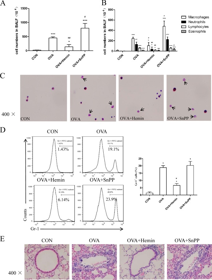
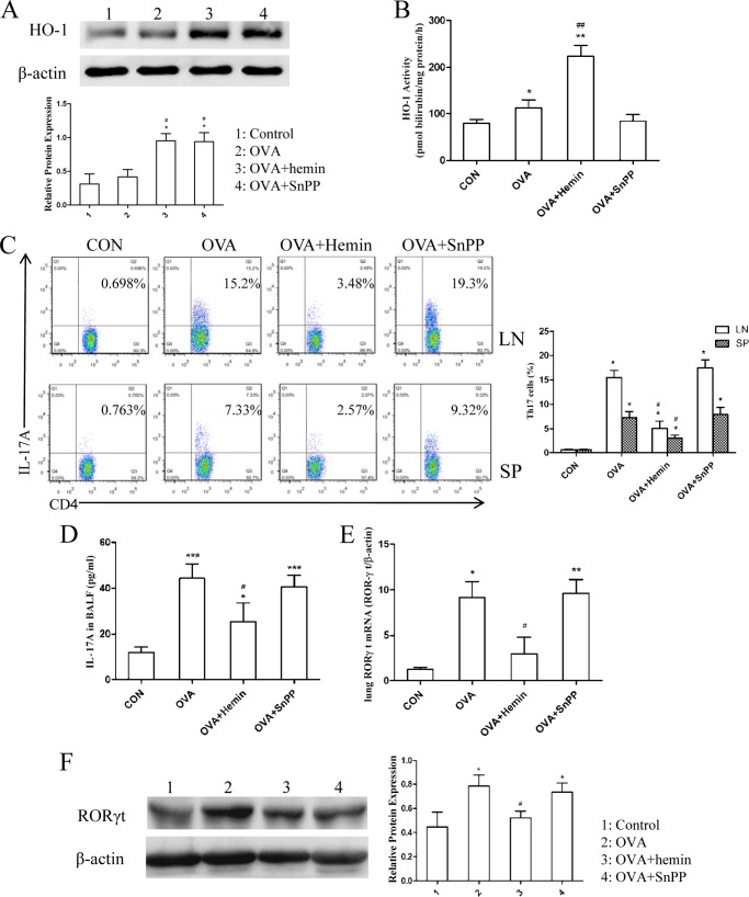
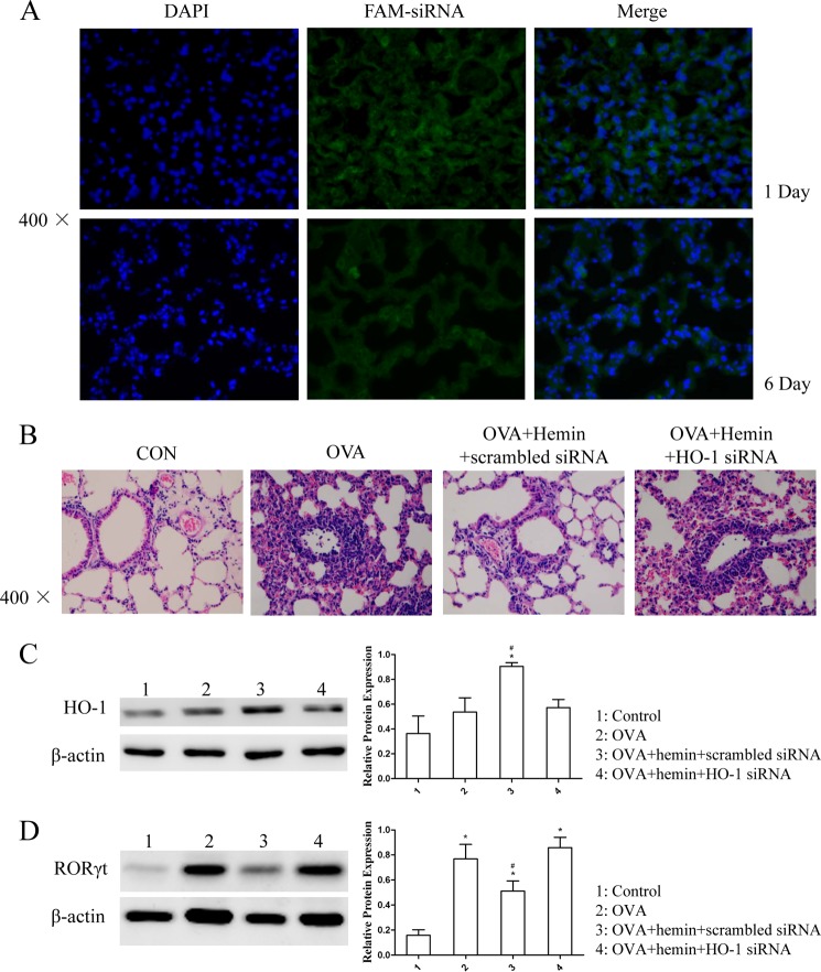
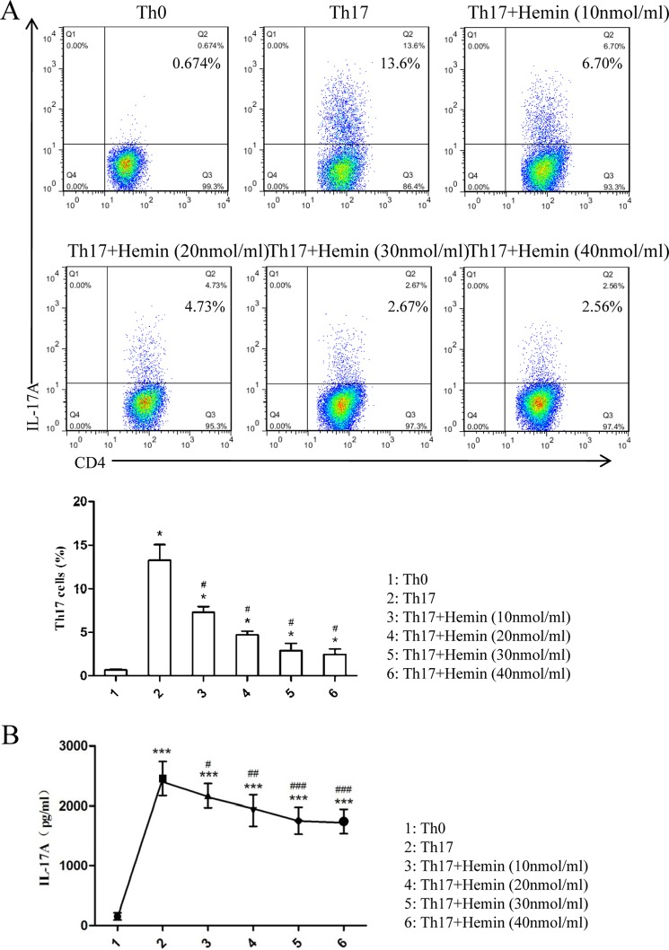
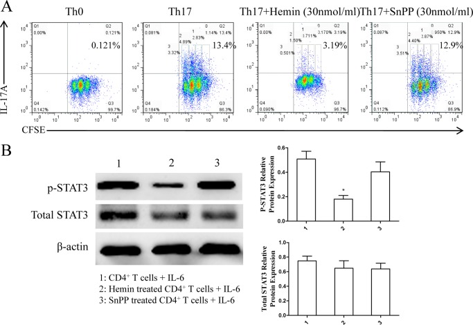
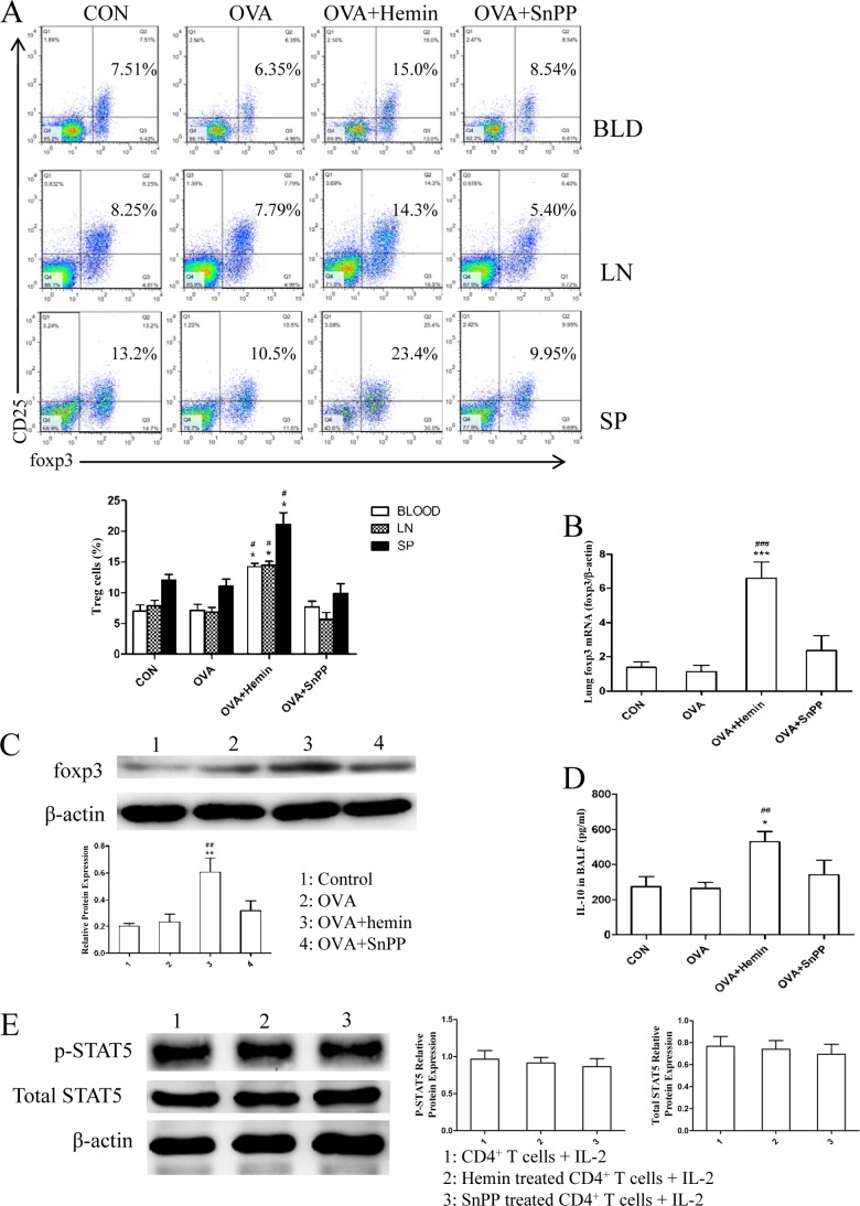
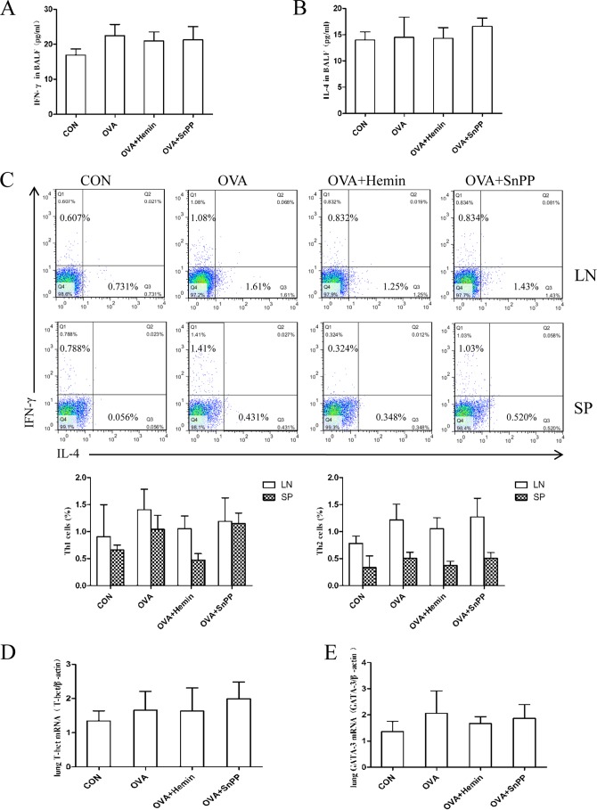
Allergic asthma is conventionally considered as a Th2 immune response characterized by eosinophilic inflammation. Recent investigations revealed that Th17 cells play an important role in the pathogenesis of non-eosinophilic asthma (NEA), resulting in steroid-resistant neutrophilic airway inflammation. Heme oxygenase-1 (HO-1) has anti-inflammation, anti-oxidation, and anti-apoptosis functions. However, its role in NEA is still unclear. Here, we explore the role of HO-1 in a mouse model of NEA. HO-1 inducer hemin or HO-1 inhibitor tin protoporphyrin IX was injected intraperitoneally into ovalbumin-challenged DO11.10 mice. Small interfering RNA (siRNA) was delivered into mice to knock down HO-1 expression. The results show that induction of HO-1 by hemin attenuated airway inflammation and decreased neutrophil infiltration in bronchial alveolar lavage fluid and was accompanied by a lower proportion of Th17 cells in mediastinal lymph nodes and spleen. More importantly, induction of HO-1 down-regulated Th17-related transcription factor retinoic acid-related orphan receptor γt (RORγt) expression and decreased IL-17A levels, all of which correlated with a decrease in phosphorylated STAT3 (p-STAT3) level and inhibition of Th17 cell differentiation. Consistently, the above events could be reversed by tin protoporphyrin IX. Also, HO-1 siRNA transfection abolished the effect of hemin induced HO-1 in vivo. Meanwhile, the hemin treatment promoted the level of Foxp3 expression and enhanced the proportion of regulatory T cells (Tregs). Collectively, our findings indicate that HO-1 exhibits anti-inflammatory activity in the mouse model of NEA via inhibition of the p-STAT3-RORγt pathway, regulating kinetics of RORγt and Foxp3 expression, thus providing a possible novel therapeutic target in asthmatic patients.

Keywords: Asthma; Heme Oxygenase; Immunology; Inflammation; T Cell; Th17; Treg.


Figures

FIGURE 1.
FIGURE 1.
HO-1 alleviates OVA-induced neutrophilic airway inflammation in DO11.10 mice. A, the total cell counts in BALF (* compared with the control (CON) group: *, p < 0.05; ***, p < 0.001; # with the OVA group: #, p < 0.05; ##, p < 0.01). B, the differential cell counts identified by morphologic criteria in BALF (* compared with the control group: *, p < 0.05; **, p < 0.01; ***, p < 0.001; # with the OVA group: #, p < 0.05; ##, p < 0.01). C, cytospin preparations of BALF cells from four groups stained with hematoxylin and eosin (original magnification ×400). Arrows indicate neutrophils with lobed nucleus in BALF. D, flow cytometric analysis of BALF cells from four groups. Numbers in the graph indicate the percentages of neutrophils (Gr-1+) (* compared with the control group: *p < 0.05; # with OVA group, ##p < 0.01). E, histological analysis of lung tissues isolated from DO11.10 mice in control, OVA, OVA + hemin, and OVA + SnPP groups. Paraffin-embedded lung sections were prepared 24 h after the last OVA challenge and were stained with hematoxylin and eosin to observe inflammation (original magnification ×400). Each symbol in the graph represents an individual mouse (n = 6). All results shown are representative of three independent experiments.
FIGURE 2.
FIGURE 2.
Induction of HO-1 suppresses Th17-mediated response in DO11.10 mice. A, Western blot analysis of HO-1 protein expression in lung tissues extracted from four groups. β-Actin was used as the loading control. Densitometry analysis was performed by normalizing to β-actin levels (* compared with the control group: *, p < 0.05; # with compared with the OVA group: #, p < 0.05). B, the analysis of HO-1 activity in lung tissues extracted from four groups (* compared with the control (CON) group: *, p < 0.05; **, p < 0.01; # compared with the OVA group; ##, p < 0.01). C, flow cytometric analysis of mediastinal lymph node (LN) and spleen (SP) cells isolated from DO11.10 mice in control, OVA, OVA + hemin, and OVA + SnPP groups. Numbers in the upper right quadrants indicate the percentages of Th17 (CD4+IL-17+) cells gated on CD4+ T cells. Each symbol in the graph represents an individual mouse (n = 6). D, ELISA analysis of IL-17A in BALF from four groups (* compared with the control group: *, p < 0.05; ***, p < 0.001; # compared with the OVA group: #. p < 0.05). E, real-time PCR analysis of RORγt mRNA in lung tissues isolated from four groups (* compared with the control group: *, p < 0.05; **, p < 0.01; # compared with the OVA group, #p < 0.05). F, Western blot analysis of RORγt protein expression in lung tissues extracted from four groups. β-Actin was used as the loading control. Densitometry analysis was performed by normalizing to β-actin levels (* compared with the control group: *, p < 0.05; #, compared with the OVA group: #, p < 0.05). All results shown are representative of three independent experiments.
FIGURE 3.
FIGURE 3.
Silencing of HO-1 with siRNA abolished the effect of HO-1 induction in vivo. A, the distribution of FAM-siRNA in lung tissues isolated from DO11.10 mice. OCT-embedded lung sections were prepared 24 h after transfection (day −2) and 1 day after the last OVA challenge (6 days after the transfection (day 3)) and stained with DAPI. The FAM-expressing cells in lung tissues were visualized under a fluorescence microscope (original magnification ×400). B, histological analysis of lung tissues isolated from DO11.10 mice in control (CON), OVA, OVA + hemin + scrambled siRNA, and OVA + hemin + HO-1 siRNA groups. Paraffin-embedded lung sections were prepared 1 day after the transfection (day −2) and 1 day after the last OVA challenge (6 days after the transfection (day 3)) and stained with hematoxylin and eosin to observe inflammation (original magnification ×400). Each symbol in the graph represents an individual mouse (n = 6). C, Western blot analysis of HO-1 protein expression in lung tissues was extracted from the four groups. β-Actin was used as the loading control. Densitometry analysis was performed by normalizing to β-actin levels (* compared with the control (CON) group: *, p < 0.05; #, compared with the OVA group: #, p < 0.05). D, Western blot analysis of RORγt protein expression in lung tissues extracted from the four groups. β-Actin was used as the loading control. Densitometry analysis was performed by normalizing to β-actin levels (* compared with the control group: *, p < 0.05; # compared with the OVA group: #, p < 0.05). All results shown are representative of three independent experiments.
FIGURE 4.
FIGURE 4.
Hemin inhibits Th17 cell differentiation in a dose-dependent manner in vitro. A, flow cytometric analysis of magnetically purified naïve T cells from spleens of BALB/c mice cultured under Th17-skewing conditions with or without different concentrations of hemin (10, 20, 30, 40 nmol/ml) for 3 days. Numbers in the upper right quadrants indicate the percentages of Th17 (CD4+ IL-17+) cells gated on CD4+ T cells (* compared with the control group: *, p < 0.05; #, with OVA group; #, p < 0.05). B, ELISA analysis of IL-17A in supernatants of cultured naïve T cells (* compared with the Th0 condition: *. p < 0.05; **, p < 0.01; ***, p < 0.001; # with the Th17-skewing condition: #, p < 0.05, ##, p < 0.01; ###, p < 0.001). All results shown are representative of three independent experiments.
FIGURE 5.
FIGURE 5.
Th17 cell differentiation, CD4+ T cell proliferation, and STAT3 phosphorylation are inhibited by hemin but not by SnPP. A, flow cytometric analysis of magnetically purified naïve T cells from spleens of BALB/c mice cultured under Th17-skewing conditions with or without hemin or SnPP (30 nmol/ml) for 3 days. Numbers in the upper right quadrants indicate the percentages of Th17 (CD4+IL-17+) cells as well as each generation of Th17 cells (undivided cells (0), generation 1 (1), generation 2 (2), and generation 3 (3)) gated on CD4+ T cells. B, Western blot analysis of purified CD4+ T cells isolated from DO11.10 mice pretreated with or without hemin and SnPP and cultured with IL-6 for 1 h. β-Actin was used as the loading control. Densitometry analysis of p-STAT3 and total STAT3 was performed by normalizing to β-actin levels (* compared with CD4+ T cells from untreated mice: *, p < 0.05). All results shown are representative of three independent experiments.
FIGURE 6.
FIGURE 6.
HO-1 up-regulates Tregs and enhances their functions in vivo but has no significant effect on STAT5 phosphorylation. A, flow cytometric analysis of peripheral blood (BLD), mediastinal lymph node (LN), and spleen (SP) cells isolated from DO11.10 mice in control, OVA, OVA + hemin, and OVA + SnPP groups. Numbers in the upper right quadrants indicate the percentages of Tregs (CD4+CD25+Foxp3+) gated on CD4+ T cells (* compared with control group: *, p < 0.05; #, with OVA group; #, p < 0.05). Each symbol in the graph represents an individual mouse (n = 6). B, real-time PCR analysis of Foxp3 mRNA in lung tissues isolated from four groups (* compared with the control (CON) group: ***, p < 0.001; # compared with the OVA group: ###, p < 0.001). C, Western blot analysis of Foxp3 protein expression in lung tissues extracted from four groups. β-Actin was used as the loading control. Densitometry analysis was performed by normalizing to β-actin levels (* compared with the control group: **, p < 0.01; # compared with the OVA group: ##, p < 0.01). D, ELISA analysis of IL-10 in BALF collected from four groups (* compared with the control group: *, p < 0.05; # compared with the OVA group: ##, p < 0.01). E, Western blot analysis of purified CD4+ T cells isolated from DO11.10 mice pretreated with or without hemin and SnPP and cultured with IL-2 for 1 h. β-Actin was used as loading control. Densitometry analysis of p-STAT5 and total STAT5 was performed by normalizing to β-actin levels. All results shown are representative of three independent experiments.
FIGURE 7.
FIGURE 7.
HO-1 does not noticeably affect Th1- or Th2-mediated response in DO11.10 mice. A, ELISA analysis of IFN-γ in BALF obtained from four groups. CON, control. B, ELISA analysis of IL-4 in BALF collected from four groups. C, flow cytometric analysis of mediastinal lymph node (LN) and spleen (SP) cells isolated from DO11.10 mice in control, OVA, OVA + hemin, and OVA + SnPP groups. Numbers in the upper left and lower right quadrants indicate the percentages of Th1 (CD4+IFN-γ+) cells and Th2 (CD4+IL-4+) cells gated on CD4+ T cells, respectively. Each symbol in the graph represents an individual mouse (n = 6). D, real-time PCR analysis of T-bet mRNAs in lung tissues from four groups. E, real-time PCR analysis of GATA-binding protein 3 mRNAs in lung tissues from four groups.

References

    1. Douwes J., Gibson P., Pekkanen J., Pearce N. (2002) Non-eosinophilic asthma. Importance and possible mechanisms. Thorax 57, 643–648 - PMC - PubMed
    1. Simpson J. L., Scott R., Boyle M. J., Gibson P. G. (2006) Inflammatory subtypes in asthma. Assessment and identification using induced sputum. Respirology 11, 54–61 - PubMed
    1. Kikuchi S., Nagata M., Kikuchi I., Hagiwara K., Kanazawa M. (2005) Association between neutrophilic and eosinophilic inflammation in patients with severe persistent asthma. Int. Arch. Allergy Immunol. 137, 7–11 - PubMed
    1. The ENFUMOSA Study Group (2003) The ENFUMOSA cross-sectional European multicentre study of the clinical phenotype of chronic severe asthma. Eur. Respir. J. 22, 470–477 - PubMed
    1. Oboki K., Ohno T., Saito H., Nakae S. (2008) Th17 and allergy. Allergol Int. 57, 121–134 - PubMed

Publication types

MeSH terms