. 2013 Nov 22;288(47):34131-34145.
doi: 10.1074/jbc.M113.507202. Epub 2013 Oct 4.

Structural insights into functional overlapping and differentiation among myosin V motors

Affiliations

Structural insights into functional overlapping and differentiation among myosin V motors

Andrey F Z Nascimento et al. J Biol Chem. .

Abstract

Myosin V (MyoV) motors have been implicated in the intracellular transport of diverse cargoes including vesicles, organelles, RNA-protein complexes, and regulatory proteins. Here, we have solved the cargo-binding domain (CBD) structures of the three human MyoV paralogs (Va, Vb, and Vc), revealing subtle structural changes that drive functional differentiation and a novel redox mechanism controlling the CBD dimerization process, which is unique for the MyoVc subclass. Moreover, the cargo- and motor-binding sites were structurally assigned, indicating the conservation of residues involved in the recognition of adaptors for peroxisome transport and providing high resolution insights into motor domain inhibition by CBD. These results contribute to understanding the structural requirements for cargo transport, autoinhibition, and regulatory mechanisms in myosin V motors.

Keywords: Cargo-binding Domain; Cell Signaling; Crystal Structure; Intracellular Trafficking; Molecular Motors; Molecular Plasticity; Myosin; Myosin V; Redox Dimerization.


Figures
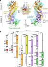
FIGURE 1.
FIGURE 1.
The molecular architecture of mammalian MyoV-CBDs. a, cartoon representation of the MyoVa-CBD showing the overall structure of human MyoV-CBDs, which are constituted by helical bundles I (helices α2–α5, orange) and II (helices α7-α10, purple), a long α-helical segment (α6 or cervical helix, yellow), a three-helical motif (helices α11-α13, green), a long C-terminal extension (surface, pale blue), and a β-sheet (β1 and β14, red). The M face (left) displays residues (represented by sticks) that interact with the MyoVa-MD according to Li et al. (21). The C face (right) comprises most of the known cargo-binding sites described for the yeast Myo2p-CBD (54). The dotted line represents the phospho loop removed by limited proteolysis. b, MyoVa-CBD topology diagram showing the structural elements that constitute lobule I (β-sheet, bundle I, first half of α6 and second half of C-terminal extension) and lobule II (second half of α6, bundle II, three helical motif, and first half of C-terminal extension). Conserved secondary structural elements are highlighted using the same color code as in a. Numbering is based on the isoform 1 of MyoVa (UNIPROT accession code Q9Y4I1).
FIGURE 2.
FIGURE 2.
Schematic representation of all constructs designed for human MyoVa-CBD (I–VII). Construct III was used to design constructs for MyoVb and MyoVc CBDs. ABCDE(F), the exon region; N, N-terminal region important for protein solubility; bd I, β1 and helical bundle I; α6, cervical helix; bd II, helical bundle II; P, phospho loop; hm, three-helical motif; C, C-terminal region, including β14. The proximal, medial, and distal tails refer to the different regions of the MyoVa tail domain.
FIGURE 3.
FIGURE 3.
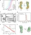
The key role of hydrophobic interactions in MyoVa-CBD stability. a, thermal shift analysis of MyoVa-CBD showing the increase of thermal stability with the increase of NaCl concentration (1–500 mm). With 500 mm NaCl, a ΔTm of ∼6 °C was observed. b, this behavior in solution is associated with a greater number of hydrophobic interactions in the inner core of the MyoVa-CBD (yellow spheres), as well as between the C-terminal extension (cyan spheres) and helices from both lobules. Residues involved in hydrophobic interactions (spheres) were defined by the Protein Interaction Calculator Server, using the interaction distance cutoff of 4 Å. c, electrophoretic pattern of human MyoVa/b/c-CBD, uncleaved and cleaved by limited proteolysis using trypsin (MyoVa-CBD), chymotrypsin (MyoVb-CBD), or chymotrypsin/thermolysin (MyoVc-CBD). The numbers below the arrows indicate the time of incubation in hours. M, molecular mass marker. N, uncleaved protein. d, analytical SEC of MyoVa-CBD before and after limited proteolysis showing that the proteolysis products elute as a single entity. e and f, SAXS analysis of the cleaved MyoVa-CBD indicating a typical scattering (e) and p(r) curve (e, inset) of an elongated-globular protein resulting in an envelope (f) into which the entire crystallographic model of the MyoVa-CBD (green) was fitted.
FIGURE 4.
FIGURE 4.
The divergent MyoVc. a, structural superposition of MyoVa-CBD (light gray or orange) and MyoVc-CBD (dark gray or blue) showing a more curved structure in MyoVc-CBD caused by angular displacement between the two lobules. In the zoom are identified the α2-α3 loop from both myosins, in which differences probably account for the observed angular shift in MyoVc. b, MyoVc crystallographic dimer suggesting that MyoVc-CBD dimerization can be regulated by a redox mechanism via Cys1600 and Cys1608 disulfide bonds. The buried area and ΔG values from the dimeric interface were calculated using the program PDBePISA (65). c, SDS-PAGE analysis of the MyoVc-CBD in nonreducing (without β-mercaptoethanol) and reducing (β-mercaptoethanol-containing) sample buffers. Monomer and dimer bands are indicated by arrowheads. M, molecular mass marker. d, analytical SEC of wild type and site-directed mutants of MyoVc-CBD showing that H2O2 induces the formation of dimers in wt (top panel) and single mutants (middle panel, C1600A or C1608A) but not in the double mutant (bottom panel, C1600A/C1608A). In reducing conditions, wild type behaved as monomer (top panel) as well as the mutants (data not shown).
FIGURE 5.
FIGURE 5.
Regulation of mammalian MyoV-CBDs. a, the phospho loop sequence from human MyoV paralogs highlighting the serine residue liable to phosphorylation (red). b, the Porod-Debye plot of MyoVa-CBD SAXS data showing a decrease of flexibility in the phospho-mimetic mutant S1652E by the formation of a plateau in comparison with the wild type.
FIGURE 6.
FIGURE 6.
Surface charge distribution in MyoV-CBDs. The positively charged clusters conserved in mammalian MyoV-CBDs are highlighted by gray and yellow ellipses, respectively. The charge distribution on the C face is variable, suggesting this region as relevant for functional divergence. Surfaces are colored by charge from red (−1 kV) to blue (+1 kV). Electrostatic potentials were calculated using PBEQ Solver (66). Myo2p-CBD is Protein Data Bank accession code 2F6H, and Myo4p-CBD is Protein Data Bank accession code 3MMI.
FIGURE 7.
FIGURE 7.
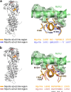
Structural (dis-)similarities between yeast and mammalian MyoV members. Myo2p-CBD (a) and Myo4p-CBD (b) superposition on the human MyoVa-CBD highlighting the major differences. Myo2p (Protein Data Bank accession code 2F6H) and Myo4p (Protein Data Bank accession code 3MMI) root mean square deviations for aligned Cα atoms are 2.5 and 4.8 Å, respectively. a, the left inset shows a zoom in to the M face illustrating the antiparallel N- and C-terminal β-sheet in mammalian MyoV-CBDs in contrast to the α-helices at the corresponding region in the yeast Myo2p. The right inset shows a zoom in to the C face highlighting the α2/α3 loop that contributes to changes in the angular curvature of Myo2p in relation to human MyoVa/Vb-CBDs. b, zoom of lobule I highlighting the structural differences among Myo4p and human MyoV-CBDs (left) and the shortened loop corresponding to the phospho loop in mammals (right). The dotted lines in all panels illustrate portions of disordered or cleaved loops absent from the crystallographic models.
FIGURE 8.
FIGURE 8.
The conservation of cargo-binding sites from yeast to human. a, the binding sites for Vac17 and Mmr1 in the yeast Myo2p are located at lobule I (M face) and poorly conserved in mammalian MyoV-CBDs. b, lobule II (C face) concentrates most of the known cargo binding sites in yeast Myo2p including for Kar9, Sec4, Ypt11, Ypt31/32, and Inp2 (54). Interestingly, all residues involved in the binding to Inp2 (highlighted with dashed lines on Myo2p), an adaptor to peroxisome transport, are conserved in mammalian MyoVa and MyoVb CBDs. In MyoVc-CBD, two of the conserved residues (Trp1713 and Tyr1721) are replaced with cysteines (Cys1600 and Cys1608), supporting the notion that the redox dimerization is a regulatory mechanism unique to this subclass.
FIGURE 9.
FIGURE 9.
PTEN-binding site analysis. The lysine cluster involved in the binding of phosphorylated PTEN (a–e) is conserved between mammalian MyoVa and MyoVb (a and c) and partially conserved in MyoVc (f and g) and in the yeast ortholog Myo2p (h and i). Surfaces (e, g, and i) are colored by charge from red (−1 kV) to blue (+1 kV). Electrostatic potentials (e, g, and i) were calculated using PBEQ Solver (65).
FIGURE 10.
FIGURE 10.
The RILPL2-binding site. Structural superposition of MyoVb (a) and MyoVc (b) with the RILPL2-MyoVa complex (Protein Data Bank accession code 4KP3). MyoVb is not able to bind RILPL2 because of the lack of a short helix at the α2-α3 link region with hydrophobic residues to mediate the contacts with helices α2 (residues numbers with asterisks) and α3N from RILPL2 (a, zoom). On the other hand, despite the greater divergence of MyoVa-CBD to MyoVc-CBD, this region is conserved with the same topological arrangements and the presence of two hydrophobic residues (Val1387 and Met1391) on the surface of the short helix within the α2-α3 link region (b, zoom).
FIGURE 11.
FIGURE 11.
High resolution analysis of the interaction between the motor and cargo-binding domains of MyoVa. a, the binding mode of MyoVa-CBD to MyoVa-MD based on docking analysis using restraints from the site-directed mutagenesis studies of Li et al. (21). MyoVa-MD subdomains (surface) were named according to Coureaux et al. (64). b, details of the interface between MyoVa-CBD and MyoVa-MD highlighting the multiple electrostatic contacts, including the interaction between Asp136 from MD and Lys1781 from CBD that are essential for stabilization of the inactive folded conformation (21, 23, 62).
FIGURE 12.
FIGURE 12.
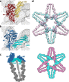
A structural model for the inhibited conformation of MyoVa. a, superposition of the CBD-MD complex (this study) on the 2DFS model proposed by Liu et al. (22) indicating a disagreement with EM experimental data (electron density map shown in gray). b, the best fit of the CBD-MD complex into the cryo-EM electron density map (gray) obtained by rigid body refinement that resembles the single-particle images (23). c, a structural model for the folded conformation of MyoVa based on the CBD-MD complex docked into the cryo-EM map in a similar orientation observed in the single-molecule studies (23). d, the fitting of the six MyoVa molecules into the rosette arrangement (upper panel) according to our results with the CBD-MD interface formed by intramolecular contacts without domain swapping, which is in agreement with the reinterpretation proposed by Sellers et al. (53). In this configuration, each molecule is tilted and laid over the adjacent molecule to form the rosette architecture as schematically represented (lower panel).

References

    1. Desnos C., Huet S., Darchen F. (2007) “Should I stay or should I go?” Myosin V function in organelle trafficking. Biol. Cell 99, 411–423 - PubMed
    1. Hammer J. A., 3rd, Sellers J. R. (2012) Walking to work. Roles for class V myosins as cargo transporters. Nat. Rev. Mol. Cell Biol. 13, 13–26 - PubMed
    1. Sellers J. R., Veigel C. (2006) Walking with myosin V. Curr. Opin. Cell Biol. 18, 68–73 - PubMed
    1. Trybus K. M. (2008) Myosin V from head to tail. Cell Mol. Life Sci. 65, 1378–1389 - PMC - PubMed
    1. Bement W. M., Hasson T., Wirth J. A., Cheney R. E., Mooseker M. S. (1994) Identification and overlapping expression of multiple unconventional myosin genes in vertebrate cell types. Proc. Natl. Acad. Sci. U.S.A. 91, 11767. - PMC - PubMed

Publication types