. 2013;9(10):e1003249.
doi: 10.1371/journal.pcbi.1003249. Epub 2013 Oct 3.

Ligand clouds around protein clouds: a scenario of ligand binding with intrinsically disordered proteins

Affiliations

Ligand clouds around protein clouds: a scenario of ligand binding with intrinsically disordered proteins

Fan Jin et al. PLoS Comput Biol. 2013.

Abstract

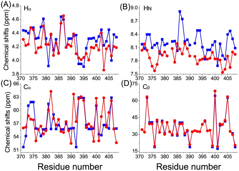
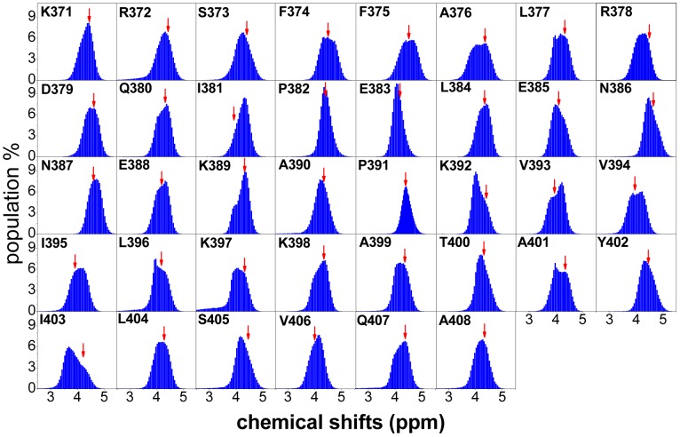
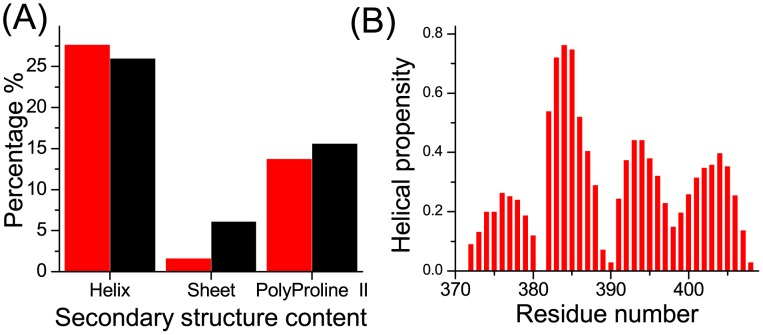
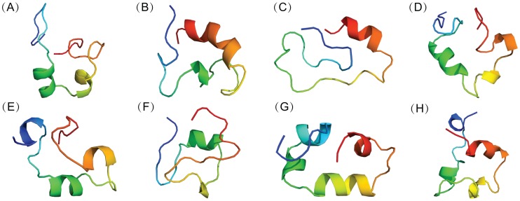
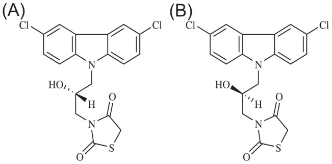
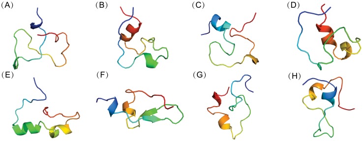
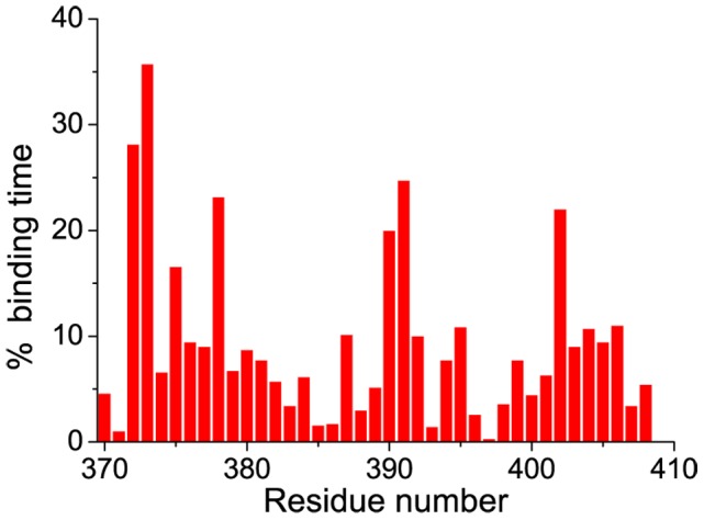
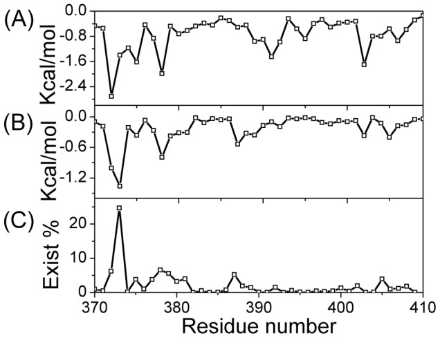
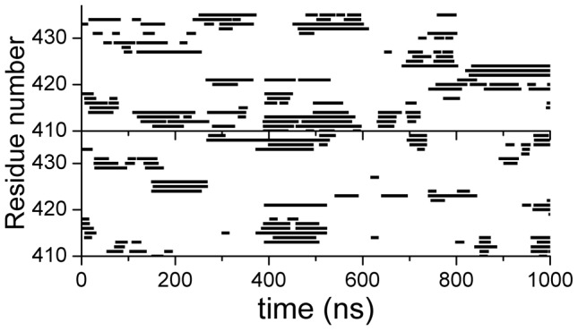
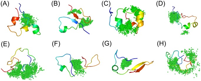
Intrinsically disordered proteins (IDPs) were found to be widely associated with human diseases and may serve as potential drug design targets. However, drug design targeting IDPs is still in the very early stages. Progress in drug design is usually achieved using experimental screening; however, the structural disorder of IDPs makes it difficult to characterize their interaction with ligands using experiments alone. To better understand the structure of IDPs and their interactions with small molecule ligands, we performed extensive simulations on the c-Myc₃₇₀₋₄₀₉ peptide and its binding to a reported small molecule inhibitor, ligand 10074-A4. We found that the conformational space of the apo c-Myc₃₇₀₋₄₀₉ peptide was rather dispersed and that the conformations of the peptide were stabilized mainly by charge interactions and hydrogen bonds. Under the binding of the ligand, c-Myc₃₇₀₋₄₀₉ remained disordered. The ligand was found to bind to c-Myc₃₇₀₋₄₀₉ at different sites along the chain and behaved like a 'ligand cloud'. In contrast to ligand binding to more rigid target proteins that usually results in a dominant bound structure, ligand binding to IDPs may better be described as ligand clouds around protein clouds. Nevertheless, the binding of the ligand and a non-ligand to the c-Myc₃₇₀₋₄₀₉ target could be clearly distinguished. The present study provides insights that will help improve rational drug design that targets IDPs.


Conflict of interest statement

The authors have declared that no competing interests exist.

Figures

Figure 1
Figure 1. Comparisons of the computed and experimental chemical shifts for apo c-Myc370–409.
The computed values are from the REMD simulations (red circles) and the experimental values are from Hammoudeh et al. (blue squares). Note that the experimental values for some residues were not available. Chemical shifts are for the atoms: A Hα, B HN, C Cα, D Cβ.
Figure 2
Figure 2. Distribution of the Hα chemical shifts for apo c-Myc370–409 determined from REMD simulations.
For comparison, the experimental values are indicated by red arrows.
Figure 3
Figure 3. Secondary structure content and helix propensity of apo c-Myc370–409.
A Secondary structure content. For the REMD simulations (red), the helix and sheet content was computed using the DSSP method; the polyproline II content was computed with the PROSS software . For the experimental data (black), the secondary structure content was estimated from the chemical shifts using δ2D . B Helix propensity from the REMD simulations using the DSSP method.
Figure 4
Figure 4. Representative conformations of apo c-Myc370–409 computed from REMD simulations.
Backbone-RMSD clustering with a cutoff of 2.0 Å of all the conformations was performed. Representative c-Myc370–409 structures (from blue at the N-terminal to red at the C-terminal) for the first eight clustering groups were displayed in cartoon. The fractional cluster populations are: A 9.5%, B 8.4%, C 7.3%, D 7.1%, E 5.8%, F 5.1%, G 4.8%, H 4.1%.
Figure 5
Figure 5. Chiral forms of the 10074-A4 ligand.
A The S form. B The R form.
Figure 6
Figure 6. Representative conformations of apo c-Myc370–409 computed from explicit solvent simulations.
Backbone-RMSD clustering with a cutoff of 2.0 Å of all the conformations was performed. Representative c-Myc370–409 structures (from blue at the N-terminal to red at the C-terminal) for the first eight clustering groups were displayed in cartoon. The fractional cluster populations are: A 10.5%, B 8.6%, C 7.8%, D 6.4%, E 6.1%, F 4.5%, G 3.5%, H 3%.
Figure 7
Figure 7. Representative conformations of holo c-Myc370–409/10074-A4 complex computed from explicit solvent simulations.
Backbone-RMSD clustering with a cutoff of 2.0 Å of all the conformations was performed. Representative c-Myc370–409 structures (from blue at the N-terminal to red at the C-terminal) for the first eight clustering groups were displayed in cartoon and 10074-A4 structures were depicted as black sticks. The fractional cluster populations are: A 14.3%, B 13.9%, C 13.7%, D 10.4%, E 7.5%, F 6.9%, G 5.4%, H 5.2%.
Figure 8
Figure 8. Binding sites determined as a function of time for two MD trajectories of holo c-Myc370–409.
MD simulations with explicit solvent simulations were performed and binding sites were determined by ΔSASA. Binding residues at any time were defined by ΔSASA values larger than 10 Å2 and are shown in squares. Continuous binding of less than 10 ns was ignored. The results for more MD trajectories are available in Figure S13.
Figure 9
Figure 9. Binding specificity of 10074-A4 with c-Myc370–409.
The binding-time percentage was computed for each residue by counting the frames with ΔSASA larger than 10 Å2. Continuous binding of less than 10 ns was ignored.
Figure 10
Figure 10. Interactions between 10074-A4 and the c-Myc370–409 peptide.
A Lennard-Jones potential. B Electrostatic potential. C Time percentage of hydrogen bonds.
Figure 11
Figure 11. Binding of c-Myc410–437 with the S (upper) and R (lower) forms of 10074-A4.
Figure 12
Figure 12. Illustration of the ligand clouds concept.
Holo conformations from the simulations were clustered and representative c-Myc370–409 structures of each clustering group were displayed in the same way as Figure 7. Ligand 10074-A4 structures from each group were depicted as green dots at the centers of mass.

References

    1. Uversky VN (2002) Natively unfolded proteins: a point where biology waits for physics. Protein Sci 11: 739–756. - PMC - PubMed
    1. Dunker AK, Brown CJ, Lawson JD, Iakoucheva LM, Obradovic Z (2002) Intrinsic disorder and protein function. Biochemistry 41: 6573–6582. - PubMed
    1. Dyson HJ, Wright PE (2005) Intrinsically unstructured proteins and their functions. Nat Rev Mol Cell Biol 6: 197–208. - PubMed
    1. Tompa P (2002) Intrinsically unstructured proteins. Trends Biochem Sci 27: 527–533. - PubMed
    1. Huang Y, Liu Z (2010) Intrinsically disordered proteins: the new sequence-structure-function relations. Acta Phys Chim Sin 26: 2061–2072.

Publication types

MeSH terms

LinkOut - more resources