. 2013;9(10):e1003641.
doi: 10.1371/journal.ppat.1003641. Epub 2013 Oct 3.

APOBEC3G polymorphism as a selective barrier to cross-species transmission and emergence of pathogenic SIV and AIDS in a primate host

Affiliations

APOBEC3G polymorphism as a selective barrier to cross-species transmission and emergence of pathogenic SIV and AIDS in a primate host

Annabel Krupp et al. PLoS Pathog. 2013.

Abstract

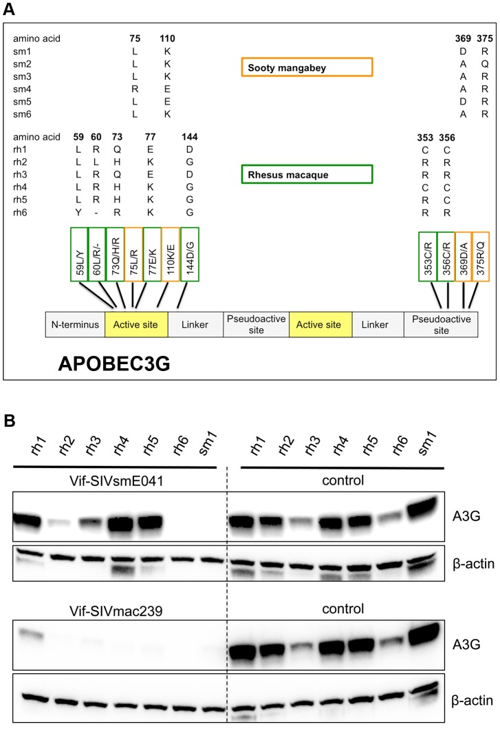
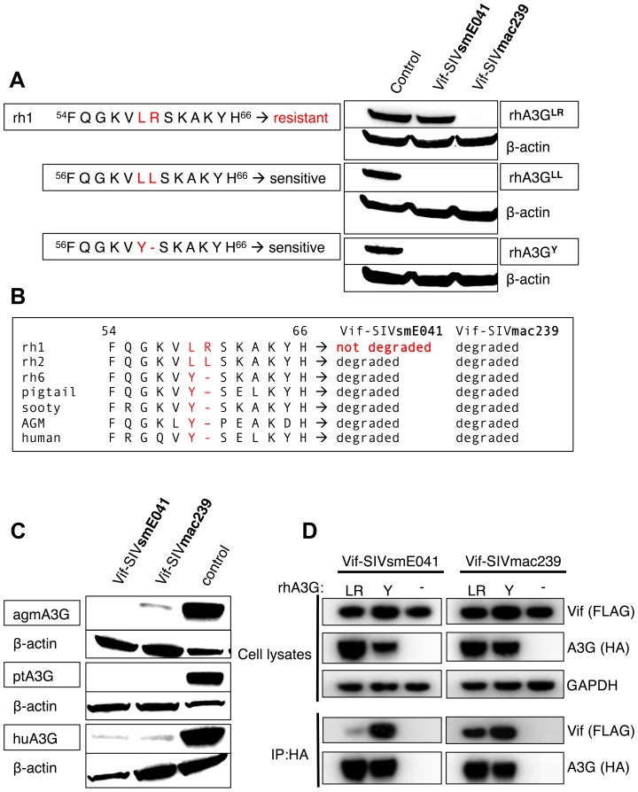
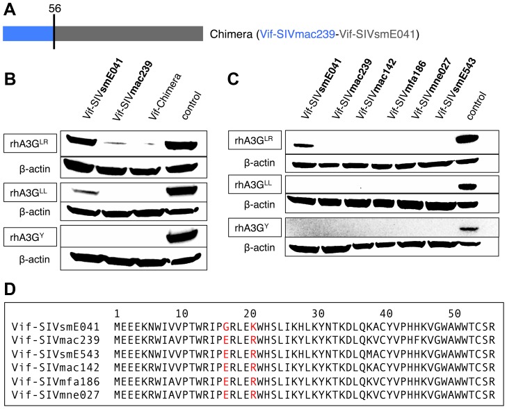
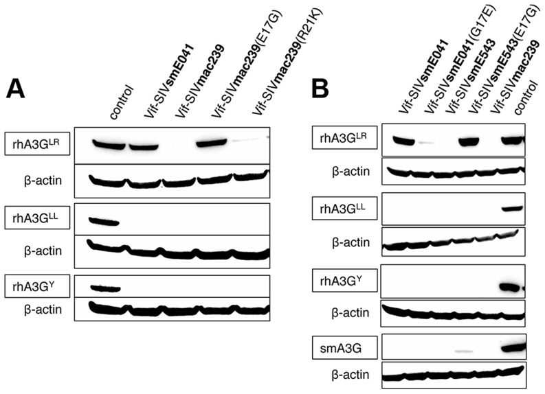
Cellular restriction factors, which render cells intrinsically resistant to viruses, potentially impose genetic barriers to cross-species transmission and emergence of viral pathogens in nature. One such factor is APOBEC3G. To overcome APOBEC3G-mediated restriction, many lentiviruses encode Vif, a protein that targets APOBEC3G for degradation. As with many restriction factor genes, primate APOBEC3G displays strong signatures of positive selection. This is interpreted as evidence that the primate APOBEC3G locus reflects a long-term evolutionary "arms-race" between retroviruses and their primate hosts. Here, we provide direct evidence that APOBEC3G has functioned as a barrier to cross-species transmission, selecting for viral resistance during emergence of the AIDS-causing pathogen SIVmac in captive colonies of Asian macaques in the 1970s. Specifically, we found that rhesus macaques have multiple, functionally distinct APOBEC3G alleles, and that emergence of SIVmac and simian AIDS required adaptation of the virus to evade APOBEC3G-mediated restriction. Our evidence includes the first comparative analysis of APOBEC3G polymorphism and function in both a reservoir and recipient host species (sooty mangabeys and rhesus macaques, respectively), and identification of adaptations unique to Vif proteins of the SIVmac lineage that specifically antagonize rhesus APOBEC3G alleles. By demonstrating that interspecies variation in a known restriction factor selected for viral counter-adaptations in the context of a documented case of cross-species transmission, our results lend strong support to the evolutionary "arms-race" hypothesis. Importantly, our study confirms that APOBEC3G divergence can be a critical determinant of interspecies transmission and emergence of primate lentiviruses, including viruses with the potential to infect and spread in human populations.


Conflict of interest statement

The authors have declared that no competing interests exist.

Figures

Figure 1
Figure 1. Rhesus macaque and sooty mangabey A3G coding sequences are highly polymorphic and differ in sensitivity to Vif-mediated degradation.
(A) Shown are nonsynonymous polymorphisms found in cDNA samples from sooty mangabeys (alleles numbered sm1-sm6) and rhesus macaques (alleles numbered rh1-rh6) and their relative distribution across the A3G protein. Sooty mangabey polymorphisms are indicated with orange boxes, rhesus macaque polymorphisms with green boxes. The numbers indicate the position of the amino acid in the protein and the letters indicate the amino acids found at that position. (B) Rhesus macaque and sooty mangabey A3G allele containing plasmids were co-transfected with plasmids expressing Vif-SIVsmE041, Vif-SIVmac239, or an empty vector (no Vif) control (indicated as “control”). The ability of Vif proteins to induce A3G degradation is reflected by a reduced or absent A3G band relative to controls. Anti-β-actin served as a protein loading control. See also Figures S1, S2 and S3.
Figure 2
Figure 2. A Tyr to Leu-Arg substitution at position 59/60 makes rhA3G resistant to Vif-SIVsmE041 induced degradation.
(A) Site-directed mutagenesis was used to mutate a resistant rhesus macaque allele (rh1) to either one of two naturally occurring sensitive alleles (rhA3GLL, rhA3GY), their ability to resist Vif-mediated degradation was visualized by western blot. We used an empty vector control to test expression in the absence of Vif (indicated as “control”). Anti-β-actin served as a protein loading control. (B) Partial alignment of A3G proteins from different species (rh1, rh2, rh6 = rhesus macaque alleles; pigtail = pigtail macaque, sooty = sooty mangabey, AGM = African green monkey). Also indicated is the observed sensitivity of the A3Gs (listed as “not degraded” or “degraded”) to either Vif-SIVsmE041 or Vif-SIVmac239 induced degradation. (C) Immunoblot showing activity of Vif-SIVsmE041, Vif-SIVmac239 and an empty vector (no Vif) control against A3G proteins from African green monkey (Haplotype 1 see [31]), pigtail macaque and human. Equal expression levels were confirmed using anti-β-actin as a loading control. (D) Co-immunoprecipitation experiment with Vif-SIVmac239 and Vif-SIVsmE041 and rhA3GLR, rhA3GY or a no A3G control. Whole-cell lysates were analyzed by immunoblotting using antibodies specific for FLAG (Vif), HA (A3G) or GAPDH (loading control). The immunoprecipitations (IP) were analyzed with antibodies specific for FLAG (Vif) or HA (A3G).
Figure 3
Figure 3. The determinant in Vif for interaction with rhA3GLR lies within the first 56 amino acids of the Vif protein.
(A) Cartoon of a chimeric protein containing the first 56 amino acids of Vif-SIVmac239 and the rest of Vif-SIVsmE041. (B) Immunoblot showing activity of the parental Vif proteins (Vif-SIVsmE041 and Vif-SIVmac239), the Vif-Chimera and an empty vector (no Vif) control against the three different rhA3G alleles (rhA3GLR, rhA3GLL and rhA3GY). Anti-β-actin served as a protein loading control. (C) Immunoblot showing activity of six Vif proteins and an empty vector (no Vif) control against the three rhA3G alleles (rhA3GLR, rhA3GLL and rhA3GY). Anti-β-actin served as a protein loading control. (D) Partial alignment of the NTD of the Vif protein from six different SIV strains as indicated. Highlighted in red are amino acids 17 and 21 which are conserved amongst Vif proteins from macaque derived SIV strains but differ in Vif from SIVsmE041. See also Figure S5.
Figure 4
Figure 4. The negatively charged glutamic acid at position 17 in the primate Vif protein allows for interaction with rhA3GLR.
(A) Immunoblot showing activity of Vif-SIVsmE041, Vif-SIVmac239, the mutants Vif-SIVmac239(E17G) and Vif-SIVmac239(R21K), and an empty vector (no Vif) control against the three rhA3G alleles (rhA3GLR, rhA3GLL and rhA3GY). Anti-β-actin served as a protein loading control. (B) Immunoblot showing activity of Vif-SIVsmE041 and Vif-SIVsmE543 and their mutants (Vif-SIVsmE041(G17E), Vif-SIVsmE543(E17)) as well as Vif-SIVmac239 and an empty vector (no Vif) control against the three rhA3G alleles and a sooty mangabey A3G. Anti-β-actin served as a protein loading control.
Figure 5
Figure 5. Mutating the negatively charged glutamic acid at position 17 to glycine in Vif-SIVmac239 reduces infectivity and increases incorporation of rhA3GLR into virions.
(A) Shown is one representative of two independent infectivity assay experiments. Infectivity was measured by titration on TZM-bl indicator cells. The virus was produced in HEK-293T/17 cells by co-transfection of a full-length SIVmac239ΔVif plasmid, a plasmid coding for one of the three A3G alleles (rhA3GLR, rhA3GLL or rhA3GY) or a no A3G control and a plasmid coding for one of three Vif proteins (Vif-SIVsmE041, Vif-SIVmac239 or Vif-SIVmac239(E17G)) or a no Vif, empty vector control. Error bars indicate the standard deviation of three replicate infections. (B) Immunoblot was used to measure packaging of rhA3G into virions using virus preparations also used in the above infectivity experiment. The amount of virus present is measured by detecting the SIV p27 core antigen with an anit-p27 antibody. The relative density of each A3G band (as compared to the corresponding p27 band) is indicated below each lane.
Figure 6
Figure 6. Model of the N-terminal domain of rhesus APOBEC3G.
A model of the N-terminal domain of rhA3GLR was generated using the PHYRE2 server using the A3C structure PDB: 3VOW as a model. (A) Position 60 is shown as a red sphere. A previously identified site of positive selection and a determinant of species tropism, K128, is shown as a violet sphere. These two independent determinants of species tropism are predicted to occur on the same face of the A3G N-terminal domain. The site of another previously identified polymorphism of rhesus macaques , D/N130, is shown as a light green sphere. Indicated in blue is an insertion found in Colobinae monkeys . Measurements using the predicted Cα backbone indicate that these residues are within 20 Å of each other. Additional A3G polymorphisms identified in this study within rhesus macaques (green spheres) or sooty mangabeys (orange spheres) are shown. (B) The same structure as in A, rotated by 270° around the Y-axis.

References

    1. VandeWoude S, Apetrei C (2006) Going wild: lessons from naturally occurring T-lymphotropic lentiviruses. Clinical microbiology reviews 19: 728–762. - PMC - PubMed
    1. Sharp PM, Hahn BH (2011) Origins of HIV and the AIDS pandemic. Cold Spring Harbor perspectives in medicine 1: a006841. - PMC - PubMed
    1. Charleston MA, Robertson DL (2002) Preferential host switching by primate lentiviruses can account for phylogenetic similarity with the primate phylogeny. Systematic biology 51: 528–535. - PubMed
    1. Hahn BH, Shaw GM, De Cock KM, Sharp PM (2000) AIDS as a zoonosis: scientific and public health implications. Science 287: 607–614. - PubMed
    1. Apetrei C, Robertson DL, Marx PA (2004) The history of SIVS and AIDS: epidemiology, phylogeny and biology of isolates from naturally SIV infected non-human primates (NHP) in Africa. Frontiers in bioscience: a journal and virtual library 9: 225–254. - PubMed

Publication types

MeSH terms