. 2013;9(10):e1003652.
doi: 10.1371/journal.ppat.1003652. Epub 2013 Oct 3.

Herpes simplex virus type 1 single strand DNA binding protein and helicase/primase complex disable cellular ATR signaling

Affiliations

Herpes simplex virus type 1 single strand DNA binding protein and helicase/primase complex disable cellular ATR signaling

Kareem N Mohni et al. PLoS Pathog. 2013.

Abstract

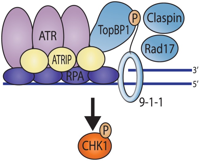
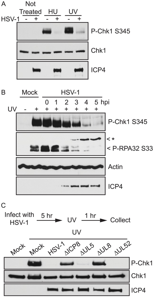
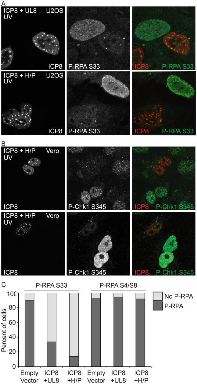
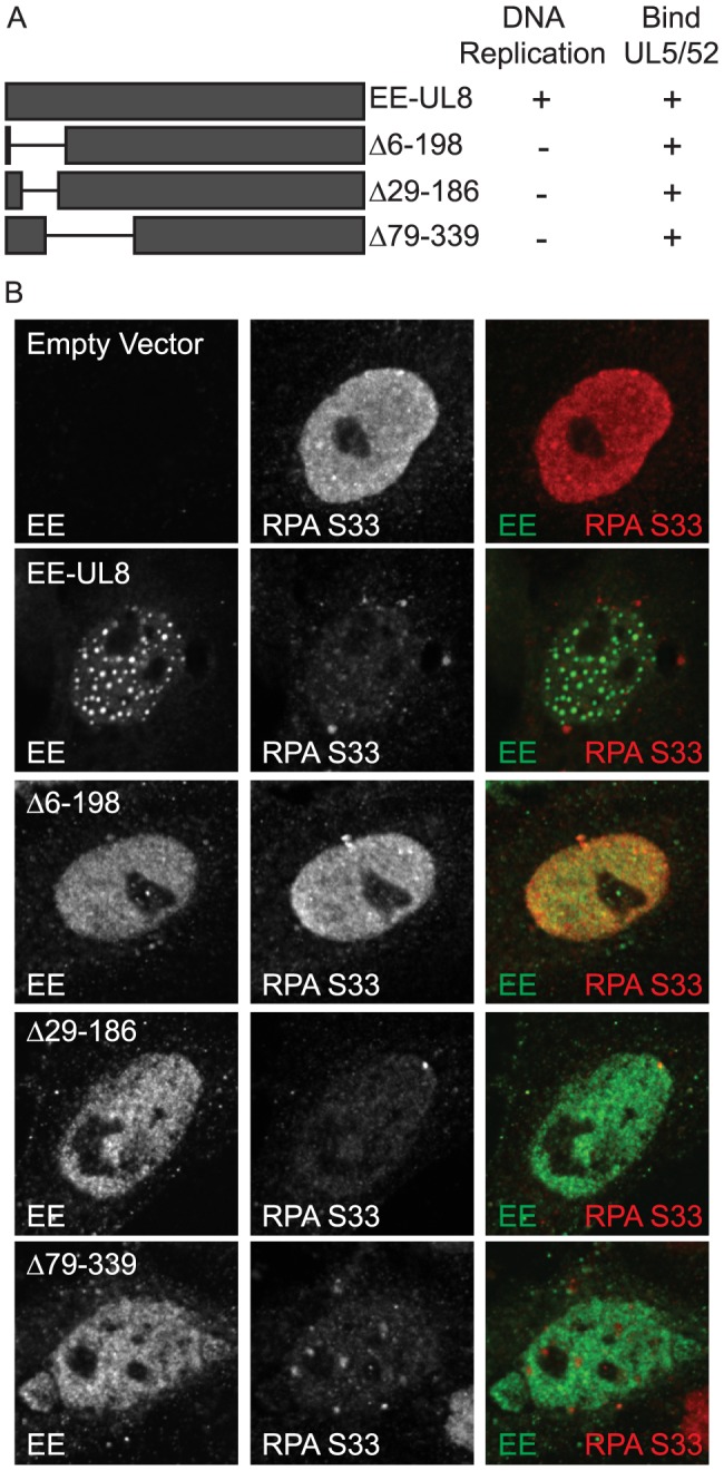
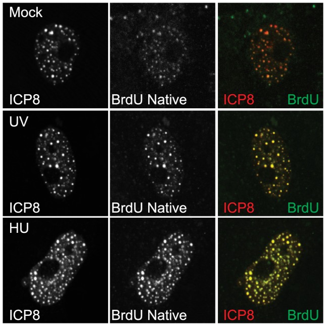
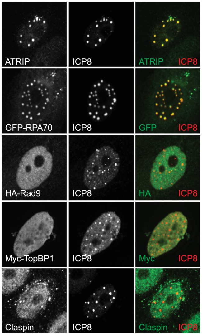
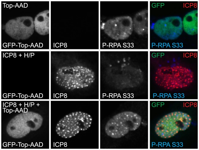
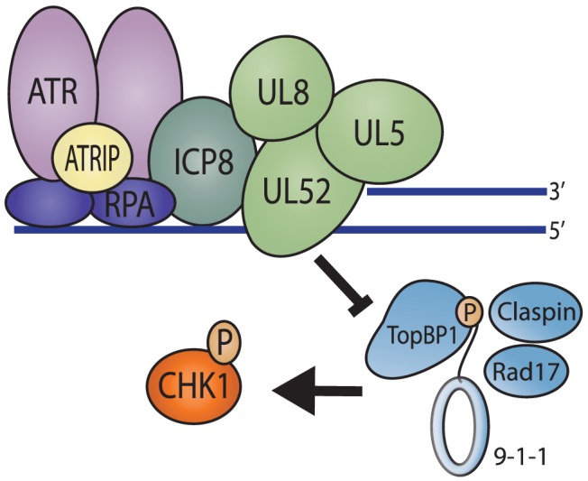
Herpes Simplex Virus type 1 (HSV-1) has evolved to disable the cellular DNA damage response kinase, ATR. We have previously shown that HSV-1-infected cells are unable to phosphorylate the ATR substrate Chk1, even under conditions in which replication forks are stalled. Here we report that the HSV-1 single stranded DNA binding protein (ICP8), and the helicase/primase complex (UL8/UL5/UL52) form a nuclear complex in transfected cells that is necessary and sufficient to disable ATR signaling. This complex localizes to sites of DNA damage and colocalizes with ATR/ATRIP and RPA, but under these conditions, the Rad9-Rad1-Hus1 checkpoint clamp (9-1-1) do not. ATR is generally activated by substrates that contain ssDNA adjacent to dsDNA, and previous work from our laboratory has shown that ICP8 and helicase/primase also recognize this substrate. We suggest that these four viral proteins prevent ATR activation by binding to the DNA substrate and obstructing loading of the 9-1-1 checkpoint clamp. Exclusion of 9-1-1 prevents recruitment of TopBP1, the ATR kinase activator, and thus effectively disables ATR signaling. These data provide the first example of viral DNA replication proteins obscuring access to a DNA substrate that would normally trigger a DNA damage response and checkpoint signaling. This unusual mechanism used by HSV suggests that it may be possible to inhibit ATR signaling by preventing recruitment of the 9-1-1 clamp and TopBP1.


Conflict of interest statement

The authors have declared that no competing interests exist.

Figures

Figure 1
Figure 1. Recruitment of ATR pathway proteins to sites of DNA damage in uninfected cells.
The ssDNA at sites of damage is coated by Replication protein A (RPA) and recruits ATR through a direct interaction with the ATR interacting protein (ATRIP). This RPA coated ssDNA also promotes the loading of the 9-1-1 (Rad9-Rad1-Hus1) checkpoint clamp by Rad17 onto the junction of the ss/dsDNA. TopBP1 is recruited via an interaction with the phosphorylated C-terminal tail of Rad9. TopBP1 then activates ATR kinase activity resulting in phosphorylation of Chk1, which is promoted by Claspin.
Figure 2
Figure 2. HSV-1 inhibition of ATR signaling requires ICP8 and UL8.
(A) Vero cells were either mock-infected or infected with HSV-1 at an MOI of 10 PFU/cell. At 5 hours post infection cells were treated with HU for 1 hour or UV and allowed to recover for 1 hour. (B) Vero cells were either mock-infected or infected with HSV-1 at an MOI of 10 PFU/cell. Cells were treated with UV at the indicated time post infection and allowed to recover for 1 hour. (C) Vero cells were either mock-infected or infected with the indicated HSV-1 mutant viruses at an MOI of 10 PFU/cell. At 5 hours post infection cells were treated with UV and allowed to recover for one hour. All cell lysates were analyzed by Western blot with the indicated antibodies. The band marked with an asterisk (*) in the P-RPA-S33 blot corresponds to a non-specific band that does not cross react with antibodies to endogenous RPA and likely represents cross-reactivity with a viral protein.
Figure 3
Figure 3. ICP8 and UL8 are sufficient to inhibit ATR signaling.
(A) U2OS or (B) Vero cells were transfected with ICP8, UL8, UL5, and UL52 (ICP8+H/P) or ICP8 and UL8 alone and then damaged with UV. Cells were fixed at 1 hour post damage and prepared for immunofluorescence as described in the materials and methods. (C) Cells were treated as in A and stained for either P-RPA-S33 or P-RPA-S4/S8. At least 100 cells were counted between two independent experiments.
Figure 4
Figure 4. UL8 mutants that do not support DNA replication still inhibit ATR signaling.
(A) Schematic of UL8 mutants used in this study. (B) Vero cells were transfected with ICP8, UL5, UL5, and the indicated UL8 mutants and then damaged with UV. Cells were fixed at 1 hour post damage and prepared for immunofluorescence as described in the materials and methods.
Figure 5
Figure 5. The four-protein complex localizes to sites of DNA damage.
Vero cells were transfected with ICP8, UL8, UL5, and UL52 and BrdU was added at the time of transfection. Cells were treated with UV or HU at 24
Figure 6
Figure 6. Essential ATR pathway proteins are excluded from the four-protein complex.
Vero cells were transfected with ICP8, UL8, UL5, and UL52 and damaged with UV and allowed to recover for 1-RPA70 or Myc-TopBP1 were cotransfected with the viral proteins. To follow HA-Rad9 Vero cells stably expressing HA-Rad9 were transfected with the viral proteins prior to UV irradiation.
Figure 7
Figure 7. ATR can be activated in cells expressing the four-protein complex and the ATR Activation Domain of TopBP1.
Vero cells were transfected with GFP-TopBP1-AAD alone or in combination with ICP8, UL8, UL5, and UL52 (ICP8+H/P). Cells were fixed at 18 hours post transfection and prepared for immunofluorescence as described in the materials and methods.
Figure 8
Figure 8. Model of DNA repair proteins at sites of DNA damage in the presence of the 4-protein complex.
In the presence of ICP8 and helicase/primase, RPA can still coat ssDNA at sites of damage and recruit ATR/ATRIP. However, these four-proteins bind to the ss/dsDNA junction that would normally serve as the loading platform for the 9-1-1 complex and exclude it from binding the DNA. This serves to prevent all of the downstream proteins from being recruited to sites of DNA damage and effectively inhibits ATR signaling.

References

    1. Cimprich KA, Cortez D (2008) ATR: an essential regulator of genome integrity. Nat Rev Mol Cell Biol 9: 616–627. - PMC - PubMed
    1. Ciccia A, Elledge SJ (2010) The DNA damage response: making it safe to play with knives. Mol Cell 40: 179–204. - PMC - PubMed
    1. Abraham RT (2004) PI 3-kinase related kinases: ‘big’ players in stress-induced signaling pathways. DNA Repair (Amst) 3: 883–887. - PubMed
    1. Nam EA, Cortez D (2011) ATR signalling: more than meeting at the fork. Biochem J 436: 527–536. - PMC - PubMed
    1. Zou L, Elledge SJ (2003) Sensing DNA damage through ATRIP recognition of RPA-ssDNA complexes. Science 300: 1542–1548. - PubMed

Publication types

MeSH terms