. 2013;9(10):e1003679.
doi: 10.1371/journal.ppat.1003679. Epub 2013 Oct 3.

Early host cell targets of Yersinia pestis during primary pneumonic plague

Affiliations

Early host cell targets of Yersinia pestis during primary pneumonic plague

Roger D Pechous et al. PLoS Pathog. 2013.

Abstract

Inhalation of Yersinia pestis causes primary pneumonic plague, a highly lethal syndrome with mortality rates approaching 100%. Pneumonic plague progression is biphasic, with an initial pre-inflammatory phase facilitating bacterial growth in the absence of host inflammation, followed by a pro-inflammatory phase marked by extensive neutrophil influx, an inflammatory cytokine storm, and severe tissue destruction. Using a FRET-based probe to quantitate injection of effector proteins by the Y. pestis type III secretion system, we show that these bacteria target alveolar macrophages early during infection of mice, followed by a switch in host cell preference to neutrophils. We also demonstrate that neutrophil influx is unable to limit bacterial growth in the lung and is ultimately responsible for the severe inflammation during the lethal pro-inflammatory phase.


Conflict of interest statement

The authors have declared that no competing interests exist.

Figures
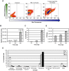
Figure 1
Figure 1. YopE-TEM translocation in the lung during pneumonic plague.
(A) Groups of mice were inoculated intranasally with 106 CFU of Y. pestis expressing YopE-TEM or PBS alone (mock), followed by staining with CCF2-AM. Lungs were harvested at 12 hpi, and live cells were gated to evaluate blue/green fluorescence by flow cytometry. Cells showing blue fluorescence have been injected with YopE-TEM. Histograms show representative data from a single mouse; (B) Total injection events (cells exhibiting blue fluorescence) in the lungs of mice inoculated with 106 CFU Y. pestis YopE-TEM; (C) The percentage of total cells in the lung injected with YopE-TEM was evaluated for samples shown in (B) at 6, 12, and 24 hpi; (D) Lung bacterial burden was evaluated in mice inoculated with 106 CFU Y. pestis YopE-TEM. (E) Live injected cells in lungs of mice inoculated with 106 CFU Y. pestis YopE-TEM were gated for identification of alveolar macrophages (F480+CD11blow/midCD11chigh), interstitial macrophages (F480+CD11bhighCD11cmid/low), F480+CD11bhighCD11chigh macrophages, monocytes (F480CD11bhighLy-6G), neutrophils (F480CD11bhighLy-6G+), and CD11b high and low dendritic cells (F480 CD11c+); All data are representative of at least three independent experiments with three to five mice at each time point. Error bars represent SEM.
Figure 2
Figure 2. Lung host cell repertoire of mice inoculated intranasally with Y. pestis.
(A) Total cell numbers of innate immune cell populations in the lungs of mice inoculated with 106 CFU Y. pestis YopE-TEM or PBS (mock) at 6, 12, and 24 hpi. (B) Y. pestis causes severe inflammation in the lungs that is visible by 48 hpi. Mice were inoculated with 106 CFU Y. pestis and lungs were inflated with 10% formalin at 24 and 48 hpi prior to H and E staining. Images are representative of experiments done a minimum of three times. Data are representative of at least three independent experiments with five mice at each time point. Error bars represent SEM.
Figure 3
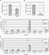
Figure 3. Pre-treatment of mice with clodrosome or neutrophil-depleting antibody α-Ly-6G.
(A) Total YopE-TEM injection events 6 hpi in mice inoculated with 106 CFU Y. pestis YopE-TEM with and without prior alveolar macrophage depletion with clodrosome. (B) Total YopE-TEM injection events 24 hpi in Y. pestis-infected mice with and without pre-treatment with neutrophil-depleting antibody. (C) Identity of injected cells 6 hpi in mice with and without prior clodrosome treatment. (D) Identity of injected cells 24 hpi with and without pre-treatment with neutrophil-depleting antibody. Data are representative of at least three independent experiments with three to five mice at each time point. Error bars represent SEM for all graphs.
Figure 4
Figure 4. Evaluating the progression of pneumonic plague in mice pre-treated with clodrosome or neutrophil-depleting antibody α-Ly-6G.
(A) Lung bacterial burden in mice with and without pre-treatment with clodrosome or neutrophil-depleting antibody and after inoculation with 104 CFU Y. pestis; (B) Lung histology 48 hpi in mice inoculated with 104 CFU Y. pestis with and without prior treatment with clodrosome or neutrophil-depleting antibody. Bar graph represents experiments repeated at least three times with three to five mice. Error bars represent SEM. Histopathology shows representative images from experiments repeated at least twice.

References

    1. Inglesby TV, Dennis DT, Henderson DA, Bartlett JG, Ascher MS, et al. (2000) Plague as a biological weapon: medical and public health management. Working Group on Civilian Biodefense. JAMA: the journal of the American Medical Association 283: 2281–2290. - PubMed
    1. Perry RD, Fetherston JD (1997) Yersinia pestis–etiologic agent of plague. Clinical microbiology reviews 10: 35–66. - PMC - PubMed
    1. Stenseth NC, Atshabar BB, Begon M, Belmain SR, Bertherat E, et al. (2008) Plague: past, present, and future. PLoS medicine 5: e3. - PMC - PubMed
    1. Lathem WW, Crosby SD, Miller VL, Goldman WE (2005) Progression of primary pneumonic plague: a mouse model of infection, pathology, and bacterial transcriptional activity. Proceedings of the National Academy of Sciences of the United States of America 102: 17786–17791. - PMC - PubMed
    1. Krishna G, Chitkara RK (2003) Pneumonic plague. Seminars in respiratory infections 18: 159–167. - PubMed

Publication types