. 2013;9(10):e1003849.
doi: 10.1371/journal.pgen.1003849. Epub 2013 Oct 3.

A genetic RNAi screen for IP₃/Ca²⁺ coupled GPCRs in Drosophila identifies the PdfR as a regulator of insect flight

Affiliations

A genetic RNAi screen for IP₃/Ca²⁺ coupled GPCRs in Drosophila identifies the PdfR as a regulator of insect flight

Tarjani Agrawal et al. PLoS Genet. 2013.

Abstract

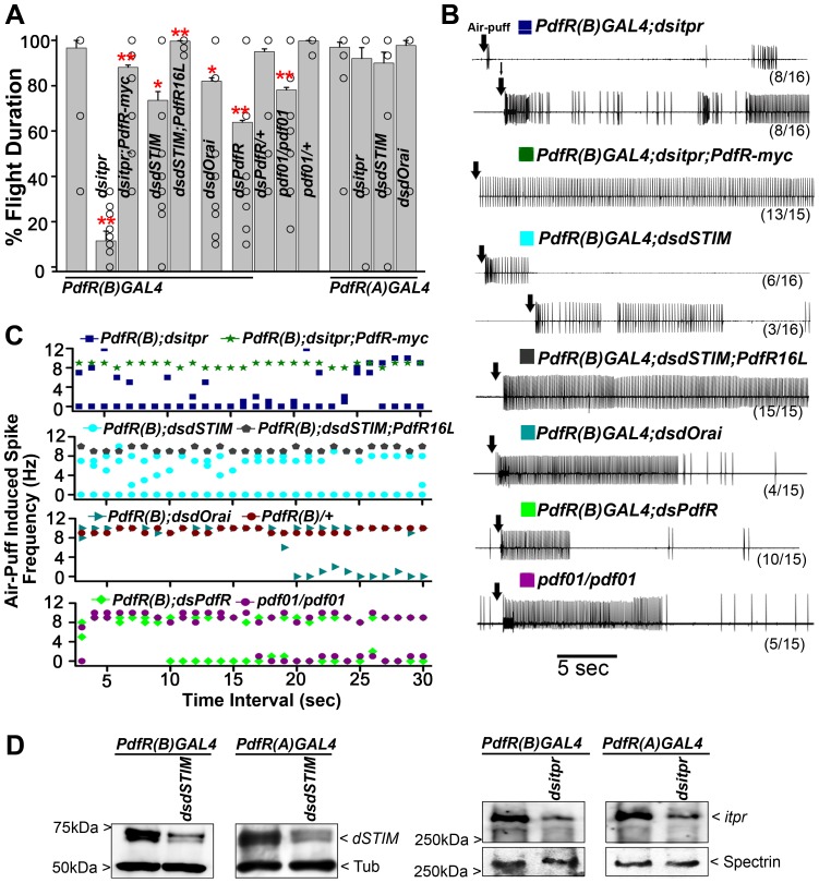
Insect flight is regulated by various sensory inputs and neuromodulatory circuits which function in synchrony to control and fine-tune the final behavioral outcome. The cellular and molecular bases of flight neuromodulatory circuits are not well defined. In Drosophila melanogaster, it is known that neuronal IP3 receptor mediated Ca²⁺ signaling and store-operated Ca²⁺ entry (SOCE) are required for air-puff stimulated adult flight. However, G-protein coupled receptors (GPCRs) that activate intracellular Ca²⁺ signaling in the context of flight are unknown in Drosophila. We performed a genetic RNAi screen to identify GPCRs that regulate flight by activating the IPIP₃ receptor. Among the 108 GPCRs screened, we discovered 5 IPIP₃/Ca²⁺ linked GPCRs that are necessary for maintenance of air-puff stimulated flight. Analysis of their temporal requirement established that while some GPCRs are required only during flight circuit development, others are required both in pupal development as well as during adult flight. Interestingly, our study identified the Pigment Dispersing Factor Receptor (PdfR) as a regulator of flight circuit development and as a modulator of acute flight. From the analysis of PdfR expressing neurons relevant for flight and its well-defined roles in other behavioral paradigms, we propose that PdfR signaling functions systemically to integrate multiple sensory inputs and modulate downstream motor behavior.


Conflict of interest statement

The authors have declared that no competing interests exist.

Figures

Figure 1
Figure 1. A genetic RNAi screen for G-protein coupled receptors that regulate flight in Drosophila.
A) A schematic of how GPCR activation can stimulate the IP3R mediated Ca2+ signaling pathway and Store-operated Ca2+ entry (SOCE) through STIM and Orai. Gq is a heterotrimeric G-protein that acts downstream of IP3R linked GPCRs and activates PLCβ upon ligand-binding to the GPCR. STromal Interacting Molecule (encoded by dSTIM in Drosophila) is an ER membrane protein that can sense reduced Ca2+ in the ER store upon IP3R-mediated Ca2+ release and subsequently activates SOCE from plasma-membrane localized Orai channels. B) A schematic representation of the screening strategy for identifying G-protein coupled receptors (GPCRs) required in Drosophila flight. Flies (n≥10) with pan-neuronal (Elav C155 GAL4) knockdown of individual GPCR were collected and tested for flight. Non-stop flight for 30 sec after a gentle air-puff was taken as 100% flight. C) Mean percentage time of flight for each genotype tested (open circles) is shown in increasing order. The average of all mean percentage flight times is shown as a red box. Average of mean percentage flight time for genotypes above and below 80% flight time are shown as blue boxes, and were found to be significantly different from each other (P<0.005). Therefore, flight time of 80% was considered as the significant cut-off for identifying putative GPCRs affecting flight. Grey bars show the number of RNAi strains lying within the indicated intervals of 10% of flight time. D) Individual GPCR RNAi strains identified with a mean percentage flight time of less than 80%. Each strain has been referred to by its CG number and the individual RNAi number (described in Table S1). Open circles within the bars show percentage flight times for each fly. Where flight times overlap a single open circle is shown. All RNAi heterozygotes (Table S1) and the pan-neuronal GAL4 used in these experiments (column on extreme left) showed normal flight durations.
Figure 2
Figure 2. Flight-deficits in flies with pan-neuronal knockdown of the IP3R can be rescued by expression of AcGq and dSTIM+.
A) Pan-neuronal expression of either AcGq or dSTIM+ suppresses wing posture defects in flies with pan-neuronal knockdown of the IP3R (dsitpr; dicer). All 100 flies analyzed showed normal wing posture. B) Air-puff stimulated tethered flight was compromised by pan-neuronal knockdown of the IP3R. Significant rescue of the mean percentage flight time was observed by expression of either AcGq or dSTIM+. Results are expressed as mean ± SEM of the flight time of 30 flies tested individually for flight. Open circles within the bars show the percentage flight times for individual flies. Pan-neuronal knockdown of the IP3R was compared with the pan-neuronal GAL4 control; and AcGq and dSTIM + rescues were compared to the itpr knockdown (one-way ANOVA, **P<0.01). C) Electrophysiological recordings from the DLMs of air-puff stimulated (arrows) tethered flies are shown. The genotypes are indicated above the traces, and the numbers indicate the number of flies with the observed flight pattern over the number of flies tested. All control flies show rhythmic firing throughout flight. Complete loss of electrical activity was seen in flies with pan-neuronal expression of dsitpr. Expression of either AcGq with dsitpr (ElavC155GAL4; dsitpr; AcGq) or dSTIM+ with dsitpr (ElavC155GAL4; dsitpr; dSTIM+) restored electrical firing from the DLMs to varying extents, which is shown as two categories below the indicated genotype. D) Quantification of spontaneous firing from DLMs of the indicated genotypes. Average spontaneous firing was restored to normal upon expression of AcGq or dSTIM+ in pan-neuronal knockdown of IP3R. Results are shown as mean ± SEM. Open circles within the bars are the average spontaneous firing quantified for individual flies. Pan-neuronal knockdown of the IP3R (dsitpr) was compared to pan-neuronal GAL4 controls and rescues were compared to the knockdown (**P<0.01, one-way ANOVA).
Figure 3
Figure 3. G-protein coupled receptors that regulate flight in Drosophila through IP3R mediated Ca2+ signaling.
A) Percentage flight times are shown. The bars represent RNAi heterozygotes (grey), pan-neuronal RNAi knockdown (red), pan-neuronal RNAi knockdown plus AcGq (green) and pan-neuronal knockdown plus dSTIM+ (blue). Mean flight times (± SEM) were obtained by measuring tethered flight in three batches of 10 tethered flies of each genotype after an air-puff stimulus. Open circles within the bars indicate percentage flight times for each fly. Pan-neuronal knockdown of GPCRs (red) was compared to pan-neuronal GAL4 controls (grey) and the rescues (blue, green) were compared to the knockdown (red; *P<0.05, one-way ANOVA). B) Pan-neuronal knockdown of the SiFamide receptor (10823-1) results in pupal lethality (shown as a skull in A). Lethality was suppressed by pan-neuronal over-expression of dSTIM+ but not AcGq.
Figure 4
Figure 4. IP3R function is required from 16 to 32 hours after puparium formation to regulate flight.
A) Quantification of wing posture defects by knockdown of the IP3R (dsitpr) at specific stages during pupal development indicated above the bars using the GAL4/GAL80ts system (TARGET), are shown. Mixed populations of normal (grey bar), mild wing posture defect (pink bar) and severe wing posture defective (blue bar) flies were obtained when dsitpr is expressed from 16 h, 24 h or 32 h after puparium formation (APF) to adulthood, but not when expression is induced at 48 h APF and onwards. Animals were either moved from 18°C to 29°C, at the appropriate pupal phases to induce RNAi expression or maintained at a constant temperature post egg-laying (PEL) as indicated. Percentage of animals with varying wing posture defects is shown as a stacked histogram for males (M) and females (F). B) Quantification of flight duration in single flight assays (mean ± SEM) from female animals of the indicated genotypes. Open circles within each bar represent percentage flight time for each fly. Mean flight times (± SEM) were obtained for three batches of 10 tethered flies of each genotype after an air-puff stimulus. All the knockdowns were compared to 29°C dsitpr/+ control (**p<0.01, one-way ANOVA). Color codes of the histograms are the same as in (A) above. C) Representative traces of electrophysiological recordings from DLMs of the indicated genotypes in response to a manual air-puff stimulus (arrow). The number of flies that showed the given response upon the number of flies tested is given below each trace. Adult female flies were sorted on the basis of the severity of their wing defects as shown. Loss of rhythmic flight patterns accompanied by severe wing posture defects is observed due to pan-neuronal depletion of IP3R during early pupal development (16 h APF). Knockdown at later time points shows reduced flight duration in flies with mild wing posture defect and normal flight in animals with normal wings. D) Quantification of the frequency of spontaneous firing as recorded from DLMs of the indicated genotypes. As with flight deficits and air-puff induced electrical firing patterns, spontaneous firing frequencies are higher in animals with wing posture defects (color coded as in A) and the time of transfer to 29°C. (**p<0.01, *p<0.05, one-way ANOVA, N = 15). Firing frequencies were calculated by counting the number of spikes over 2 min. Individual data points are represented by open circles. E) Representative traces for (D) showing increased spontaneous firing in animals with severe wing posture defect due to early knockdown of the IP3R.
Figure 5
Figure 5. Adult flight deficits result from RNAi knockdown of specific GPCRs during pupal development.
A) Percentage flight time of RNAi heterozygotes (grey bars) and pan-neuronal knockdown of GPCR RNAi strains (dsFz-2R in red, dsmAcR in purple, dsCCH1aR in blue, dsSIFaR in green) at specific developmental stages, as indicated above the bars using the GAL4/GAL80ts system (TARGET), are shown. Open circles within each bar represent percentage flight time for each fly. Mean flight times (± SEM) were obtained for three batches of 10 tethered flies of each genotype after an air-puff stimulus. Pupal knockdowns at 29°C were compared with the specific RNAi control at 18°C; post egg-laying (PEL) knockdowns were compared with their specific RNAi controls (**P<0.01, *P<0.05; one-way ANOVA). B) Electrophysiological recordings from the DLMs of air-puff stimulated tethered flies with pan-neuronal knockdown of RNAi (dsFz-2R in red, dsmAcR in purple, dsCCH1aR in blue). The temporal stage at which knockdown was initiated is indicated above each trace. Numbers in brackets below each trace is the number of flies with the given response upon the number of flies tested. The remaining flies in each case showed a normal firing response. All flies maintained at 18°C show rhythmic firing throughout flight. Flight pattern durations were reduced in flies either with pupal knockdowns (PUPAL 29°C) or kept at 29°C post egg-laying (PEL).
Figure 6
Figure 6. RNAi mediated knockdown of specific GPCRs during development and in adults result in flight deficits.
A) Percentage flight times of GPCR RNAi heterozygotes (grey) and pan-neuronal knockdowns of RNAi strains (dsFmrfR in red, ds43795 in green) were obtained after knockdown from specific developmental stages as indicated above the bars. Open circles within the bars represent percentage flight times for individual flies. Mean flight times (± SEM) were obtained by measuring tethered flight in three batches of 10 tethered flies of each genotype after an air-puff stimulus. Pupal and adult knockdowns at 29°C were compared with the specific RNAi control at 18°C; post egg-laying (PEL) knockdowns were compared with their specific RNAi controls at 29°C (**P<0.01, *P<0.05; one-way ANOVA). B) Electrophysiological recordings from the DLMs of air-puff stimulated tethered flies of the indicated genotypes. The temporal stages at which knockdowns were initiated are indicated above each trace. Also shown below each trace is the number of flies that showed the given response upon the number of flies tested. In each case the remaining flies showed normal firing responses. All control flies at 18°C showed rhythmic firing throughout flight. C) Percentage flight time (mean ± SEM) of flies with pan-neuronal knockdown of the PdfR. Knockdown was initiated from different developmental stages at 29°C as indicated. Open circles within the bars show percentage flight times for each fly. Pan-neuronal knockdown of the PdfR, either in adults or during development (larval + pupal), results in significant flight deficits (*P<0.05, as compared to 18°C PdfR knockdown control, by one way ANOVA). Strongest flight deficits are observed upon PdfR knockdown throughout development by shifting to 29°C post egg-laying (PEL; **P<0.01, when compared to RNAi heterozygotes at 29°C post egg-laying). D) Snapshots of single flight assay video recordings and electrophysiological recordings from DLMs of animals with pan-neuronal knockdown of the PdfR at the indicated temperatures and developmental stages. The number of flies that showed the given response upon the number of flies tested is shown below each trace. The remaining animals showed normal flight and firing responses as seen in the 18°C PEL control on top.
Figure 7
Figure 7. Depletion of IP3R, SOCE and PdfR in a specific sub-domain of PdfR expressing neurons affects flight duration.
A) Significant flight deficits were obtained upon knockdown of the IP3R (dsitpr), the SOCE components, dSTIM (dsdSTIM) and dOrai (dsdOrai) by the PdfR(B)GAL4 strain (**P<0.01 and *P<0.05; compared to PdfR(B)GAL4 heterozygote controls). Flight deficits induced by dsitpr in PdfR(B)GAL4 expressing neurons were rescued by the PdfR-myc genomic construct (**P<0.01 compared to dsitpr knockdown). Similarly, the flight phenotype in PdfR(B)GAL4;dsdSTIM animals could be rescued by over-expression of PdfR (UAS-PdfR16L; **P<0.01 as compared to dsdSTIM knockdown). Knockdown of the PdfR in PdfR(B)GAL4 expressing neurons also resulted in significant flight deficits (**P<0.05; dsPdfR/PdfR(B)GAL4 compared with dsPdfR/+). Flight duration in the PDF null allele (pdf01/pdf01) was significantly reduced as compared with pdf01 heterozygotes (**P<0.01). All significance values were obtained by one-way ANOVA tests. No flight defects were observed upon knockdown of the IP3R (dsitpr), dSTIM (dsdSTIM) and dOrai (dsdOrai) in PdfR(A)GAL4 expressing cells. B) Representative electrophysiological recordings from DLMs of tethered flies are shown after an air-puff stimulus (arrows). Genotypes are indicated above the traces. Shown below each trace is the number of flies in which the given response was elicited from amongst the total number of flies tested. In each case normal patterns and durations were observed in the remaining flies (not shown). Significant loss of rhythmic flight patterns were observed upon knockdown of the IP3R (dark blue) shown in two categories, dSTIM (light blue) shown in two categories, dOrai (dark cyan) and PdfR (light green) in the PdfR(B)GAL4 domain. Normal firing patterns were restored by expression of PdfR-myc (green) and PdfR16L (dark grey) in flies with IP3R (dsitpr) and dSTIM knockdowns respectively. PDF null mutants (pdf01/pdf01; magenta) also showed a reduced duration of rhythmic firing patterns. PdfR(B)GAL4 heterozygotes and RNAi heterozygotes showed rhythmic firing throughout flight (data not shown). C) Quantification of the spike frequency for electrophysiological traces (as shown in C) for the indicated genotypes. D) Western blots from protein extracts of adult brains and thoracic ganglia. Expression of dSTIM and the IP3R is reduced, upon their knockdown with specific RNAi expression in PdfR(B)GAL4(2) and PdfR(A)GAL4(2) strains.
Figure 8
Figure 8. Differential expression of PdfRGAL4 strains in neurons of the adult AMMC, SOG and thoracic ganglia.
Expression patterns of the indicated PdfRGAL4 strains in the sub-esophageal ganglion, SOG (A, B), thoracic region 1 and 2, T1–T2 (C, D) and abdominal regions, Ab (E, F) are shown. A schematic of these regions of the Drosophila central nervous system is given in Figure S4. GFP expression is reduced in the SOG, TI, T2 and abdominal region in PdfR(A)GAL4. Expression of GFP in left (G, I) and right (H, J) ventrolateral protocerebrum (VLP) and antennal mechanosensory and motor complex (AMMC) using PdfR(B)GAL4 (G, H) and PdfR(A)GAL4 (I, J) driver is shown. White arrows indicate neuron/cluster of neurons which are positive for GFP expression. GFP expression is reduced in the AMMC when using PdfR(A)GAL4 driver as compared to PdfR(B)GAL4. Expression pattern of GFP in medial neurosecretory region, mNSC (K, L) using PdfR(B)GAL4 (K) and PdfR(A)GAL4 (L) drivers are shown. GFP expression is similar in medial neurosecretory region in PdfR(B)GAL4 and PdfR(A)GAL4. M) Summary table of expression patterns of PdfR(B)GAL4 and PdfR(A)GAL4. The areas that were examined: DLP: dorsolateral protocerebrum, OPTU: optic tubercle, MB: mushroom body, SDFP: superior dorsofrontal protocerebrum, MED: medulla. Ticks indicate the presence of expression. Two ticks indicate high expression. Red ticks represent areas that have been shown in high magnification. For each genotype, 5 brain samples were analyzed.
Figure 9
Figure 9. A summary of GPCRs that regulate flight in Drosophila.
Classification of GPCRs that were used for the primary screen, the secondary suppressor screen and finally validated as activators of IP3/Ca2+ signaling in the context of Drosophila flight.

References

    1. Taghert Paul H, Nitabach Michael N (2012) Peptide Neuromodulation in Invertebrate Model Systems. Neuron 76: 82–97. - PMC - PubMed
    1. Marder E (2012) Neuromodulation of Neuronal Circuits: Back to the Future. Neuron 76: 1–11. - PMC - PubMed
    1. Banerjee S, Lee J, Venkatesh K, Wu CF, Hasan G (2004) Loss of flight and associated neuronal rhythmicity in inositol 1,4,5-trisphosphate receptor mutants of Drosophila. J Neurosci 24: 7869–7878. - PMC - PubMed
    1. Venkiteswaran G, Hasan G (2009) Intracellular Ca2+ signaling and store-operated Ca2+ entry are required in Drosophila neurons for flight. Proc Natl Acad Sci U S A 106: 10326–10331. - PMC - PubMed
    1. Banerjee S, Joshi R, Venkiteswaran G, Agrawal N, Srikanth S, et al. (2006) Compensation of inositol 1,4,5-trisphosphate receptor function by altering sarco-endoplasmic reticulum calcium ATPase activity in the Drosophila flight circuit. J Neurosci 26: 8278–8288. - PMC - PubMed

Publication types

MeSH terms

Substances

LinkOut - more resources