. 2013 Oct 3;8(10):e75374.
doi: 10.1371/journal.pone.0075374. eCollection 2013.

Specific increase in MDR1 mediated drug-efflux in human brain endothelial cells following co-exposure to HIV-1 and saquinavir

Affiliations

Specific increase in MDR1 mediated drug-efflux in human brain endothelial cells following co-exposure to HIV-1 and saquinavir

Upal Roy et al. PLoS One. .

Abstract

Persistence of HIV-1 reservoirs within the Central Nervous System (CNS) remains a significant challenge to the efficacy of potent anti-HIV-1 drugs. The primary human Brain Microvascular Endothelial Cells (HBMVEC) constitutes the Blood Brain Barrier (BBB) which interferes with anti-HIV drug delivery into the CNS. The ATP binding cassette (ABC) transporters expressed on HBMVEC can efflux HIV-1 protease inhibitors (HPI), enabling the persistence of HIV-1 in CNS. Constitutive low level expression of several ABC-transporters, such as MDR1 (a.k.a. P-gp) and MRPs are documented in HBMVEC. Although it is recognized that inflammatory cytokines and exposure to xenobiotic drug substrates (e.g HPI) can augment the expression of these transporters, it is not known whether concomitant exposure to virus and anti-retroviral drugs can increase drug-efflux functions in HBMVEC. Our in vitro studies showed that exposure of HBMVEC to HIV-1 significantly up-regulates both MDR1 gene expression and protein levels; however, no significant increases in either MRP-1 or MRP-2 were observed. Furthermore, calcein-AM dye-efflux assays using HBMVEC showed that, compared to virus exposure alone, the MDR1 mediated drug-efflux function was significantly induced following concomitant exposure to both HIV-1 and saquinavir (SQV). This increase in MDR1 mediated drug-efflux was further substantiated via increased intracellular retention of radiolabeled [(3)H-] SQV. The crucial role of MDR1 in (3)H-SQV efflux from HBMVEC was further confirmed by using both a MDR1 specific blocker (PSC-833) and MDR1 specific siRNAs. Therefore, MDR1 specific drug-efflux function increases in HBMVEC following co-exposure to HIV-1 and SQV which can reduce the penetration of HPIs into the infected brain reservoirs of HIV-1. A targeted suppression of MDR1 in the BBB may thus provide a novel strategy to suppress residual viral replication in the CNS, by augmenting the therapeutic efficacy of HAART drugs.


Conflict of interest statement

Competing Interests: The authors have declared that no competing interests exist.

Figures

Figure 1
Figure 1. Effect of HIV-1 exposure on MDR1 gene expression and efflux function in HBMVEC.
(A) MDR-1, MRP-1 and MRP-2 gene expression following exposure to HIV-1 (0.2 and 1.0 MOI) within 6 hrs post exposure was monitored by qRT-PCR. Data obtained in each sample was normalized to GAPDH mRNA levels (internal control). Fold change in ABC-transporter gene expression with respect to that observed with MDR1 in untreated cells (1.0) was calculated under each treatment condition and for each transporter. Data represent three independent experiments (n = 3) where standard deviation (error bars) were calculated from triplicate samples and significant differences from controls are shown (*, p<0.05 and ** p<0.01). (B) MDR1 efflux function following exposure to HIV-1 (0.2 and 1.0 MOI) for 24 hrs. Verapamil and MK-571 were used as MDR1 and MRPs blocker, respectively. Intracellular fluorescence was normalized to total protein content in cell lysates and change in calcein-AM retention is shown in the bar graphs (*, p<0.05). (C) Comparative effects of conditioned medium (CM) from HIV-1-infected or PMA-stimulated HuT-78 T-cells on MDR1 gene expression. The RT-PCR products from HBMVEC control (1); Hut-78 CM (2); CM from HuT-78 cells treated with PMA (3); or CM from virus producing HTLV-IIIB cells (4), are shown. (D) Effect of live HIV-1 (HTLV-IIIB CM) and heat inactivated HIV-1 (HTLV-IIIB CM-HI) on MDR1 gene expression. The respective fold change in MDR1 gene expression was calculated with respect to GAPDH (internal control). A representative data from three independent experiments, is shown.
Figure 2
Figure 2. Effect of HIV-1 and SQV coexposure on MDR1 expression and function in HBMVEC.
(A) MDR1 gene expression in presence of HIV-1 (0.2 MOI) and increasing concentration of SQV (0.3 and 1.0 µM). (B) Densitometric analysis of mRNA expression from Fig. 2A. (C) A representative of western blot analysis data of MDR1 protein expression in untreated (1), HIV-1 (2), SQV (3) and HIV-1+ SQV (4) exposed cells. Beta (β)-actin was used as internal control. The fold change analysis was done from three sets of independent western immunodetection studies. MDR1 protein expressions are normalized with β-actin expression in respective samples. (D) MDR1 mediated calcein-AM efflux in presence of HIV-1 (0.2 MOI) and/or SQV (0.3 µM). Grey bars indicate calcein retenition without inhibitor and black bars indicate calcein retention in presence of 50 µM verapamil. Data shown are mean of three independent experiments (*, p<0.05 and **, p<0.01).
Figure 3
Figure 3. Localization of MDR-1 (P-gp) protein in HIV-1 and SQV coexposed HBMVEC.
A representative immunofluorescence microscopy data of MDR-1 protein in HBMVEC following exposure to HIV-1 or SQV for 24 hrs is shown. Green punctate MDR-1 staining appeared in (A) untreated cells, (B) cells exposed to SQV (1 µM), (C) cells exposed to HIV-1 (0.2 MOI) only, and (D) combined exposure of SQV and HIV-1. DAPI (blue) staining of the nucleus and WGA (red) staining of cell plasma membrane are also shown. Image Magnification: 630X, scale bar: 21 µm.
Figure 4
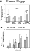
Figure 4. A comparative analysis of 3H-SQV efflux in HBMVECs exposed to HIV-1 and/or SQV in presence of verapamil or PSC-833.
(A) Effect of verapamil (50 µM) on 3H-SQV retention in HBMVEC exposed to HIV-1 and/or SQV for 24 hrs. Under each treatment condition, intracellular 3H-SQV was measured in presence of verapamil and compared with control (without inhibitor). The radioactivity in cell lysates were normalized to total cell protein content (CPM/ug of protein) and data from three independent experiments are presented as fold changes. (B) Effect of increasing concentrations (5–50 µM) of verapamil or PSC-833 on 3H-SQV retention in HBMVEC exposed to HIV-1 and SQV for 24 hrs. Data are representative of three separate independent experiments carried out in triplicates (*, p<0.05 and **, p<0.01).
Figure 5
Figure 5. Effect of MDR1 specific siRNA on HIV-1 and SQV induced drug efflux in HBMVEC.
(A) Fold change in 3H-SQV retention (CPM/µg of protein) in HBMVEC are shown in control (untreated) and HIV-1 (0.2 MOI) and SQV (0.3 µM) exposed cells. Data from non-transfected, and in cells transfected with either a random (NS) or an MDR1 specific siRNA, are shown. Data are representative of three independent experiments carried out in triplicates (*, p<0.05 and **, p<0.001). (B) Relative inhibition of MDR1 gene expression in MDR1 siRNA transfected and control (random) siRNA transfected cells are shown by semi-quantitative RT-PCR. Change in MDR1 gene expression following treatment with HIV-1 (0.2 MOI) and HIV-1+SQV, in control and siRNA transfected cells, are shown. Changes in band intensities of PCR products for MDR1 mRNA were normalized to the GAPDH mRNA levels, in respective samples, and fold changes obtained compared to control (1.0) are shown.

References

    1. Zastre JA, Chan GN, Ronaldson PT, Ramaswamy M, Couraud PO, et al. (2009) Up-regulation of P-glycoprotein by HIV protease inhibitors in a human brain microvessel endothelial cell line. J Neurosci Res 87: 1023–1036. - PubMed
    1. Ahn M, Ghaemmaghami S, Huang Y, Phuan PW, May BC, et al. (2012) Pharmacokinetics of quinacrine efflux from mouse brain via the P-glycoprotein efflux transporter. PLoS One 7: e39112. - PMC - PubMed
    1. Pal D KD, Minocha M, Paturi DK, Budda B, Mitra AK (2011) Efflux transporters- and cytochrome P-450-mediated interactions between drugs of abuse and antiretrovirals. Life Sciences 88: 959–971. - PMC - PubMed
    1. Dohgu S, Fleegal-DeMotta MA, Banks WA (2011) Lipopolysaccharide-enhanced transcellular transport of HIV-1 across the blood-brain barrier is mediated by luminal microvessel IL-6 and GM-CSF. J Neuroinflammation 8: 167. - PMC - PubMed
    1. Potschka H (2010) Targeting regulation of ABC efflux transporters in brain diseases: a novel therapeutic approach. Pharmacol Ther 125: 118–127. - PubMed

Publication types

MeSH terms

Substances