. 2013 Oct 2;8(10):e76176.
doi: 10.1371/journal.pone.0076176. eCollection 2013.

Differential binding of IgG and IgA to mucus of the female reproductive tract

Affiliations

Differential binding of IgG and IgA to mucus of the female reproductive tract

Kelly M Fahrbach et al. PLoS One. .

Abstract

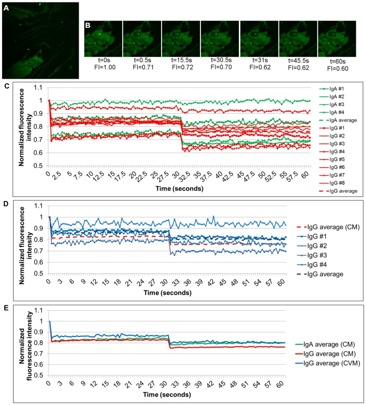
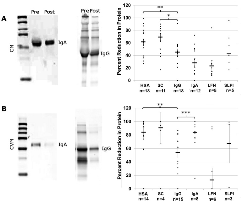
Cells of the endocervix are responsible for the secretion of mucins, which provide an additional layer of protection to the female reproductive tract (FRT). This barrier is likely fortified with IgA as has previously been shown in the gastrointestinal tract and lungs of mice. Mucus associated IgA can facilitate clearance of bacteria. While a similar function for IgG has been proposed, an association with mucus has not yet been demonstrated. Here we find that IgA and IgG are differentially associated with the different types of mucus of the FRT. We observed that while both IgA and IgG are stably associated with cervical mucus, only IgG is associated with cervicovaginal mucus. These findings reveal that antibodies can bind tightly to mucus, where they can play a significant role in the fortification of the mucus barriers of the FRT. It may be possible to harness this interaction in the development of vaccines designed to protect the FRT mucosal barriers from sexually transmitted diseases such as HIV.


Conflict of interest statement

Competing Interests: The authors have declared that no competing interests exist.

Figures

Figure 1
Figure 1. Immunofluorescence of mucins in CM and CVM.
CM (A) and CVM (B) were frozen, cryosetioned, and fixed for immunofluorescent staining. All sections were stained for WGA (blue), mucin5AC (green), and mucin5B (red). Upper panels are images acquired with a 20X (A) or 10X (B) lens. Lower panels were acquired with a 100X lens.
Figure 2
Figure 2. Ig expression in CM.
(A) CM was stained for IgA (green) and IgG (red). Left column shows Ig expression with WGA (blue). White pixels highlight regions of A-hi/G-lo (middle column) and G-hi/A-lo (right column) staining. (B) Graph of total area of highlighted pixels for IgG and IgA dense regions.
Figure 3
Figure 3. Immunofluorescence of IgG and IgA in CVM.
Frozen CVM was stained for IgG (red) and IgA (green) and imaged with a 100X lens. (A) CVM also stained with WGA (blue). (B) Same field of view in (A) with nuclei now shown in blue. (C) Zoom in of white boxes in (B) to show separated IgA (top) and IgG (bottom) stains.
Figure 4
Figure 4. Detection of immunogloblulins in endocervical explants.
Sectioned explants of endocervix were stained with antibodies to detect IgA (green) and IgG (red). Cytokeratin 7 (blue) was also stained to identify the layer of columnar cells. Each row represents a different donor sample. Images were acquired with a 100X lens as a panel and then stitched together. Second and third columns have red or green color removed, respectively, from image in first column.
Figure 5
Figure 5. FRAP in mucus.
(A) 100X image of fluorescently labeled IgG in CM. (B) Zoomed in ROI from (A) at select timepoints. Images were acquired every 0.50ms over the course of 1 minutes, with a rebleach after 30 seconds. Fluorescence intensities (FI) after normalization are shown. (C) Graph of normalized intensities of photobleached ROIs in IgG or IgA labeled CM. (D) Graph of normalized intensities of photobleached ROIs in IgG labeled CVM. CM average from (C) is included for comparison. (E) Average values from other graphs redisplayed.
Figure 6
Figure 6. Dialysis of CM and CVM.
CM (A) and CVM (B) were dialyzed through a 700 kDa filter (Post) and loaded onto gels for western blotting with pre-dialyzed mucus (Pre). Blots were stained for IgA or IgG. Various proteins known to be components of mucus were identified pre-and post- dialysis, the intensities of bands quantified as described, and the percent reduction graphed. P-value <0.01 indicated by a *, <0.005 by a **, and <0.001 by a ***. Abbreviations: HSA: human serum albumin, SC: secretory component, LFN: lactoferrin, SLPI: secretory leukocyte protease inhibitor.

References

    1. Linford E (1974) Cervical mucus: an agent or a barrier to conception? J Reprod Fertil 37: 239–250. - PubMed
    1. Andersch-Bjorkman Y, Thomsson KA, Holmen Larsson JM, Ekerhovd E, Hansson GC (2007) Large scale identification of proteins, mucins, and their O-glycosylation in the endocervical mucus during the menstrual cycle. Mol Cell Proteomics 6: 708–716. - PubMed
    1. Gipson IK (2001) Mucins of the human endocervix. Front Biosci 6: D1245–1255. - PubMed
    1. Barros C, Arguellos B, Jedlicki A, Vigil P, Herrera E (1985) Scanning Electron Microscope Study of Human Cervical Mucus. Gamete Research 12: 85–89.
    1. Bonilla-Musoles F (1983) Scanner electron microscopy of the cervical mucus. Clin Exp Obstet Gynecol 10: 151–156. - PubMed

Publication types