

Downregulated expression of peroxiredoxin 4 in granulosa cells from polycystic ovary syndrome
- PMID: 24098506
- PMCID: PMC3789707
- DOI: 10.1371/journal.pone.0076460
Downregulated expression of peroxiredoxin 4 in granulosa cells from polycystic ovary syndrome
Abstract
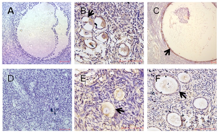
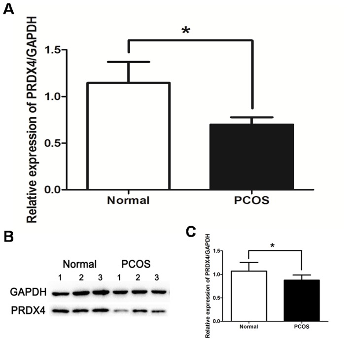
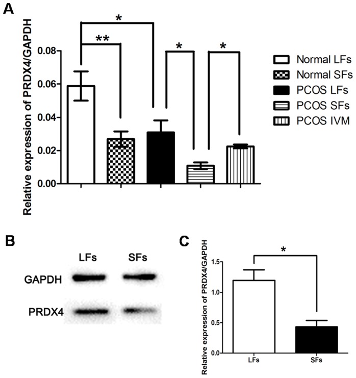
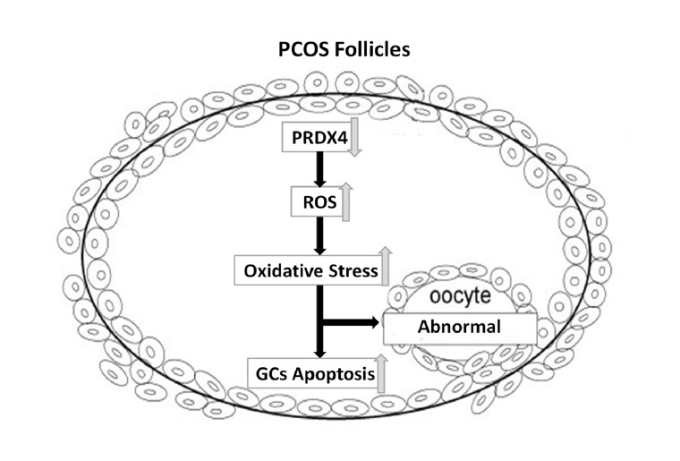
Peroxiredoxin 4 (PRDX4), a member of Peroxiredoxin (PRDX) family, is a typical 2-Cys PRDX. PRDX4 monitors the oxidative burden within cellular compartment and reduces hydrogen peroxide and alkyl hydroperoxide related to oxidative stress and apoptosis. Antioxidant, like PRDX4, may promote follicle development and participate in the pathophysiology of PCOS. In our previous study, we found that PRDX4 was expressed in mice oocyte cumulus oophorus complex, and that PRDX4 could be associated with follicle development. In this study, we explored the expression of PRDX4 in human follicles and possible role of PRDX4 in PCOS pathophysiology. Our data showed that PRDX4 was mainly expressed in granulosa cells in human ovaries. When compared to control group, both PRDX4 mRNA level and protein level decreased in PCOS group. The lowered levels of PRDX4 may relate to oxidative stress in the pathophysiologic progress of PCOS. Furthermore, expression of PRDX4 in the granulosa cells of in vivo or in vitro matured follicles was higher than that in immatured follicles, which suggested that PRDX4 may have a close relationship with follicular development. Altogether, our findings may provide new clues of the pathophysiologic mechanism of PCOS and potential therapeutic strategy using antioxidant, like PRDX4.
Conflict of interest statement
Figures
Similar articles
-
Implication of Differential Peroxiredoxin 4 Expression with Age in Ovaries of Mouse and Human for Ovarian Aging.Curr Mol Med. 2016;16(3):243-51. doi: 10.2174/1566524016666160225151647. Curr Mol Med. 2016. PMID: 26917265
-
Peroxiredoxin 4 protects against ovarian ageing by ameliorating D-galactose-induced oxidative damage in mice.Cell Death Dis. 2020 Dec 11;11(12):1053. doi: 10.1038/s41419-020-03253-8. Cell Death Dis. 2020. PMID: 33311472 Free PMC article.
-
Peroxiredoxin 4 deficiency induces accelerated ovarian aging through destroyed proteostasis in granulosa cells.Biochim Biophys Acta Mol Basis Dis. 2024 Oct;1870(7):167334. doi: 10.1016/j.bbadis.2024.167334. Epub 2024 Jul 5. Biochim Biophys Acta Mol Basis Dis. 2024. PMID: 38971505
-
MMPS and TIMPS in ovarian physiology and pathophysiology.Front Biosci. 2004 Sep 1;9:2474-83. doi: 10.2741/1409. Front Biosci. 2004. PMID: 15353300 Review.
-
Long non-coding RNAs in ovarian granulosa cells.J Ovarian Res. 2020 Jun 5;13(1):63. doi: 10.1186/s13048-020-00663-2. J Ovarian Res. 2020. PMID: 32503679 Free PMC article. Review.
Cited by
-
Peroxiredoxin 4, a new oxidative stress marker in follicular fluid, may predict in vitro fertilization and embryo transfer outcomes.Ann Transl Med. 2020 Sep;8(17):1049. doi: 10.21037/atm-20-397. Ann Transl Med. 2020. PMID: 33145268 Free PMC article.
-
The Emerging Key Role of Klotho in the Hypothalamus-Pituitary-Ovarian Axis.Reprod Sci. 2021 Feb;28(2):322-331. doi: 10.1007/s43032-020-00277-5. Epub 2020 Aug 11. Reprod Sci. 2021. PMID: 32783104 Review.
-
A Brief Review on the Regulatory Roles of MicroRNAs in Cystic Diseases and Their Use as Potential Biomarkers.Genes (Basel). 2022 Jan 22;13(2):191. doi: 10.3390/genes13020191. Genes (Basel). 2022. PMID: 35205236 Free PMC article. Review.
-
Transcripts encoding free radical scavengers in human granulosa cells from primordial and primary ovarian follicles.J Assist Reprod Genet. 2018 Oct;35(10):1787-1798. doi: 10.1007/s10815-018-1240-3. Epub 2018 Jun 29. J Assist Reprod Genet. 2018. PMID: 29959620 Free PMC article.
-
Zearalenone exposure differentially affects the ovarian proteome in pre-pubertal gilts during thermal neutral and heat stress conditions.J Anim Sci. 2024 Jan 3;102:skae115. doi: 10.1093/jas/skae115. J Anim Sci. 2024. PMID: 38666409 Free PMC article.
References
-
- Azziz R, Woods KS, Reyna R, Key TJ, Knochenhauer ES, et al. (2004) The prevalence and features of the polycystic ovary syndrome in an unselected population. J Clin Endocrinol Metab 89: 2745–2749. - PubMed
-
- Franks S (1989) Polycystic ovary syndrome: a changing perspective. Clin Endocrinol (Oxf) 31: 87–120. - PubMed
-
- Balen AH, Conway GS, Kaltsas G, Techatrasak K, Manning PJ, et al. (1995) Polycystic ovary syndrome: the spectrum of the disorder in 1741 patients. Hum Reprod 10: 2107–2111. - PubMed
-
- Hughesdon PE (1982) Morphology and morphogenesis of the Stein-Leventhal ovary and of so-called “hyperthecosis”. Obstet Gynecol Surv 37: 59–77. - PubMed
-
- Webber LJ, Stubbs S, Stark J, Trew GH, Margara R, et al. (2003) Formation and early development of follicles in the polycystic ovary. Lancet 362: 1017–1021. - PubMed
Publication types
MeSH terms
Substances
LinkOut - more resources
Full Text Sources
Other Literature Sources
Medical
Research Materials
Miscellaneous
