. 2013 Oct 1;8(10):e77619.
doi: 10.1371/journal.pone.0077619. eCollection 2013.

Melanotic pathology and vertical transmission of the gut commensal Elizabethkingia meningoseptica in the major malaria vector Anopheles gambiae

Affiliations

Melanotic pathology and vertical transmission of the gut commensal Elizabethkingia meningoseptica in the major malaria vector Anopheles gambiae

Idir G Akhouayri et al. PLoS One. .

Abstract

Background: The resident gut flora is known to have significant impacts on the life history of the host organism. Endosymbiotic bacterial species in the Anopheles mosquito gut are potent modulators of sexual development of the malaria parasite, Plasmodium, and thus proposed as potential control agents of malaria transmission.

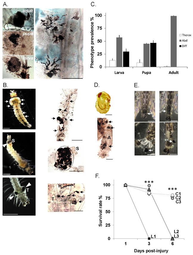
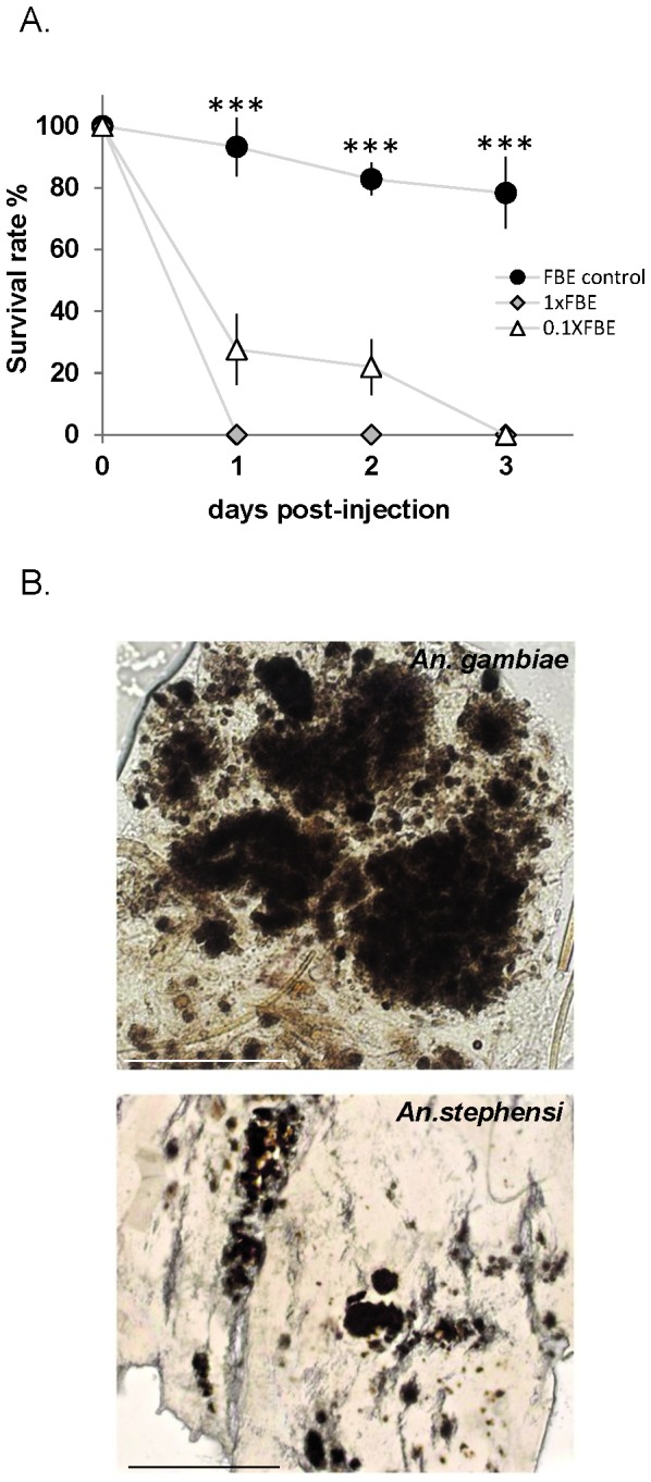
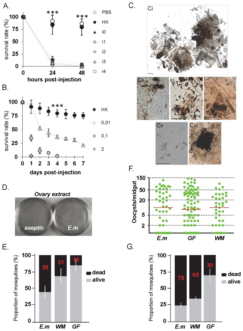
Results: Here we report a melanotic pathology in the major African malaria vector Anopheles gambiae, caused by the dominant mosquito endosymbiont Elizabethkingiameningoseptica. Transfer of melanised tissues into the haemolymph of healthy adult mosquitoes or direct haemolymph inoculation with isolated E. meningoseptica bacteria were the only means for transmission and de novo formation of melanotic lesions, specifically in the fat body tissues of recipient individuals. We show that E. meningoseptica can be vertically transmitted from eggs to larvae and that E. meningoseptica-mono-associated mosquitoes display significant mortality, which is further enhanced upon Plasmodium infection, suggesting a synergistic impact of E. meningoseptica and Plasmodium on mosquito survival.

Conclusion: The high pathogenicity and permanent association of E. meningoseptica with An. Gambiae through vertical transmission constitute attractive characteristics towards the potential design of novel mosquito/malaria biocontrol strategies.


Conflict of interest statement

Competing Interests: The authors have declared that no competing interests exist.

Figures

Figure 1
Figure 1. Melanotic phenotypes throughout An gambiae developmental stages.
(A) Melanotic lesions in the mosquito fat body throughout the developmental stages (left panels; scale bar ~1mm). Variation in shape and size of lesions with clusters and multiple melanised cells in a trail-like progression (right panel; scale bar ~0.5mm) (B) Phenotypic larval lesions. Light micrographs (left panels) and fixed samples (right panels). Melanotic lesions diffused throughout the larval body (upper panels; arrows). Lesions specific to abdominal segments (middle left panel; white arrow head) visible through the cuticle below the siphon (S). Fixed abdominal segments showing complete melanisation of the 8th segment (middle left panel; dotted area). Thoracic melanotic lesions (T) (lower left panel; white arrows), and a fixed larva with four lesions (lowest right panel) (black white arrows); H, head; T, thorax. Scale bar ~1mm. (C) Prevalence of melanotic phenotypes throughout the mosquito development is indicated as bars corresponding to the mean value +/- SD of 3 experiments. Abdominal, Abd; Diffused, Diff; Thoracic, Thorax. (D) Diffused phenotype in pupae. A light micrograph of a non-affected pupa (upper panel) and a fixed affected pupa with the diffused phenotype (lowest panel) showing melanotic lesions throughout the thorax (T) and abdomen (A) (black arrowheads). Scale bar ~1mm. (E) Lesions in adult mosquitoes. Melanotic abdominal lesions are observed at the junction between the 7th and 8th abdominal segments of female and male mosquitoes (upper panels; white arrows). Partially dissected abdominal segments from the same specimens (lowest panels) revealing melanotic aggregates (white arrows). Scale bar ~1mm. (F) Lesion-bearing mosquitoes survive poorly after thoracic injury. Groups of 40-60 female mosquitoes with lesions (L1, L2, L3) and corresponding controls without lesions (C1, C2, C3) were injured in the thorax below the base of the wing. Survival rate was monitored at 1, 3 and 6 days post-injury. Points indicated by *** are statistically very highly significant (P<0.05) between L and C.
Figure 2
Figure 2. Induction of lesions by injection of melanotic tissues in Anopheles species.
(A) Melanotic fat body tissues induce lethality in injected mosquitoes. Adult females were injected with abdomen-derived fat body tissues of melanotic lesion-bearing or of control adult females. Melanotic fat body extracts (FBE) were injected using two dilutions (1XFBE crude extract or 10-fold diluted extract 0.1XFBE). Survival rate was monitored daily, and represented as bars with standard error, as a percentage for each group. Three biologically independent injection assays were performed with 15-20 mosquitoes per condition. Points indicated by *** are very highly significantly different (P<0.05) between FBE control and 1XFBE or 0.1XFBE values.(B) Light micrographs of fat body tissues derived from FBE-injected female mosquitoes. Melanotic lesions in An. gambiae (upper panel; scale bar 0.05mm), and An. stephensi (lower panel; scale bar 0.5mm) mosquito species, resembling melanotic aggregates in the fat body of the affected ‘donor’ mosquito (see Figure 1).
Figure 3
Figure 3. Pathogenicity, lesion induction and vertical transmission of E . meningoseptica .
(A) Virulence of E . meningoseptica toward mosquitoes. Independent isolates of E meningoseptica (isolates 0 to 4) were injected in adult females. Survival rate is shown as percentage with standard error (n=120 per isolate) from 3 independent assays. Sterile PBS and heat-killed (HK) bacterial cultures were injected as controls. Points indicated by *** are statistically very highly significant (P<0.05) between HK and isolate injected samples. (B) Mosquito survival rate after injection of serial dilutions of E . meningoseptica (isolate 0) as indicated; n=180 from 3 independent assays. Values of HK and live bacterial samples were very highly significantly different (P<0.05). (C) Induced melanotic lesions in the fat body by E . meningoseptica ; (Ci) Partially dissected 6-8th abdominal segments after bacterial injection showing massive melanisation of fat body tissues; (Cii) Dissemination of melanotic spots throughout the abdominal fat body; (Ciii) Trail-like melanisation; (Civ) An aggregate with disseminated spots; (Cv) Melanised individual fat body cells (Cvi). A major melanotic thoracic aggregate. Scale bar 0.5mm (Ci, Ciii-Cvi), 0.2mm (Cii). (D) Enrichment of E . meningoseptica in An. gambiae ovaries. E . meningoseptica fed to germ-free females shows colonization of ovaries (E.m), whereas aseptic adults did not show any colony. (E) Mosquito lethality upon bacterial recolonisation. E . meningoseptica (E.m) or whole microbiota (WM) suspensions were fed to germ-free mosquitoes (GF) and survival was assessed 24h later. Non-supplemented GF mosquitoes were used as control. Bars show distribution as percentage (mean value in red with standard error) of dead (black area) and alive (grey area) mosquitoes (n=80) from 2 independent experiments. (F) Plasmodium berghei development is not affected by bacterial reconstitution. E.m, WM and GF mosquitoes were infected with malaria 24h post-reconstitution, and oocyst load monitored 7 days post-infection, represented as oocyst number per midgut (green dot) with mean values as red dotted bar. Mosquitoes were blood-fed on the same infective host (n=120) from 2 independent experiments. No statistical difference was found in between all oocyst load mean values. (G) Synergistic effect on mosquito survival upon bacterial reconstitution and Plasmodium infection. Mosquito survival of malaria infected-Em, -WM and -GF was assessed 24h post-infection.

References

    1. Backhed F, Ley RE, Sonnenburg JL, Peterson DA, Gordon JI (2005) Host-bacterial mutualism in the human intestine. Science 307: 1915-1920. doi:10.1126/science.1104816. PubMed: 15790844. - DOI - PubMed
    1. Ley RE, Peterson DA, Gordon JI (2006) Ecological and evolutionary forces shaping microbial diversity in the human intestine. Cell 124: 837-848. doi:10.1016/j.cell.2006.02.017. PubMed: 16497592. - DOI - PubMed
    1. Pumpuni CB, Beier MS, Nataro JP, Guers LD, Davis JR (1993) Plasmodium falciparum: inhibition of sporogonic development in Anopheles stephensi by gram-negative bacteria. Exp Parasitol 77: 195-199. doi:10.1006/expr.1993.1076. PubMed: 8375488. - DOI - PubMed
    1. Beier MS, Pumpuni CB, Beier JC, Davis JR (1994) Effects of para-aminobenzoic acid, insulin, and gentamicin on Plasmodium falciparum development in anopheline mosquitoes (Diptera: Culicidae). J Med Entomol 31: 561-565. PubMed: 7932602. - PubMed
    1. Demaio J, Pumpuni CB, Kent M, Beier JC (1996) The midgut bacterial flora of wild Aedes triseriatus, Culex pipiens, and Psorophora columbiae mosquitoes. Am J Trop Med Hyg 54: 219-223. PubMed: 8619452. - PubMed

Publication types

MeSH terms

LinkOut - more resources