. 2013 Sep 30;8(9):e75137.
doi: 10.1371/journal.pone.0075137. eCollection 2013.

Increased protein stability of CDKN1C causes a gain-of-function phenotype in patients with IMAGe syndrome

Affiliations

Increased protein stability of CDKN1C causes a gain-of-function phenotype in patients with IMAGe syndrome

Naoki Hamajima et al. PLoS One. .

Abstract

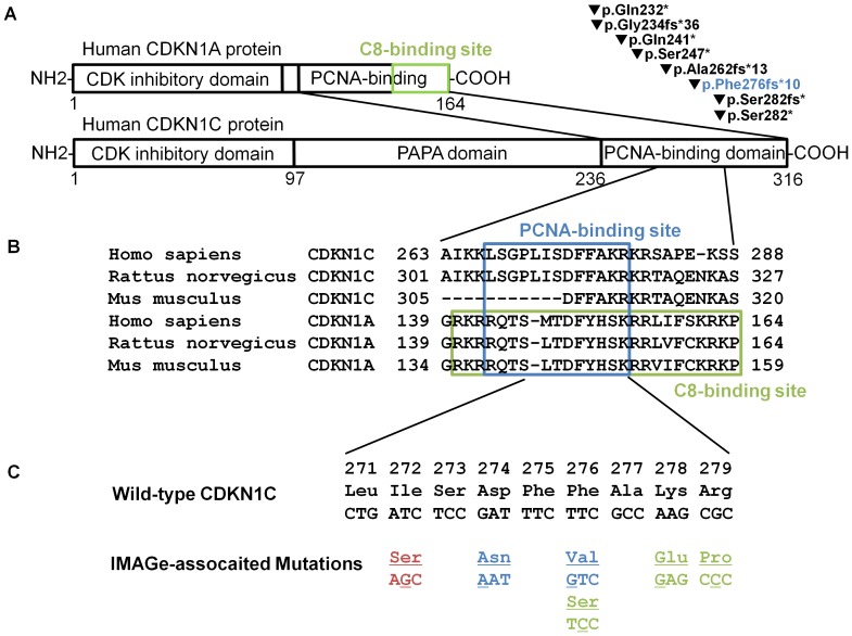
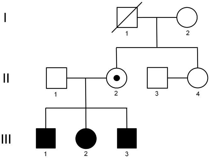
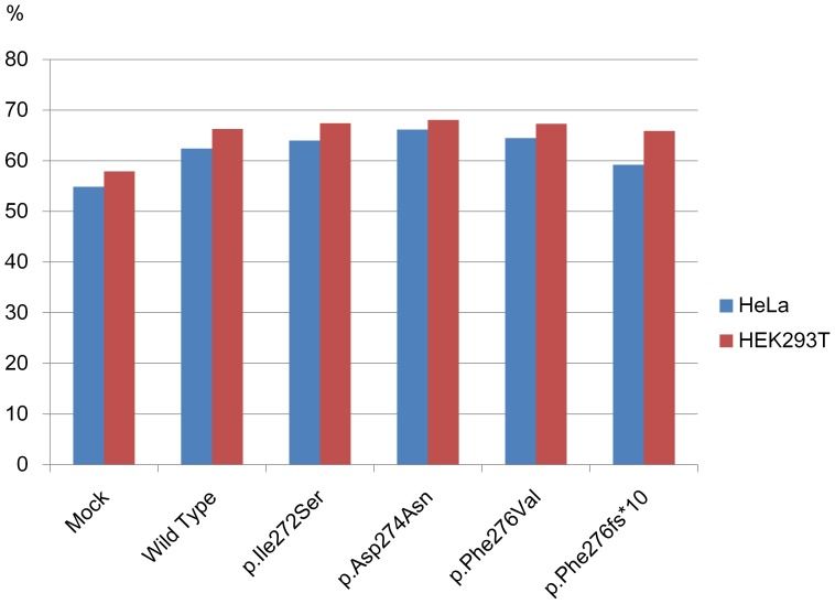
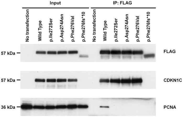
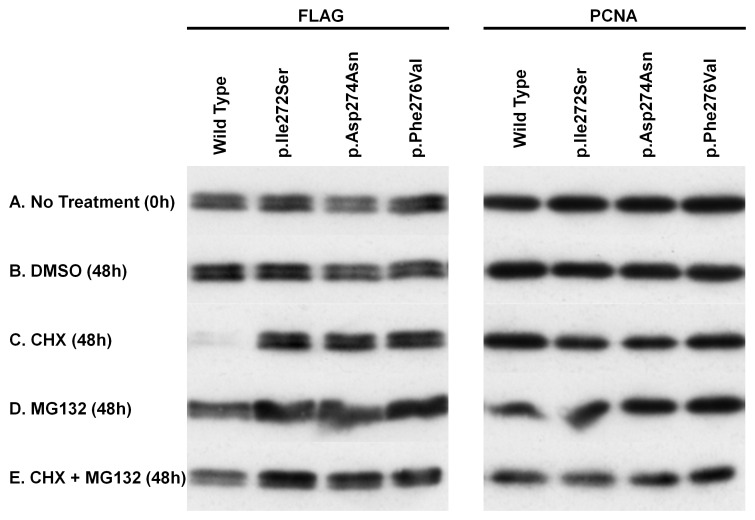
Mutations in the proliferating cell nuclear antigen (PCNA)-binding domain of the CDKN1C gene were recently identified in patients with IMAGe syndrome. However, loss of PCNA binding and suppression of CDKN1C monoubiquitination by IMAGe-associated mutations hardly explain the reduced-growth phenotype characteristic of IMAGe syndrome. We demonstrate here that IMAGe-associated mutations in the CDKN1C gene dramatically increased the protein stability. We identified a novel heterozygous mutation, c.815T>G (p.Ile272Ser), in the CDKN1C gene in three siblings manifesting clinical symptoms associated with IMAGe syndrome and their mother (unaffected carrier). PCNA binding to CDKN1C was disrupted in the case of p.Ile272Ser, and for two other IMAGe-associated mutations, p.Asp274Asn and p.Phe276Val. Intriguingly, the IMAGe-associated mutant CDKN1C proteins were fairly stable even in the presence of cycloheximide, whereas the wild-type protein was almost completely degraded via the proteasome pathway, as shown by the lack of degradation with addition of a proteasome inhibitor, MG132. These results thus suggested that the reduced-growth phenotype of IMAGe syndrome derives from CDKN1C gain-of-function due to IMAGe-associated mutations driving increased protein stability.


Conflict of interest statement

Competing Interests: The authors have declared that no competing interests exist.

Figures

Figure 1
Figure 1. Structure of CDKN1C and CDKN1A proteins and IMAGe-associated mutations.
(A) Schematic representation for the structures of human CDKN1C and CDKN1A proteins and for the BWS-associated truncation mutations in the PCNA-binding domain of CDKN1C. The green closed square represents the C8-binding site . Numbers below the schemas represent the locations of amino acid residues. Filled inverted triangles denote the truncation mutants in the PCNA-binding domain of CDKN1C reported in patients with BWS , . The blue characters represent the mutation analyzed in this article (p.Phe276fs*10). (B) Alignment of amino acid sequences around the PCNA- and C8-binding sites in human, rat, and mouse CDKN1C and CDKN1A. The numbers above the set of sequences represent the amino acid residues. The blue closed square represents the PCNA-binding site and the green closed square represents the C8-binding site . Multiple sequence alignment was performed by using ClustalW (http://www.genome.jp/tools/clustalw/). Accession numbers of the amino acid sequences described here are as follows: NP_000067.1, Homo sapiens CDKN1C; NP_001028929.1, Rattus norvegicus CDKN1C; NP_001155096.1, Mus musculus CDKN1C; AAH13967, H. sapiens CDKN1A; AAI00621, R. norvegicus CDKN1A; and AAH02043, M. musculus CDKN1A. (C) Amino acid and nucleotide sequences of the PCNA-binding domain in the wild-type and IMAGE-associated mutant CDKN1C genes in human. Numbers on the top line represent amino residues of the CDKN1C protein based on accession number NM_000076.2. Red characters represent the mutation reported in this article (c.815T>G and p.Ile272Ser). Blue and green characters represent mutations described in the previous report : blue characters represent mutations analyzed in this article (p.Asp274Asn and p.Phe276Val). Underlined characters represent substituted residues and nucleotides.
Figure 2
Figure 2. Pedigree of the family with IMAGe syndrome.
Filled squares and circles represent the male and female patients, respectively. Closed squares and circles represent the male and female unaffected individuals, respectively. Small filled circles with large closed circles or squares represent unaffected carriers. Diagonal lines represent deceased individuals.
Figure 3
Figure 3. Cell cycle analysis.
HeLa and HEK293T cells were transfected with plasmids expressing wild-type or one of four mutant CDKN1C proteins, p.Ile272Ser, p.Asp274Asn, p.Phe276Val, and p.Phe276fs*10. Mock transfections were performed by using pCMV-3Tag-1B vector alone. At 48 h after transfection, cells were fixed in 70% ethanol, stained by propidium iodide, and then subjected to cell cycle analysis by FACSCanto II. Percentages of the cells in G1 phase are presented by bar graphs: blue bars, HeLa cells; red bars, HEK293T cells.
Figure 4
Figure 4. Western blot analysis.
HEK293T cells were transiently transfected with plasmids expressing FLAG-tagged wild-type or one of four mutant CDKN1C proteins, p.Ile272Ser, p.Asp274Asn, p.Phe276Val, and pPhe276fs*10. Forty-eight hours after transfection, cell lysates were prepared and immunoprecipitated by anti-FLAG antibody. Both input and immunoprecipitated (IP) samples were subjected to Western blot analysis with antibodies against FLAG, CDKN1C, and PCNA.
Figure 5
Figure 5. Protein stability assay.
HEK293T cells were transiently transfected with plasmids expressing FLAG-tagged wild-type or one of three mutant CDKN1C proteins, p.Ile272Ser, p.Asp274Asn, and p.Phe276Val. At 48 h after transfection, cells were treated with DMSO, cycloheximide (CHX), or MG132 for 48 h. Cell lysates were prepared at 0 h (no treatment) or at 48 h (DMSO, CHX, MG132, CHX+MG132), and subjected to Western blot analysis using anti-FLAG antibody for detecting the CDKN1C proteins and anti-PCNA antibody as a loading control.

References

    1. Vilain E, Le Merrer M, Lecointre C, Desangles F, Kay MA, et al. (1999) IMAGe, a new clinical association of intrauterine growth retardation, metaphyseal dysplasia, adrenal hypoplasia congenita, and genital anomalies. J Clin Endocrinol Metab 84: 4335–4340. - PubMed
    1. Bergadá I, Del Rey G, Lapunzina P, Bergadá C, Fellous M, et al. (2005) Familial occurrence of the IMAGe association: additional clinical variants and a proposed mode of inheritance. J Clin Endocrinol Metab 90: 3186–3190. - PubMed
    1. Hutz JE, Krause AS, Achermann JC, Vilain E, Tauber M, et al. (2006) IMAGe association and congenital adrenal hypoplasia: no disease-causing mutations found in the ACD gene. Mol Genet Metab 88: 66–70. - PubMed
    1. Lienhardt A, Mas JC, Kalifa G, Chaussain JL, Tauber M (2002) IMAGe association: additional clinical features and evidence for recessive autosomal inheritance. Horm Res 57: 71–78. - PubMed
    1. Pedreira CC, Savarirayan R, Zacharin MR (2004) IMAGe syndrome: a complex disorder affecting growth, adrenal and gonadal function, and skeletal development. J Pediatr 144: 274–277. - PubMed

MeSH terms

Substances

Supplementary concepts