

Formation of impeller-like helical DNA-silica complexes by polyamines induced chiral packing
- PMID: 24098845
- PMCID: PMC3438577
- DOI: 10.1098/rsfs.2011.0119
Formation of impeller-like helical DNA-silica complexes by polyamines induced chiral packing
Abstract
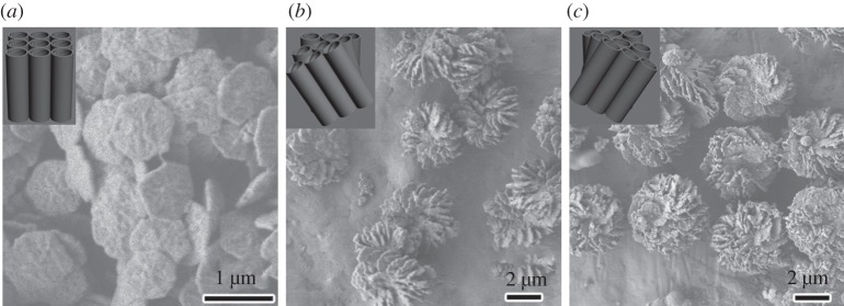
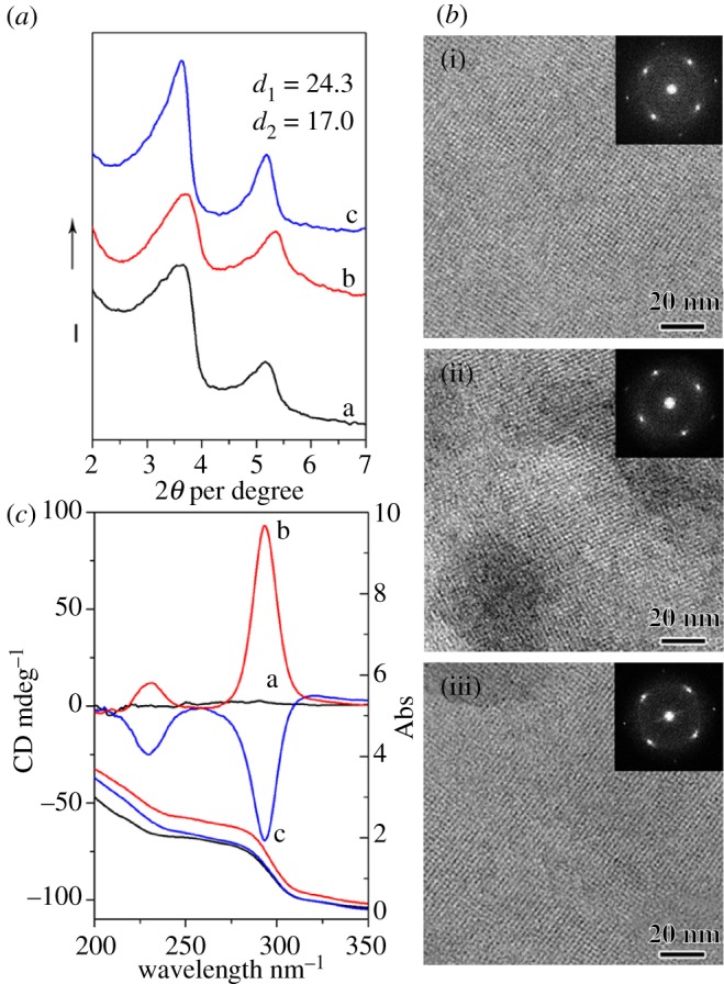
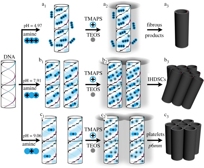
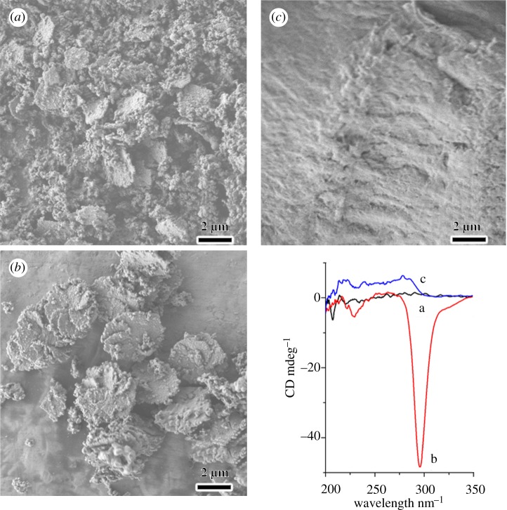
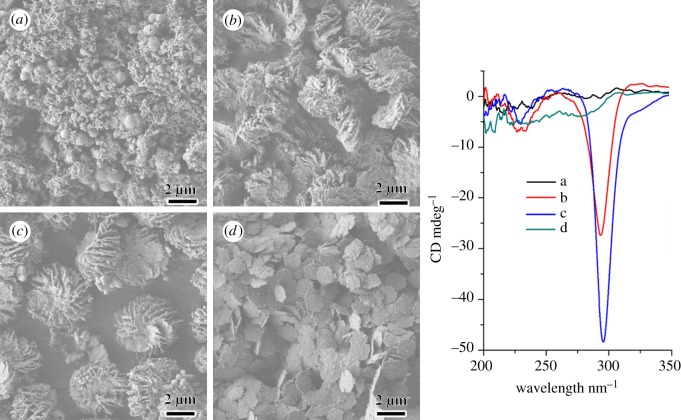
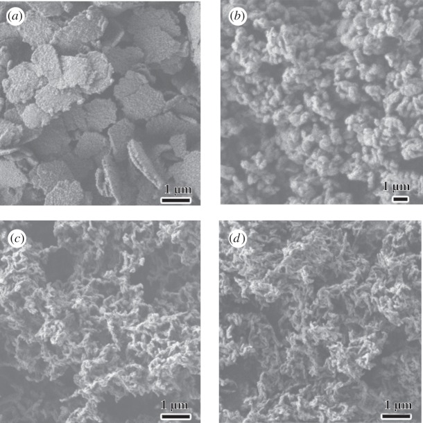
The helicity of DNA and its long-range chiral packing are widespread phenomena; however, the packing mechanism remains poorly understood both in vivo and in vitro. Here, we report the extraordinary DNA chiral self-assembly by silica mineralization, together with circular dichroism measurements and electron microscopy studies on the structure and morphology of the products. Mg(2+) ion and diethylenetriamine were found to induce right- and left-handed chiral DNA packing with two-dimensional-square p4mm mesostructures, respectively, to give corresponding enantiomeric impeller-like helical DNA-silica complexes. Moreover, formation of macroscopic impeller-like helical architectures depends on the types of polyamines and co-structure-directing agents and pH values of reaction solution. It has been suggested that interaction strength between negatively charged DNA phosphate strands and positively charged counterions may be the key factor for the induction of DNA packing handedness.
Keywords: biomolecule; chirality; helical architecture; liquid crystal; polyamine, DNA–silica complex; self-assemble.
Figures
References
-
- Cölfen H., Mann S. 2003. Higher-order organization by mesoscale self-assembly and transformation of hybrid nanostructures. Angew. Chem. Int. Ed. 42, 2350–236510.1002/anie.200200562 (doi:10.1002/anie.200200562) - DOI - DOI - PubMed
-
- Strzelecka T. E., Davidson M. W., Rill R. L. 1988. Multiple liquid crystal phases of DNA at high concentrations. Nature 331, 457–46010.1038/331457a0 (doi:10.1038/331457a0) - DOI - DOI - PubMed
-
- Bloomfield V. A. 1996. DNA condensation. Curr. Opin. Struc. Biol. 6, 334–34110.1016/S0959-440X(96)80052-2 (doi:10.1016/S0959-440X(96)80052-2) - DOI - DOI - PubMed
-
- Livolant F., Leforestier A. 1996. Condensed phases of DNA: structures and phase transitions. Prog. Polym. Sci. 21, 1115–116410.1016/S0079-6700(96)00016-0 (doi:10.1016/S0079-6700(96)00016-0) - DOI - DOI
-
- Livolant F., Levelut A., Doucet J., Benoit J. 1989. The highly concentrated liquid-crystalline phase of DNA is columnar hexagonal. Nature 339, 724–72610.1038/339724a0 (doi:10.1038/339724a0) - DOI - DOI - PubMed
LinkOut - more resources
Full Text Sources
