Ensemble Semi-supervised Frame-work for Brain Magnetic Resonance Imaging Tissue Segmentation
- PMID: 24098863
- PMCID: PMC3788199
Ensemble Semi-supervised Frame-work for Brain Magnetic Resonance Imaging Tissue Segmentation
Abstract
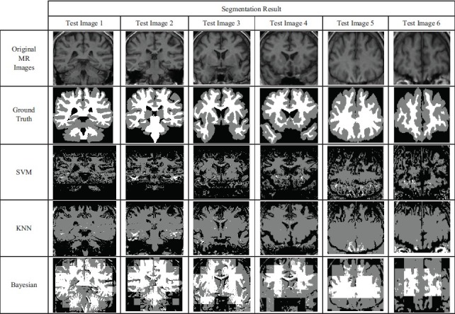
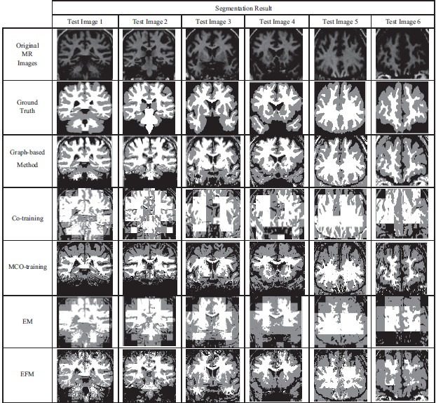
Brain magnetic resonance images (MRIs) tissue segmentation is one of the most important parts of the clinical diagnostic tools. Pixel classification methods have been frequently used in the image segmentation with two supervised and unsupervised approaches up to now. Supervised segmentation methods lead to high accuracy, but they need a large amount of labeled data, which is hard, expensive, and slow to obtain. Moreover, they cannot use unlabeled data to train classifiers. On the other hand, unsupervised segmentation methods have no prior knowledge and lead to low level of performance. However, semi-supervised learning which uses a few labeled data together with a large amount of unlabeled data causes higher accuracy with less trouble. In this paper, we propose an ensemble semi-supervised frame-work for segmenting of brain magnetic resonance imaging (MRI) tissues that it has been used results of several semi-supervised classifiers simultaneously. Selecting appropriate classifiers has a significant role in the performance of this frame-work. Hence, in this paper, we present two semi-supervised algorithms expectation filtering maximization and MCo_Training that are improved versions of semi-supervised methods expectation maximization and Co_Training and increase segmentation accuracy. Afterward, we use these improved classifiers together with graph-based semi-supervised classifier as components of the ensemble frame-work. Experimental results show that performance of segmentation in this approach is higher than both supervised methods and the individual semi-supervised classifiers.
Keywords: Brain magnetic resonance image tissue segmentation; MCo_Training classifier; ensemble semi-supervised frame-work; expectation filtering maximization classifier.
Conflict of interest statement
Figures


References
-
- Limperopoulos C, Clouchoux C. Advancing fetal brain MRI: Targets for the future. Semin Perinatol. 2009;33:289–98. - PubMed
-
- Duta N, Sonka M. Segmentation and interpretation of MR brain images: An improved active shape model. IEEE Trans Med Imaging. 1998;17:1049–62. - PubMed
-
- Li H, Yezzi A, Cohen LD. Fast 3D brain segmentation using dual-front active contours with optional user-interaction. Proc. International Workshop on Computer Vision for Biomedical Image Applications, Lecture Notes in Computer Science. 2005;3765:335–45.
-
- Pohle R, Toennies KD. Segmentation of medical images using adaptive region growing. Proc SPIE Medical Imaging. 2001;4322:1337–46.
-
- Sammouda R, Niki N, Nishitani H. A Comparison of hopfield neural network and Boltzmann machine in segmenting MR images of the brain. IEEE Trans Nucl Sci. 1996;43:3361–9.
LinkOut - more resources
Full Text Sources
Other Literature Sources