Review
. 2013 Dec;1828(12):2873-85.
doi: 10.1016/j.bbamem.2013.03.032.

Biochemical and structural insights into intramembrane metalloprotease mechanisms

Affiliations
Review

Biochemical and structural insights into intramembrane metalloprotease mechanisms

Lee Kroos et al. Biochim Biophys Acta. 2013 Dec.

Abstract

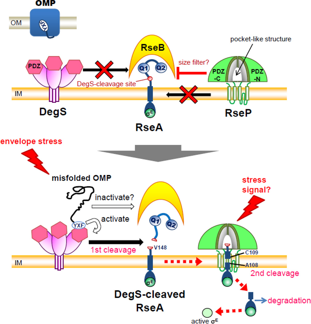
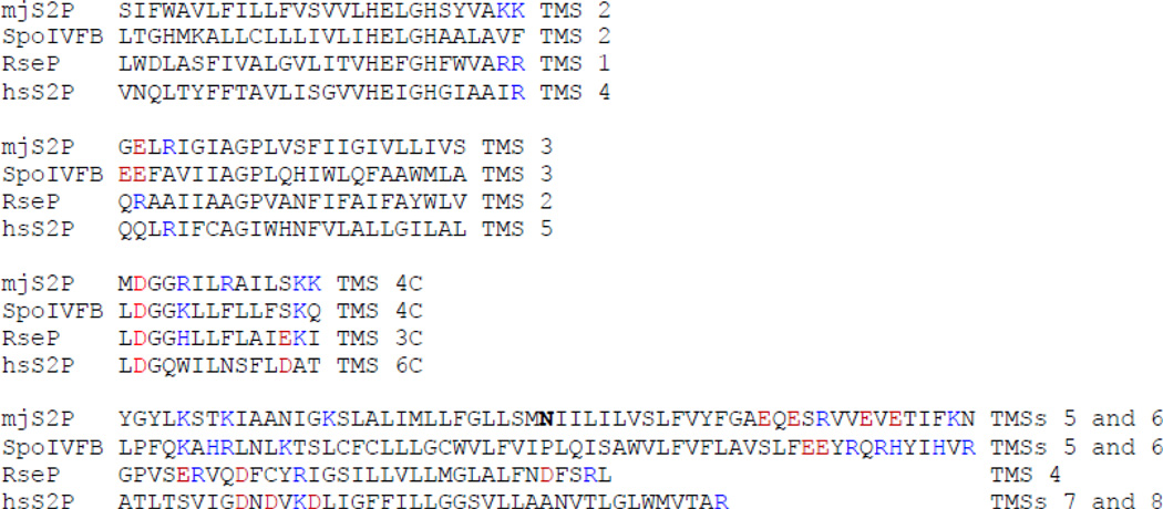
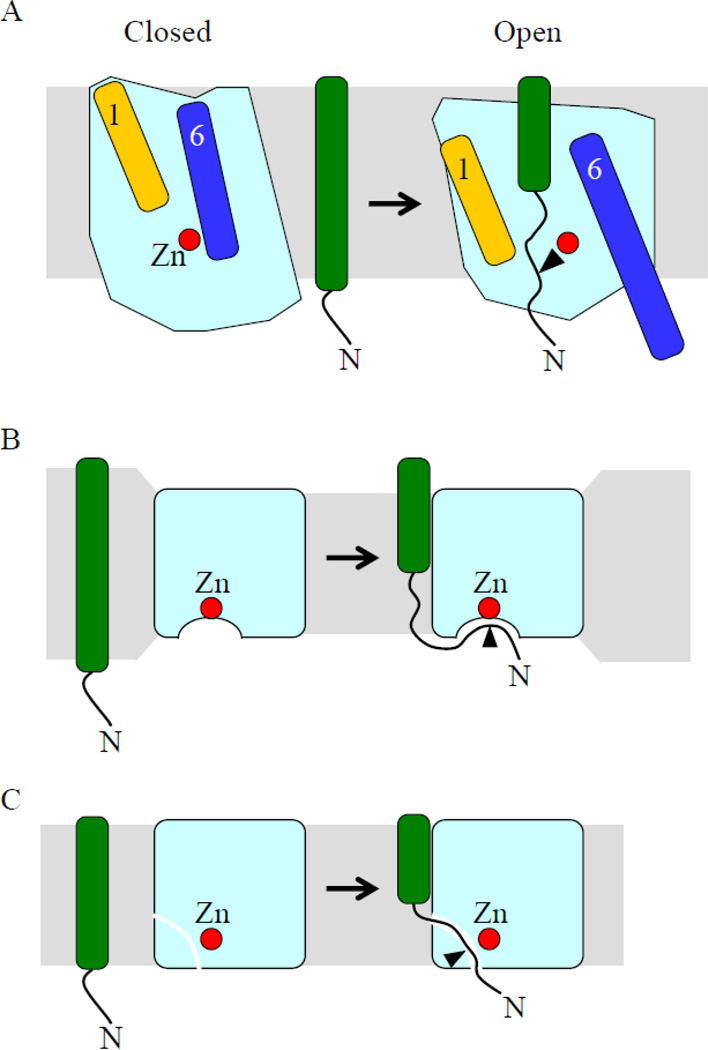
Intramembrane metalloproteases are nearly ubiquitous in living organisms and they function in diverse processes ranging from cholesterol homeostasis and the unfolded protein response in humans to sporulation, stress responses, and virulence of bacteria. Understanding how these enzymes function in membranes is a challenge of fundamental interest with potential applications if modulators can be devised. Progress is described toward a mechanistic understanding, based primarily on molecular genetic and biochemical studies of human S2P and bacterial SpoIVFB and RseP, and on the structure of the membrane domain of an archaeal enzyme. Conserved features of the enzymes appear to include transmembrane helices and loops around the active site zinc ion, which may be near the membrane surface. Extramembrane domains such as PDZ (PSD-95, DLG, ZO-1) or CBS (cystathionine-β-synthase) domains govern substrate access to the active site, but several different mechanisms of access and cleavage site selection can be envisioned, which might differ depending on the substrate and the enzyme. More work is needed to distinguish between these mechanisms, both for enzymes that have been relatively well-studied, and for enzymes lacking PDZ and CBS domains, which have not been studied. This article is part of a Special Issue entitled: Intramembrane Proteases.

Keywords: (PSD-95, DLG, ZO-1); (cystathionine-β-synthase); CBS; CBS domain; GFP; IMMP(s); IP(s); Intramembrane metalloprotease; OMP(s); PDZ; PDZ domain; RNA polymerase; RNAP; RseP; S1P; S2P; SCAP; SREBP(s); SREBP-cleavage-activating protein; SpoIVFB; TMS(s); green fluorescent protein; intramembrane metalloprotease(s); intramembrane protease(s); outer membrane protein(s); site-1 protease; site-2 protease; sterol-regulatory element-binding protein(s); transmembrane segment(s).


Figures

Figure 1
Figure 1
Cellular location, membrane topology, and extramembrane domains of S2P, SpoIVFB, and RseP. A) Human S2P is located in the membranes of the Golgi apparatus and has 8 predicted TMSs of which TMSs 4–6 make up the conserved core (orange). The positions of a Ser-rich loop, the HEXXH motif, the lumenal PDZ domain with its Cys-rich insert, and D467 in TMS 6 are shown. When substrate SREBPs are transported from the endoplasmic reticulum to the Golgi apparatus, S1P cleaves a lumenal loop, allowing S2P to cleave TMS 1 and release the N-terminal basic helix-loop-helix (bHLH) leucine-zipper (zip) domain into the cytosol. The bHLH-zip domain enters the nucleus and activates transcription. The regulatory domain (Reg.) interacts with SCAP (not shown). B) SpoIVFB is located in the outermost membrane surrounding the forespore during B. subtilis sporulation. Features analogous to those noted above in S2P are indicated. The CBS domain is in the mother cell, as is most of Pro-σK, whose pro-sequence is depicted to interact peripherally and loop into the membrane, although this is speculative. SpoIVFB cleaves Pro-σK, releasing σK into the mother cell to direct transcription. C) RseP is located in the E. coli inner membrane with its tandem PDZ domains in the periplasm. DegS cleaves the periplasmic C-terminal domain of RseA, then RseP cleaves the TMS, releasing the RseA N-terminal domain in complex with σE into the cytosol, where ClpXP degrades the rest of RseA, releasing σE to direct transcription.
Figure 2
Figure 2
Regulation of SpoIVFB activity. During B. subtilis sporulation, the forespore is surrounded by two membranes upon the completion of engulfment (top part). The bottom parts depict a series of proteolytic cleavages. First, σG RNAP in the forespore causes expression of serine proteases SpoIVB and CtpB (also expressed under σE RNAP control in the mother cell), which are translocated into the intermembrane space, where they cleave the C-terminal domain of SpoIVFA to initiate its degradation (dashes). SpoIVFA is in complex with BofA and SpoIVFB in the outermost membrane surrounding the forespore, after these proteins are expressed in the mother cell under σE RNAP control. In the second step, CtpB and one or more other proteases (not shown) cleave BofA to initiate its degradation (dashes). BofA is the primary inhibitor of SpoIVFB. Finally, SpoIVFB cleaves Pro-σK, releasing σK into the mother cell.
Figure 3
Figure 3
A model for ATP transport and accumulation during B. subtilis sporulation, and localization of the SpoIVFB complex with channel and engulfment complexes. A complex of SpoIIM, SpoIIP, and SpoIID proteins (red) interacts with the cell wall and causes the mother cell membrane to engulf the forespore (top left). During engulfment, channels (green) composed of SpoIIQ expressed in the forespore and SpoIIIA proteins expressed in the mother cell are formed. The channels span the intermembrane space and have been proposed to allow small molecules like ATP to move from the mother cell into the forespore. Upon completion of engulfment, the channels undergo reorganization and some components are degraded (top right). We propose that the ATP concentration rises in the mother cell and this is sensed by the CBS domain of SpoIVFB. SpoIVFA facilitates assembly of SpoIVFB with its inhibitor BofA and localizes the complex (magenta) to foci that during engulfment include the channel and engulfment complexes (bottom part), although whether SpoIVFA interacts directly with a protein(s) in the other complexes or interacts indirectly is unknown (dashed arrows).
Figure 4
Figure 4
A model for RseP function in transmembrane signaling of the σE stress response. Upper part) Under resting conditions, DegS remains inactive as to RseA cleavage. Cleavage of intact RseA by RseP is prevented by several negative regulators including RseB, the Gln-rich sequences (Q1 and Q2) in RseA, and the PDZ domains (PDZ-N and PDZ-C) of RseP. The RseP PDZ domains could act as a size-exclusion filter to block the access of RseB-bound intact RseA into the active site of RseP. Lower part) Extracytoplasmic stresses cause accumulation of misfolded OMPs, which trigger DegS-catalyzed cleavage of the RseA periplasmic region through activation of DegS and inactivation of RseB. This first cleavage releases negative regulation and allows intramembrane cleavage of DegS-processed RseA by RseP. σE is finally activated in the cytoplasm and promotes transcription of stress-responsive genes. Some stress signal(s) might be directly recognized by the RseP PDZ domains to induce cleavage of intact RseA, resulting in σE activation.
Figure 5
Figure 5
Residues in SREBP-2 important for cleavage by S2P and a model for partial α-helix unwinding of SREBPs. A) Effect of substitutions in TMS 1 of SREBP-2 on cleavage by S2P. Residues 478–502 include four residues (blue) preceding TMS 1 (black). The cleavage site is indicated by an arrow. Single-residue substitutions having no effect (green) or reducing cleavage (orange) are shown immediately above and below the sequence, respectively. Multi-residue substitutions having no effect (green) or abolishing cleavage (red) are separated from the sequence by lines that indicate which residues were substituted. B) Model for partial α-helix unwinding of SREBPs. Left) S1P cleaves the lumenal loop of an SREBP (see Fig. 1A for domain abbreviations). Right) Separation of the two TMSs is proposed to allow the N-terminal part of TMS 1 to unwind, exposing the cleavage site to the cytosolic face of the membrane for cleavage by S2P.
Figure 6
Figure 6
Structure of the membrane domain of an archaeal IMMP. The 6-TMS domain of the M. jannaschii enzyme, referred to as mjS2P, is shown in the open conformation with each α-helix a different color and other parts blue (PDB ID: 3B4R). The three residues that coordinate a zinc atom, which together with E55 activates a water molecule to catalyze hydrolysis of a substrate peptide bond, are shown. Part of the loop connecting TMSs 2 and 3 is predicted to enter the membrane and a 6-residue loop interrupts TMS 4. These loops are the basis for predicting analogous loops in S2P, SpoIVFB, and RseP as depicted in Figure 1.
Figure 7
Figure 7
Residues with charged side chains near the cytosolic ends of predicted TMSs in IMMPs. The indicated TMSs of mjS2P (numbered as in Fig. 6) are aligned with corresponding predicted TMSs of B. subtilis SpoIVFB, E. coli RseP, and human S2P (hsS2P). Residues with positively (blue) or negatively (red) charged side chains that are near the cytosolic ends of predicted TMSs are shown in color. The Asn residue at the turn separating mjS2P TMSs 5 and 6 is shown in bold.
Figure 8
Figure 8
Models for substrate access to the active sites of IMMPs. A) Based on closed and open conformations observed in crystals of mjS2P, TMSs 1 and 6 have been proposed to function as lateral gates that in the open conformation allow substrate access to the active site (red zinc atom). The substrate presumably must be unwound in the vicinity of the cleavage site in order for cleavage to occur (arrowhead). B) The membrane is proposed to be compressed around the enzyme so that when substrate interacts with the enzyme, a portion of the target TMS unwinds, allowing it to access the enzyme active site from the solvent-exposed juxtamembrane region. C) When substrate interacts with the enzyme, a portion of the target TMS unwinds, allowing it to access the active site via a lateral opening in the enzyme.

Similar articles

Cited by

References

    1. Rawson R, Zelenski N, Nijhawan D, Ye J, Sakai J, Hasan M, Chang T, Brown M, Goldstein J. Complementation cloning of SP2, a gene encoding a putative metalloprotease required for intramembrane cleavage of SREBPs. Mol. Cell. 1997;1:47–57. - PubMed
    1. Lewis A, Thomas P. A novel clan of zinc metallopeptidases with possible intramembrane cleavage properties. Protein Sci. 1999;8:439–442. - PMC - PubMed
    1. Rudner D, Fawcett P, Losick R. A family of membrane-embedded metalloproteases involved in regulated proteolysis of membrane-associated transcription factors. Proc. Natl. Acad. Sci. USA. 1999;96:14765–14770. - PMC - PubMed
    1. Yu Y-TN, Kroos L. Evidence that SpoIVFB is a novel type of membrane metalloprotease governing intercompartmental communication during Bacillus subtilis sporulation. J. Bacteriol. 2000;182:3305–3309. - PMC - PubMed
    1. Alba BM, Leeds JA, Onufryk C, Lu CZ, Gross CA. DegS and YaeL participate sequentially in the cleavage of RseA to activate the σE-dependent extracytoplasmic stress response. Genes Dev. 2002;16:2156–2168. - PMC - PubMed

Publication types

MeSH terms

LinkOut - more resources