Review
. 2013:29:551-69.
doi: 10.1146/annurev-cellbio-101011-155838. Epub 2013 May 31.

Viral membrane scission

Affiliations
Review

Viral membrane scission

Jeremy S Rossman et al. Annu Rev Cell Dev Biol. 2013.

Abstract

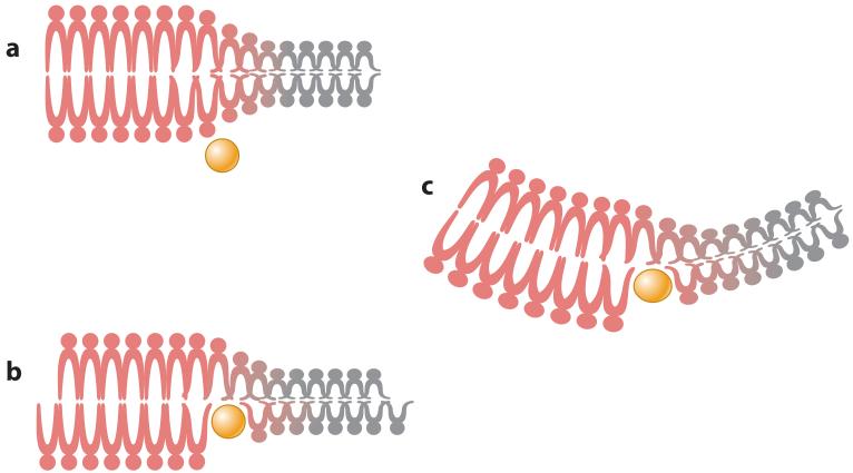
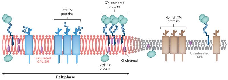
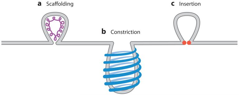
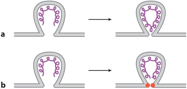
Virus budding is a complex, multistep process in which viral proteins make specific alterations in membrane curvature. Many different viral proteins can deform the membrane and form a budding virion, but very few can mediate membrane scission to complete the budding process. As a result, enveloped viruses have developed numerous ways of facilitating membrane scission, including hijacking host cellular scission machinery and expressing their own scission proteins. These proteins mediate scission in very different ways, though the biophysical mechanics underlying their actions may be similar. In this review, we explore the mechanisms of membrane scission and the ways in which enveloped viruses use these systems to mediate the release of budding virions.


Figures

Figure 1
Figure 1
Induction of membrane curvature by protein insertion. (a) Amphipathic proteins (yellow) can bind to and insert into lipid bilayers, occasionally at the junction between two lipid phases (liquid-ordered- or raft-phase lipids, shown in red, and liquid-disordered- or bulk-phase plasma membrane lipids, shown in gray). (b) Protein insertion expands one leaflet of the lipid bilayer, which places the membrane under strain. (c) Membrane strain can be resolved by inducing curvature. Peptide insertion at the lipid phase boundary can also alter membrane curvature by modifying the line tension force between the two lipid phases.
Figure 2
Figure 2
Organization of lipid phases in the plasma membrane. Depiction of the lipid bilayer showing the differences between the raft and nonraft phases. Unsaturated glycophospholipids (GPL) represent the bulk (liquid-disordered) phase of the plasma membrane and contain mainly nonraft transmembrane (TM) proteins. Saturated GPL and sphingomyelin (SM) represent the raft (liquid-ordered) phase of the plasma membrane and are associated with higher levels of cholesterol and GPI-anchored, acetylated, and raft-localized TM proteins. The ordered packaging of the saturated lipids causes the raft phase to be thicker than the bulk plasma membrane phase, which leads to line tension between the lipid phases.
Figure 3
Figure 3
Examples of the different types of membrane scission. (a) Assembly of scaffolding proteins on the inside of the membrane can deform the membrane sufficiently to cause membrane scission. (b) Constriction is accomplished by the formation of protein rings around the outside of a budding vesicle. Contraction of the rings then constricts the budding neck, which causes scission. (c) Lipid insertion can cause membrane scission after a bud has formed by protein insertion into the inner membrane leaflet (in the case of the influenza virus budding) or the outer membrane leaflet (in the case of cellular budding events) at the neck of the membrane bud. Stacking defects caused by the insertion event then alter membrane curvature, which leads to scission.
Figure 4
Figure 4
Capacity for scaffold proteins to cause membrane scission during virus budding. (a) In vitro viral matrix protein assembly can cause both membrane budding and scission; however, (b) in vivo matrix assembly may be sufficient to deform the membrane but could require a dedicated scission protein (red) to mediate membrane scission.

References

    1. Adell MA, Teis D. Assembly and disassembly of the ESCRT-III membrane scission complex. FEBS Lett. 2011;585:3191–96. - PMC - PubMed
    1. Ai E, Skop AR. Endosomal recycling regulation during cytokinesis. Commun. Integr. Biol. 2009;2:444–47. - PMC - PubMed
    1. Amorim MJ, Bruce EA, Read EK, Foeglein A, Mahen R, et al. A Rab11- and microtubule-dependent mechanism for cytoplasmic transport of influenza A virus viral RNA. J. Virol. 2011;85:4143–56. - PMC - PubMed
    1. Babst M, Sato TK, Banta LM, Emr SD. Endosomal transport function in yeast requires a novel AAA-type ATPase, Vps4p. EMBO J. 1997;16:1820–31. - PMC - PubMed
    1. Babst M, Wendland B, Estepa EJ, Emr SD. The Vps4p AAA ATPase regulates membrane association of a Vps protein complex required for normal endosome function. EMBO J. 1998;17:2982–93. - PMC - PubMed

Publication types

MeSH terms

Substances

LinkOut - more resources