

Silver nanowire exposure results in internalization and toxicity to Daphnia magna
- PMID: 24099093
- PMCID: PMC3912856
- DOI: 10.1021/nn4034103
Silver nanowire exposure results in internalization and toxicity to Daphnia magna
Abstract
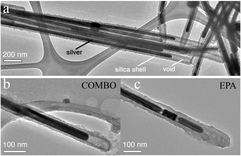
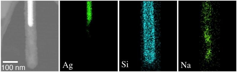
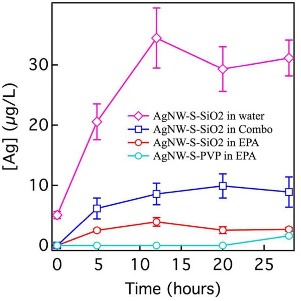
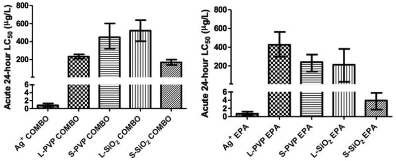
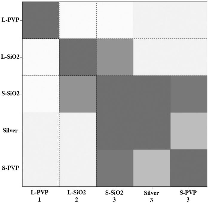
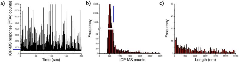
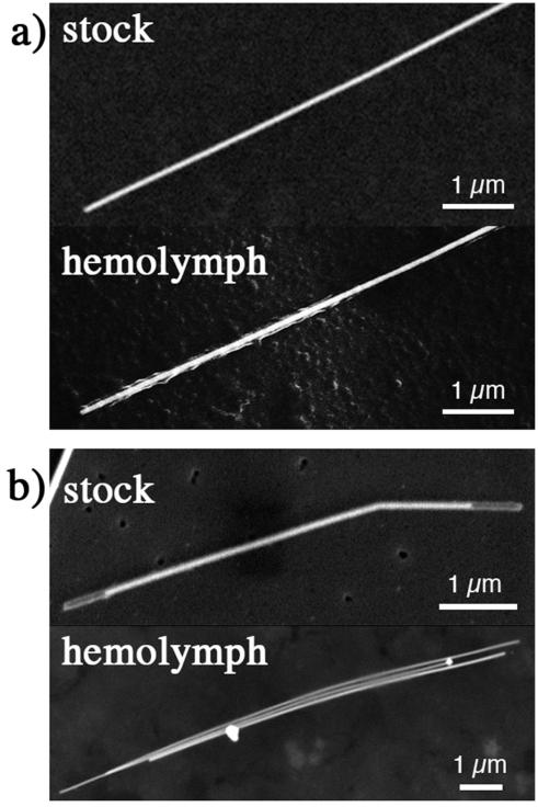
Nanowires (NWs), high-aspect-ratio nanomaterials, are increasingly used in technological materials and consumer products and may have toxicological characteristics distinct from nanoparticles. We carried out a comprehensive evaluation of the physicochemical stability of four silver nanowires (AgNWs) of two sizes and coatings and their toxicity to Daphnia magna . Inorganic aluminum-doped silica coatings were less effective than organic poly(vinyl pyrrolidone) coatings at preventing silver oxidation or Ag(+) release and underwent a significant morphological transformation within 1 h following addition to low ionic strength Daphnia growth media. All AgNWs were highly toxic to D. magna but less toxic than ionic silver. Toxicity varied as a function of AgNW dimension, coating, and solution chemistry. Ag(+) release in the media could not account for observed AgNW toxicity. Single-particle inductively coupled plasma mass spectrometry distinguished and quantified dissolved and nanoparticulate silver in microliter-scale volumes of Daphnia magna hemolymph with a limit of detection of approximately 10 ppb. The silver levels within the hemolymph of Daphnia exposed to both Ag(+) and AgNW met or exceeded the initial concentration in the growth medium, indicating effective accumulation during filter feeding. Silver-rich particles were the predominant form of silver in hemolymph following exposure to both AgNWs and Ag(+). Scanning electron microscopy imaging of dried hemolymph found both AgNWs and silver precipitates that were not present in the AgNW stock or the growth medium. Both organic and inorganic coatings on the AgNW were transformed during ingestion or absorption. Pathway, gene ontology, and clustering analyses of gene expression response indicated effects of AgNWs distinct from ionic silver on Daphnia magna .
Figures
References
-
- Afal A, Coskun S, Emrah Unalan H. All Solution Processed, Nanowire Enhanced Ultraviolet Photodetectors. Appl. Phys. Lett. 2013;102:043503–043503.
-
- Kelly KL, Coronado E, Zhao LL, Schatz GC. The Optical Properties of Metal Nanoparticles: The Influence of Size, Shape and Dielectric Environment. J. Phys. Chem. B. 2003;107:668–677.
-
- Yiin-Kuen F, Li-Chih L. Pattern Transfer of Aligned Metal Nano/microwires as Flexible Transparent Electrodes Using an Electrospun Nanofiber Template. Nanotechnology. 2013;24:055301–055301. - PubMed
-
- Hu J, Liu Y, Ning CZ, Dutton R, Kang SM. Fringing Field Effects on Electrical Resistivity of Semiconductor Nanowire-Metal Contacts. Appl. Phys. Lett. 2008;92:083503–083505.
-
- Law M, Greene LE, Johnson JC, Saykally R, Yang P. Nanowire Dye-Sensitized Solar Cells. Nat. Mater. 2005;4:455–459. - PubMed
Publication types
MeSH terms
Substances
Grants and funding
LinkOut - more resources
Full Text Sources
Other Literature Sources
Molecular Biology Databases
Miscellaneous
