. 2013 Nov;184(2):335-44.
doi: 10.1016/j.jsb.2013.09.020. Epub 2013 Oct 4.

Common mechanistic themes for the powerstroke of kinesin-14 motors

Affiliations

Common mechanistic themes for the powerstroke of kinesin-14 motors

Miguel A Gonzalez et al. J Struct Biol. 2013 Nov.

Abstract

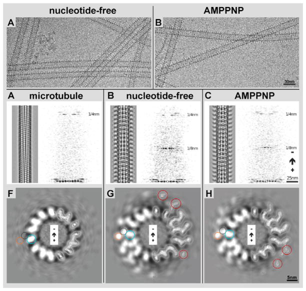
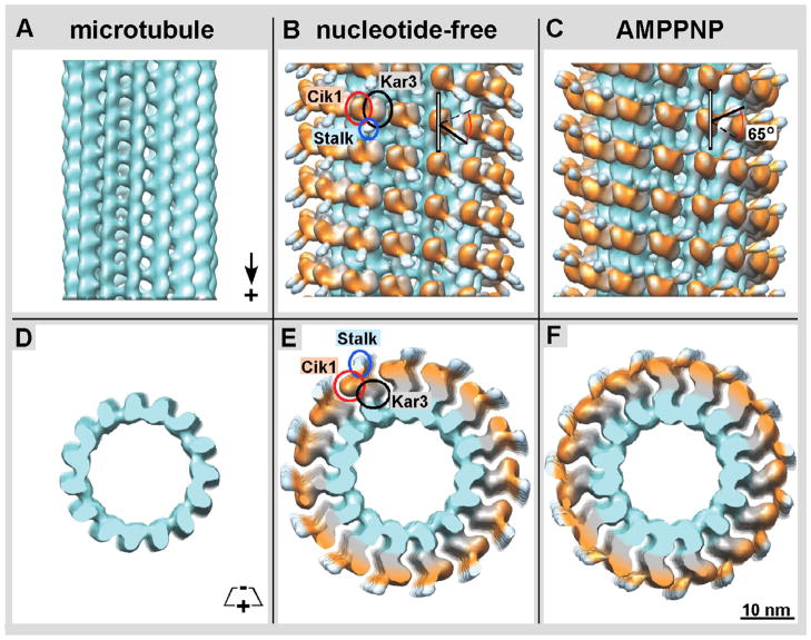
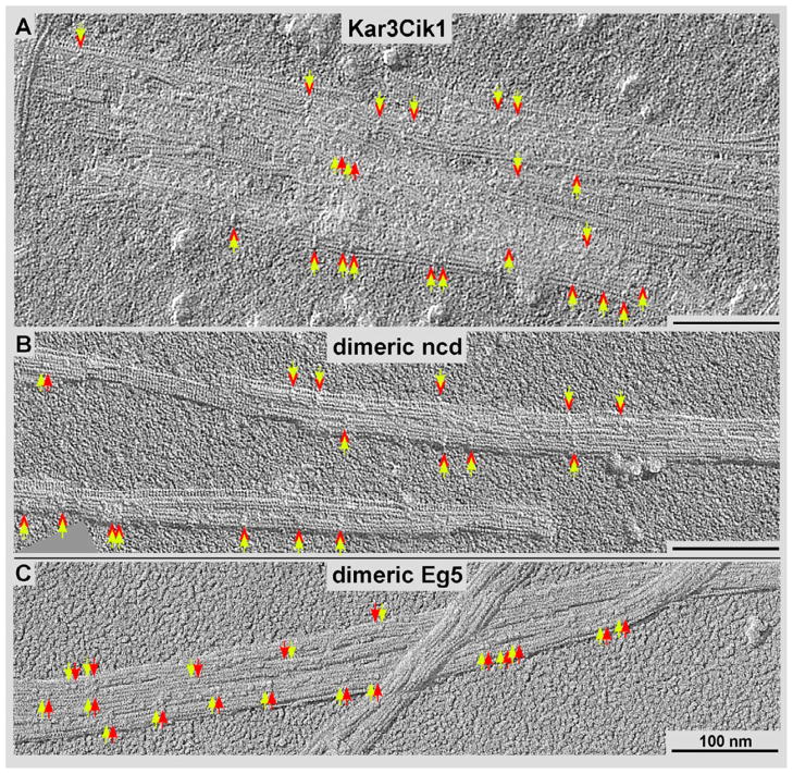
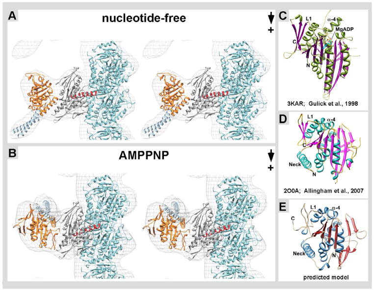
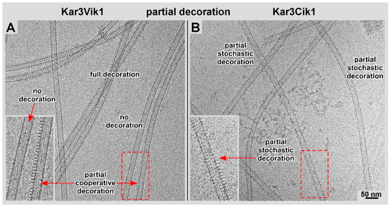
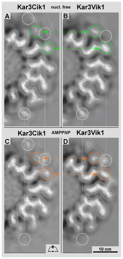
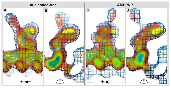
Kar3Cik1 is a heterodimeric kinesin-14 from Saccharomyces cerevisiae involved in spindle formation during mitosis and karyogamy in mating cells. Kar3 represents a canonical kinesin motor domain that interacts with microtubules under the control of ATP-hydrolysis. In vivo, the localization and function of Kar3 is differentially regulated by its interacting stoichiometrically with either Cik1 or Vik1, two closely related motor homology domains that lack the nucleotide-binding site. Indeed, Vik1 structurally resembles the core of a kinesin head. Despite being closely related, Kar3Cik1 and Kar3Vik1 are each responsible for a distinct set of functions in vivo and also display different biochemical behavior in vitro. To determine a structural basis for their distinct functional abilities, we used cryo-electron microscopy and helical reconstruction to investigate the 3-D structure of Kar3Cik1 complexed to microtubules in various nucleotide states and compared our 3-D data of Kar3Cik1 with that of Kar3Vik1 and the homodimeric kinesin-14 Ncd from Drosophila melanogaster. Due to the lack of an X-ray crystal structure of the Cik1 motor homology domain, we predicted the structure of this Cik1 domain based on sequence similarity to its relatives Vik1, Kar3 and Ncd. By molecular docking into our 3-D maps, we produced a detailed near-atomic model of Kar3Cik1 complexed to microtubules in two distinct nucleotide states, a nucleotide-free state and an ATP-bound state. Our data show that despite their functional differences, heterodimeric Kar3Cik1 and Kar3Vik1 and homodimeric Ncd, all share striking structural similarities at distinct nucleotide states indicating a common mechanistic theme within the kinesin-14 family.

Keywords: Cryo-electron microscopy; Helical 3-D analysis; Kar3Cik1; Kinesin-14; Microtubules; Molecular docking.


Figures

Figure 1
Figure 1. 3-D helical reconstruction of decorated and undecorated microtubules
Microtubule/Kar3Cik1 solutions were incubated with apyrase (A) or AMPPNP (B) to generate the nucleotide-free and ATP states respectively. In both states, Kar3Cik1 heterodimers can be seen decorating the microtubules although structural differences between the states cannot be detected. C) 3D helical reconstruction of a plain microtubule. The same result was obtained by incubating Kar3Cik1 motors with ADP prior to addition to microtubules, but where no decoration was detected. A Fourier transform reveals only the 4 nm layer line, which corresponds to the tubulin repeats, illustrating only sparse decoration with motor complexes. D) Reconstruction of a Kar3Cik1 decorated microtubule in the nucleotide-free state. Two distinct densities are visible at each motor subunit corresponding to Kar3 and Cik1. The Fourier transform reveals a strong 4 nm layer line (tubulin repeats) as well as an 8 nm layer line that corresponds to the motor repeat every 8 nm along the microtubule. E) Reconstruction of the ATP state using the non-hydrolyzable ATP analog AMPPNP. The microtubule is fully decorated with Kar3Cik1 as in (D) illustrated by the strong 4 nm and 8 nm layer lines. Corresponding cross-sectional views of the reconstructed microtubules are shown in F) undecorated microtubule, G) nucleotide-free state, and H) AMPPNP state. On the right half of each image, contour lines are shown depicting densities observed from tubulin and Kar3Cik1. Circles corresponding to the position of tubulin (blue), Kar3 (purple), and Cik1 (orange) are shown as well as red circles identifying differences in density between the nucleotide-free and AMPPNP states
Figure 2
Figure 2. Isosurface rendering of density maps obtained by helical reconstruction
A) Isosurface of a naked 15-protofilament microtubule showing the orientation of tubulin (turquoise). B) The nucleotide-free Kar3Cik1 is in complex with the microtubule. It appears that Kar3 (grey / black rings) is in contact with the microtubule while Cik1 (orange / red rings) is oriented away from the microtubule. The coiled coil stalk (light blue / blue rings) connecting Kar3 and Cik1 is pointed toward the microtubule plus end. C) In the ATP state Kar3Cik1 is also in complex with the microtubule with Kar3 in contact with the microtubule and Cik1 oriented away. Interestingly, there is a change in Kar3Cik1’s conformation showing a ~65° rotation of the stalk that causes it to point toward the minus end of the microtubule (black arrows). Other subtle differences can be seen when comparing the orange globular region corresponding to Cik1 (red rings). A cross-sectional view of D) a naked microtubule, E) the nucleotide-free, and F) ATP states shows how Kar3Cik1 (grey and orange) is in contact with tubulin (turquoise).
Figure 3
Figure 3. High-resolution surface metal shadowing of kinesin motors
Kinesin motors bound to ADP were incubated with microtubules at sub-stoichiometric levels and shadowed with tantalum/tungsten. Dimeric motor constructs are identified by a yellow arrow pairs in A) Kar3Cik1, B) dimeric Ncd, and C) dimeric Eg5 (a kinesin-5). The kinesin-14 motors appear to bind sparsely along the microtubules and span adjacent protofilaments (red relative to yellow arrows in A and B) while dimeric kinesin-5s appear to bind to adjacent tubulin subunits along the same protofilament (red relative to yellow arrows in C).
Figure 4
Figure 4. Docking of near-atomic structures into cryo-EM derived isosurfaces
A) In the nucleotide-free state, the structure of tubulin (turquoise, PDB: 1JFF) is shown and the Kar3 structure (grey, PDB: 3KAR) is positioned in the globular region in contact with β-tubulin. Kar3’s helix α-4 (red) is at the interface of where Kar3 contacts the microtubule. As there is currently no known structure for Cik1, a predicted structure was obtained using Phyre 2 (Kelley & Sternberg 2009). This predicted Cik1 structure (orange) is positioned in the globular region oriented away from the microtubule. The structure of the GCN4 leucine zipper (light blue, PDB: 2ZTA) is docked into the stalk region. In the nucleotide-free state the stalk is pointing toward the plus end of the microtubule. B) In the ATP state, there is a conformational change in Kar3Cik1 that leads to a ~65° rotation resulting in the stalk pointing toward the minus end of the microtubule. Phyre2 used structural information from C) Kar3, D) Vik1, and other proteins to generate the predicted model of E) Cik1.
Figure 5
Figure 5. Kar3Vik1/Kar3Cik1 binding cooperativity
Kar3Vik1 appears to bind more cooperatively than Kar3Cik1. A) Kar3Vik1 exhibits high cooperativity. Microtubules that are completely decorated, partially decorated, and undecorated can be seen in the same image area (see also: Cope et al., 2010). B) In contrast, Kar3Cik1 exhibits a more stochastic pattern, illustrated by scattered motor binding with numerous gaps appearing in between the motors. The difference in the Kar3Vik1 and Kar3Cik1 binding patterns is most easily visualized along the sidewalls of the microtubule projections (insets in A and B).
Figure 6
Figure 6. Structural Comparison of Kar3Cik1 and Kar3Vik1
By comparing the density map of a single 3.8 nm slice of A) Kar3Cik1 and B) Kar3Vik1 in the nucleotide-free state, a difference in the density of the motors is detected (green circles). Cik1 appears to have less density present than Vik1, which may suggest that Cik1 is more mobile than Vik1. The same observation is seen when comparing C) Kar3Cik1 and D) Kar3Vik1 in the ATP state. Cik1 again seems to have less density (and thus perhaps more flexibility) than Vik1 (orange circles). White circles show differences in Kar3Cik1 and Kar3Vik1 between their own nucleotide-free and ATP states. Since these 15-protofilament microtubules exhibit a dominant 2-start short-pitched helix (Bessel order -2), a thin slice (~4nm) of a half cross-section, as shown here, represents the complete density distribution of a single motor from these maps.
Figure 7
Figure 7. 3-D structural comparison of Kar3Cik1 and Kar3Vik1
The 3-D electron density map of Kar3Cik1 was docked into the isosurface mesh representation of the 3-D electron density map of Kar3Vik1. Longitudinal, A) and cross-sectional B) views of the nucleotide-free state show that Kar3Cik1 docks into the Kar3Vik1 map extremely well demonstrating that Kar3Cik1 adopts the same pre-powerstroke configuration as Kar3Vik1. Similarly, longitudinal C) and cross-sectional D) views of the AMPPNP state confirm that Kar3Cik1 fits excellently into the Kar3Vik1 structure showing a nearly identical post-powerstroke position following uptake of ATP. Difference mapping (yellow) reveals locations of density differences between Kar3Vik1 and Kar3Cik1 with a significance of >95%. Interestingly, these results show that overall, Kar3Cik1 and Kar3Vik1 appear structurally identical at this resolution, and it seems that they utilize the same mechanism of movement to perform different functions in the cell. Wire mesh: Kar3Vik1; red/green/blue diffuse density: Kar3Cik1; solid yellow density: difference map at >95% significance.

Similar articles

Cited by

References

    1. Allingham JS, Sproul LR, Rayment I, Gilbert SP. Vik1 modulates microtubule-Kar3 interactions through a motor domain that lacks an active site. Cell. 2007;128:1161–1172. - PMC - PubMed
    1. Arnal I, Metoz F, DeBonis S, Wade RH. Three-dimensional structure of functional motor proteins on microtubules. Curr Biol. 1996;6:1265–70. - PubMed
    1. Beuron F, Hoenger A. Structural analysis of the microtubule-kinesin complex by cryo-electron microscopy. Methods Mol Biol. 2001;164:235–54. - PubMed
    1. Case RB, Pierce DW, Hom-Booher N, Hart CL, Vale RD. The directional preference of kinesin motors is specified by an element outside of the motor catalytic domain. Cell. 1997;90:959–966. - PubMed
    1. Chen CJ, Rayment I, Gilbert SP. Kinesin Kar3Cik1 ATPase pathway for microtubule cross-linking. J Biol Chem. 2011;286:29261–72. - PMC - PubMed

MeSH terms

Substances

LinkOut - more resources