. 2013 Dec;33(24):4766-78.
doi: 10.1128/MCB.00647-13. Epub 2013 Oct 7.

Androgen induces a switch from cytoplasmic retention to nuclear import of the androgen receptor

Affiliations

Androgen induces a switch from cytoplasmic retention to nuclear import of the androgen receptor

Li Ni et al. Mol Cell Biol. 2013 Dec.

Abstract

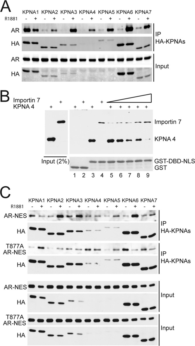
The androgen receptor (AR) has critical functions as a transcription factor in both normal and cancer cells, but the specific mechanisms that regulate its nuclear localization are not well defined. We found that an AR mutation commonly reported in prostate cancer generates an androgen-independent gain of function for nuclear import. The substitution, Thr877Ala, is within the ligand-binding domain, but the nuclear import gain of function is mediated by the bipartite nuclear localization signal (NLS) spanning the DNA-binding domain (DBD) and hinge region. Bipartite NLS activity depends on the structure provided by the DBD, and protein interactions with the bipartite NLS are repressed by the hinge region. The bipartite NLS is recognized by importin 7, a nuclear import receptor for several proteins. Importin 7 binding to AR, however, inhibits import by shielding the bipartite NLS. Androgen binding relieves the inhibition by inducing a switch that promotes exchange of importin 7 for karyopherin alpha import receptors. Importin 7 contributes to the regulation of AR import by restraining import until androgen is detected in the cytoplasm.


Figures

Fig 1
Fig 1
The nuclear import gain of function provided by T877A is mediated by the bipartite NLS in AR. (A) Localization of endogenous AR (T877A) in LNCaP cells and transfected AR (WT and T877A) in PC3 cells. Cells were treated with vehicle (0.1% ethanol) or synthetic androgen (1 nM R1881) for 2 h and processed for IF microscopy. (B) Immunoblotting AR from nuclear (N) and cytoplasmic (C) fractions from LNCaP cells with or without R1881. To separate nuclear and cytoplasmic fractions, cell pellets were permeabilized on ice for 6 min in the same buffer described in a digitonin-permeabilized cell import assay. After a brief centrifugation step, the supernatants were treated as cytoplasmic fractions, and the pellets were treated as nuclear fractions. (C) Immunoblotting WT and T877A forms of AR in nuclear and cytoplasmic fractions from transfected PC3 cells ± R1881. (D) Localization of WT AR and AR containing LBD and NLS mutations in PC3 cells with or without R1881. (E) Nuclear/cytoplasmic (N/C) ratios of AR proteins with or without R1881. The data are presented as means ± the standard deviations (SD). *, P < 0.01.
Fig 2
Fig 2
DBD structure is important for activity of the bipartite NLS in AR. (A) Ribbon model of the AR DBD. Amino acids that comprise the first element of the bipartite NLS (R617, K618) are shown as a wire-frame diagram. The protein sequence of the second element and the hinge region is shown but was not part of the crystal structure. The model was generated with PyMOL using the AR DBD structure (1R4I) solved by the Gewirth laboratory (22). (B) Fluorescence images (GFP) showing nuclear import of GST-GFP-DBD-NLS reconstituted in the digitonin-permeabilized cell import assay. The assay used HeLa cells and conditions indicated in the figure, including with (+) or without (−) an energy-regenerating system (ERS). (C) Quantitation of nuclear import in the digitonin-permeabilized cell import assay. Nuclear levels of GST-GFP-DBD-NLS and the control protein GST-GFP were measured by microscopy (n = number of cells measured). (D) Quantitation of GST-GFP-DBD-NLS import in the digitonin-permeabilized cell import assay. Including histone H1 in the reaction inhibits GST-GFP-DBD-NLS import. (E) Mutation of a lysine (K618E) in the first element of the bipartite NLS of AR inhibits nuclear import in vitro. Nuclear import of GST-GFP-DBD-NLS containing the K618E mutation was analyzed in the permeabilized cell import assay, quantified, and compared to the WT protein. (F) The bipartite NLS of AR requires the DBD for import activity. Sequence encoding AR amino acids 617 to 633 were engineered as a GST-GFP fusion protein and compared to GST-GFP-DBD-NLS for import activity in the permeabilized cell import assay. (G) Reticulocyte lysate (RL) has opposite effects on import of AR DBD-NLS and SV40 NLS. RL addition to the digitonin-permeabilized cell import assay inhibited nuclear import of GST-GFP-DBD-NLS and stimulated nuclear import of GST-GFP-SV40 NLS. **, P < 0.001.
Fig 3
Fig 3
Importin 7 and importin β bind the bipartite NLS of AR. (A and B) Isolation of importin 7 and importin β from RL using GST-DBD-NLS immobilized on glutathione beads. The beads were eluted with a step gradient of MgCl2, and the fractions were precipitated and analyzed by SDS-PAGE and silver staining (A) and by immunoblotting (B) with antibodies to importin 7 and importin β. Two arrows in panel A indicate the positions of importin 7 (upper) and importin β (lower), respectively. (C) Importin 7 and importin β binding to the DBD-NLS requires a functional NLS. WT (left panels) and mutant (K633E) forms of GST-DBD-NLS (right panels) were used to enrich for binding factors from RL. The beads were eluted with MgCl2 and analyzed by SDS-PAGE and immunoblotting.
Fig 4
Fig 4
The hinge region in AR represses importin 7 binding to the NLS. (A) Importin 7 binding to the AR bipartite NLS. Importin 7 was translated as [35S]methionine-labeled proteins and used in binding assays with GST fusion proteins immobilized on glutathione beads. (B) The DBD and hinge regions affect importin 7 binding to the bipartite NLS. Binding assays (see panel A) were performed in duplicate, and bound fractions were measured by densitometry of X-ray films. (C) Quantitation of importin 7 binding to the bipartite NLS in AR. Interactions between importin 7 with the bipartite AR NLS requires the DBD, since only background levels of binding are observed with GST-NLS (amino acids 617 to 633). The hinge region strongly represses importin 7 binding the bipartite AR NLS. The data are presented as means ± the SD. *, P < 0.01. (D) Immunoblotting with an anti-AR NLS antibody. Affinity-purified antibody was used to probe the indicated GST fusion proteins. A duplicate gel was stained with Coomassie blue. (E) The hinge region reduces anti-AR NLS antibody binding measured by ELISA. GST-DBD-NLS and GST-DBD-NLS-hinge fusion proteins were adsorbed to microtiter wells and incubated with a dilution series of antibody. (F) Inhibitory effect of the hinge region on nuclear import. Nuclear import of GST-GFP-DBD-NLS and GST-GFP-DBD-NLS-hinge was measured in digitonin-permeabilized HeLa cells. **, P < 0.001.
Fig 5
Fig 5
Importin 7 can negatively regulate AR and GR import. (A) Effect of importin 7 cotransfection on AR localization, assayed with or without R1881. Wild-type AR was coexpressed with empty vector (left panels) or HA-importin 7 (middle and right panels) in Cos7 cells. Cells were treated with 1 nM R1881 for 30 min (lower panels). The samples were examined by IF microscopy. (B) N/C ratios of AR (from panel A) were quantified and plotted by box plot. **, P < 0.001. (C) AR interaction with importin 7. Cos7 cells were cotransfected with Flag-tagged AR (Flag-AR) and HA-tagged importin 7 (HA-Imp7). After the cells were treated with 1 nM R1881 for 30 min, AR complexes were isolated by IP using anti-Flag–agarose beads, and immunoblotted for AR and HA. (D) Importin 7 binds to AR via the bipartite NLS, and complexes formed in vitro can be dissociated by androgen. WT and K633E mutant AR were translated as [35S]methionine-labeled proteins and used in GST-importin 7 fusion protein binding assays. (E) GFP-tagged AR, GR, and ERβ coexpressed with empty vector (first column) or HA-tagged importin 7 (middle and left panels) in Cos7 cells and visualized by IF microscopy.
Fig 6
Fig 6
AR domains that contribute to cytoplasmic retention by importin 7. (A) Full-length WT AR, AR ΔAF1, and AR ΔLBD were coexpressed with empty vector or HA-tagged importin 7 in Cos7 cells. Cells were stained with antibody against AR hinge (14) and processed for IF microscopy. (B) N/C ratios of AR (from panel A) were quantified and displayed as box plots. **, P < 0.001. (C) Full-length, AF1, and LBD domains of AR were translated as [35S]methionine-labeled proteins and used in GST-importin 7 fusion protein binding assays with or without R1881. (D) WT AR and T877A AR was coexpressed with empty vector or HA-tagged importin 7 in Cos7 cells in the absence of androgen. (E) N/C ratios of AR (from panel D) were quantified and displayed as box plots. **, P < 0.001.
Fig 7
Fig 7
Importin 7 negatively regulates AR-dependent transcription. (A) AR transcription activity was measured by using a PSA-luciferase reporter gene in Cos7 cells. WT AR and T877A AR was cotransfected with empty vector or HA-importin 7. Cells were treated with 1 nM R1881 for 18 h prior to harvest and luciferase assay. F/R, firefly/Renilla. (B) Real-time PCR to measure endogenous androgen-responsive genes in LAPC4 cells. LAPC4 cells were transfected with siRNA against importin 7 with or without 1 nM R1881 for 18 h before RNA isolation. The transcript levels of PSA, KLK2, and importin 7 were measured by real-time PCR.
Fig 8
Fig 8
Importin 7 and KPNA proteins compete for binding AR. (A) AR interactions with KPNA proteins in response to androgen treatment. Cos7 cells were cotransfected with AR and HA-tagged KPNAs (HA-KPNAs). After the cells were treated with 1 nM R1881 for 30 min, protein complexes were isolated using HA-agarose beads and immunoblotted for AR and HA. (B) Importin 7 and KPNA4 can compete for binding to AR NLS. Recombinant importin 7 and KPNA4 (each His-tagged) were combined with GST-DBD-NLS immobilized on glutathione beads, and bound fractions were examined by immunoblotting. Increasing amounts of importin 7 were used to compete with a constant amount of KPNA4. (C) Comparison of WT and T877A mutant AR binding to KPNAs. Nuclear export signal (NES) fusions of WT and T877A mutant were cotransfected with HA-KPNAs in Cos7 cells. Immunoprecipitation and immunoblotting were performed using the same methods as panel A.
Fig 9
Fig 9
Summary of the proposed AR import mechanism. After its translation in the cytoplasm, AR associates with importin 7, which shields the bipartite NLS and causes cytoplasmic retention. Androgen binding induces a switch in protein structure that results in importin 7 dissociation and binding of KPNA/importin β receptors. The AR/KPNA/importin β complex translocates into the nucleus where it is disassembled via RanGTP binding to importin β. The switch is presumed to involve a change in structure that increases importin 7 dissociation from AR, but it could also reflect a change that is necessary for or enhances KPNA binding.

Similar articles

Cited by

References

    1. Lange A, Mills RE, Lange CJ, Stewart M, Devine SE, Corbett AH. 2007. Classical nuclear localization signals: definition, function, and interaction with importin alpha. J. Biol. Chem. 282:5101–5105 - PMC - PubMed
    1. Pemberton LF, Paschal BM. 2005. Mechanisms of receptor-mediated nuclear import and nuclear export. Traffic 6:187–198 - PubMed
    1. Harel A, Forbes DJ. 2004. Importin beta: conducting a much larger cellular symphony. Mol. Cell 16:319–330 - PubMed
    1. Goldfarb DS, Corbett AH, Mason DA, Harreman MT, Adam SA. 2004. Importin alpha: a multipurpose nuclear-transport receptor. Trends Cell Biol. 14:505–514 - PubMed
    1. Gorlich D, Vogel F, Mills AD, Hartmann E, Laskey RA. 1995. Distinct functions for the two importin subunits in nuclear protein import. Nature 377:246–248 - PubMed

Publication types

MeSH terms

LinkOut - more resources