

Bedside to bench: role of muscarinic receptor activation in ultrarapid growth of colorectal cancer in a patient with pheochromocytoma
- PMID: 24100192
- PMCID: PMC4076953
- DOI: 10.1016/j.mayocp.2013.06.023
Bedside to bench: role of muscarinic receptor activation in ultrarapid growth of colorectal cancer in a patient with pheochromocytoma
Abstract
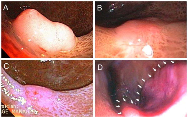
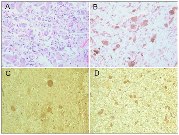
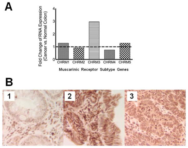
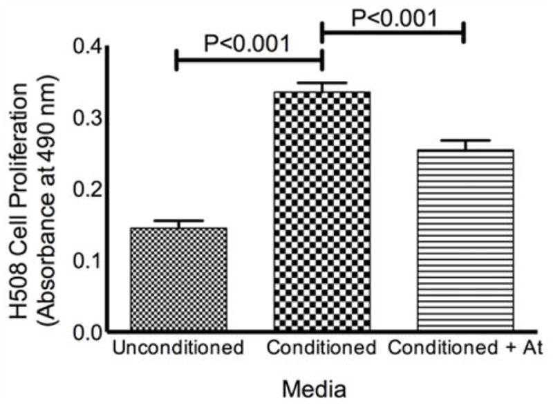
An elderly man with long-standing, nonresectable pheochromocytoma had rapid development of rectal adenocarcinoma despite close endoscopic surveillance. We determined that the patient's colorectal cancer overexpressed muscarinic receptor subtype 3, whereas his pheochromocytoma expressed choline acetyltransferase, an enzyme required to produce acetylcholine, which is a muscarinic receptor agonist. These findings suggested that acetylcholine release from the pheochromocytoma stimulated rapid growth of the rectal neoplasm. As proof of principle, we found that culture media conditioned by pheochromocytoma cells stimulates proliferation of a human colon cancer cell line, an effect attenuated by atropine, a muscarinic receptor inhibitor. Our observations provide both clinical and laboratory evidence that muscarinic receptor agonists promote the growth of colorectal neoplasia.
Keywords: ChAT; M3R; PCR; choline acetyltransferase; muscarinic receptor subtype 3; polymerase chain reaction.
Copyright © 2013 Mayo Foundation for Medical Education and Research. Published by Elsevier Inc. All rights reserved.
Figures
Similar articles
-
Overcoming Obstacles to Targeting Muscarinic Receptor Signaling in Colorectal Cancer.Int J Mol Sci. 2021 Jan 13;22(2):716. doi: 10.3390/ijms22020716. Int J Mol Sci. 2021. PMID: 33450835 Free PMC article. Review.
-
Acetylcholine release by human colon cancer cells mediates autocrine stimulation of cell proliferation.Am J Physiol Gastrointest Liver Physiol. 2008 Sep;295(3):G591-7. doi: 10.1152/ajpgi.00055.2008. Epub 2008 Jul 24. Am J Physiol Gastrointest Liver Physiol. 2008. PMID: 18653726 Free PMC article.
-
Regulation of tyrosine hydroxylase gene expression by the m1 muscarinic acetylcholine receptor in rat pheochromocytoma cells.Brain Res Mol Brain Res. 1996 Aug;40(1):42-54. doi: 10.1016/0169-328x(96)00030-7. Brain Res Mol Brain Res. 1996. PMID: 8840012
-
Postnatal development of muscarinic autoreceptors modulating acetylcholine release in the septohippocampal cholinergic system. II. Cell body region: septum.Brain Res Dev Brain Res. 1998 Jun 15;108(1-2):31-7. doi: 10.1016/s0165-3806(98)00027-3. Brain Res Dev Brain Res. 1998. PMID: 9693781
-
Cholinergic system and cell proliferation.Chem Biol Interact. 2016 Nov 25;259(Pt B):257-265. doi: 10.1016/j.cbi.2016.04.014. Epub 2016 Apr 13. Chem Biol Interact. 2016. PMID: 27083142 Review.
Cited by
-
Overcoming Obstacles to Targeting Muscarinic Receptor Signaling in Colorectal Cancer.Int J Mol Sci. 2021 Jan 13;22(2):716. doi: 10.3390/ijms22020716. Int J Mol Sci. 2021. PMID: 33450835 Free PMC article. Review.
-
Role of Muscarinic Acetylcholine Receptors in Breast Cancer: Design of Metronomic Chemotherapy.Curr Clin Pharmacol. 2019;14(2):91-100. doi: 10.2174/1574884714666181203095437. Curr Clin Pharmacol. 2019. PMID: 30501602 Free PMC article. Review.
-
The Role of M3 Muscarinic Receptor Ligand-Induced Kinase Signaling in Colon Cancer Progression.Cancers (Basel). 2019 Mar 5;11(3):308. doi: 10.3390/cancers11030308. Cancers (Basel). 2019. PMID: 30841571 Free PMC article. Review.
-
Optimal Management of a Synchronous Diagnosis of Phaeochromocytoma and Colorectal Neoplasia.Indian J Surg Oncol. 2017 Dec;8(4):622-626. doi: 10.1007/s13193-017-0627-4. Epub 2017 Feb 22. Indian J Surg Oncol. 2017. PMID: 29203998 Free PMC article.
-
Exposure to antimuscarinic medications for treatment of overactive bladder and risk of lung cancer and colon cancer.Clin Epidemiol. 2019 Jan 23;11:133-143. doi: 10.2147/CLEP.S186842. eCollection 2019. Clin Epidemiol. 2019. PMID: 30774448 Free PMC article.
References
-
- Fearon ER, Vogelstein B. A genetic model for colorectal tumorigenesis. Cell. 1990;61(5):759–767. - PubMed
-
- Flynn C, Montrose DC, Swank DL, Nakanishi M, Ilsley JN, Rosenberg DW. Deoxycholic acid promotes the growth of colonic aberrant crypt foci. Mol Carcinog. 2007;46(1):60–70. - PubMed
-
- Frucht H, Jensen RT, Dexter D, Yang WL, Xiao Y. Human colon cancer cell proliferation mediated by the M3 muscarinic cholinergic receptor. Clin Cancer Res. 1999;5(9):2532–2539. - PubMed
-
- Cheng K, Chen Y, Zimniak P, Raufman J, Xiao Y, Frucht H. Functional interaction of lithocholic acid conjugates with M3 muscarinic receptors on a human colon cancer cell line. Biochim Biophys Acta. 2002;1588(1):48–55. - PubMed
Publication types
MeSH terms
Substances
Grants and funding
LinkOut - more resources
Full Text Sources
Other Literature Sources
Medical
Research Materials
