. 2013 Dec;27(12):2013-27.
doi: 10.1210/me.2013-1161. Epub 2013 Oct 7.

Cell-specific actions of a human LHX3 gene enhancer during pituitary and spinal cord development

Affiliations

Cell-specific actions of a human LHX3 gene enhancer during pituitary and spinal cord development

Soyoung Park et al. Mol Endocrinol. 2013 Dec.

Abstract

The LIM class of homeodomain protein 3 (LHX3) transcription factor is essential for pituitary gland and nervous system development in mammals. In humans, mutations in the LHX3 gene underlie complex pediatric syndromes featuring deficits in anterior pituitary hormones and defects in the nervous system. The mechanisms that control temporal and spatial expression of the LHX3 gene are poorly understood. The proximal promoters of the human LHX3 gene are insufficient to guide expression in vivo and downstream elements including a conserved enhancer region appear to play a role in tissue-specific expression in the pituitary and nervous system. Here we characterized the activity of this downstream enhancer region in regulating gene expression at the cellular level during development. Human LHX3 enhancer-driven Cre reporter transgenic mice were generated to facilitate studies of enhancer actions. The downstream LHX3 enhancer primarily guides gene transcription in α-glycoprotein subunit -expressing cells secreting the TSHβ, LHβ, or FSHβ hormones and expressing the GATA2 and steroidogenic factor 1 transcription factors. In the developing nervous system, the enhancer serves as a targeting module active in V2a interneurons. These results demonstrate that the downstream LHX3 enhancer is important in specific endocrine and neural cell types but also indicate that additional regulatory elements are likely involved in LHX3 gene expression. Furthermore, these studies revealed significant gonadotrope cell heterogeneity during pituitary development, providing insights into the cellular physiology of this key reproductive regulatory cell. The human LHX3 enhancer-driven Cre reporter transgenic mice also provide a valuable tool for further developmental studies of cell determination and differentiation in the pituitary and nervous system.


Figures

Figure 1.
Figure 1.
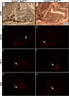
The endogenous mouse LHX3 protein and a human LHX3 gene 3′ enhancer-guided reporter gene are expressed in anterior pituitary αGSU-expressing thyrotrope and gonadotrope cells but not in somatotropes and corticotropes. A, Frozen transverse pituitary sections from newborn (P0) (panels A–J) and e17.5 (panels K–O) 3′ human LHX3 gene enhancer-HSP68-nLacZ transgenic mice were stained by immunohistochemistry for LHX3 (green) (panels A–E) or stained for transgene-derived β-galactosidase activity (blue) (panels F–O) and colabeled for hormone subunits (red or brown): GH (panels A, F, and K), ACTH (panels B, G, and L), αGSU (panels C, H, and M), TSHβ (panels D, I, and N), and LHβ (panels E, J, and O). Colocalization was highest (arrowheads) for gonadotropes and thyrotropes (panels C, D, E, H, I, J, M, N, and O) and nearly absent (arrowheads) for corticotropes and somatotropes (panels A, B, F, G, K, and L). All experiments were performed a minimum of three times. B, Schematic diagram showing generation of ELHX3Cre (human LHX3 gene 3′ enhancer-HSP68-Cre) transgenic mice and crossing to ROSA26 reporter strains to reveal enhancer-driven activities. ELHX3Cre mice were crossed to reporter mice for conditional activation of reporter genes expression including LacZ, YFP, or DTA.
Figure 2.
Figure 2.
The 3′ enhancer from the human LHX3 gene directs Cre recombinase expression in the developing and neonatal anterior pituitary and spinal cord. Frozen sagittal sections from ELHX3+/+/R26LacZ/+ (negative control) and ELHX3Cre/+/R26LacZ/+ (Cre expressing) pituitary glands were analyzed at e14.5 (panels A and B) and P0 (panels C–F) in mice carrying either LacZ (panels A–D) or YFP (panels E and F) reporter genes. Black arrows, LacZ expression in the anterior pituitary gland; P, posterior lobe of the pituitary gland; I, intermediate lobe; A, anterior lobe. G and H, X-gal-stained whole embryos at e12.5 show enhancer-driven LacZ activity in the developing pituitary and spinal cord (green arrows). G, Single heterozygous ELHX3+/+/R26LacZ/+ mice as controls (n = 3). H, Double-heterozygous ELHX3Cre/+/R26LacZ/+ mice (n = 5). Yellow arrowhead indicates ectopic expression (possibly in the facial/vestibuloacoustic ganglion complex of the nervous system). I, ELHX3Cre/+/R26LacZ/+ mice (newborn, stage P0) were stained by immunohistochemistry for LHX3 (green) and colabeled using antibodies recognizing hormone subunits (red). Arrowheads denote LHX3 protein colocalizing with αGSU, TSHβ, FSHβ and LHβ, but not with GH cells.
Figure 3.
Figure 3.
The human 3′ LHX3 enhancer is active in many αGSU-expressing anterior pituitary cells: αGSU cells are notably reduced in the developing pituitary glands of ELHX3Cre/+/R26DTA/+ mice. Frozen pituitary sections from e14.5 ELHX3+/+/R26DTA/+ (as negative controls; panels A, C, E, and G) and ELHX3Cre/+/R26DTA/+ (panels B, D, F, and H) mice were stained with antibodies specific for αGSU (panels A and B), ACTH (panels C and D), PIT-1 (panels E and F), and T-PIT (panels G and H) proteins, and expression was revealed using either DAB chemistry (panels A and B) or immunofluorescence (panels C–H). Arrows indicate positively stained cells. P, posterior lobe, I, intermediate lobe, A, caudomedial region of the anterior lobe; R, rostral tip region of the anterior lobe.
Figure 4.
Figure 4.
Thyrotrope and gonadotrope cell lineages are reduced in neonatal ELHX3Cre/+/R26DTA/+ mice. Immunostaining of transverse sections from P0 pituitaries reveals reduction of αGSU cells (panels A and B), TSHβ-positive thyrotropes (panels C and D), and gonadotropes (panels E–I) in ELHX3Cre/+/R26DTA/+ mice. FSHβ-staining gonadotropes are reduced (panels E and F) and LHβ-expressing gonadotropes are either highly reduced (panel G and H) or in some cases completely ablated (panel I). Control and enhancer-expressing ELHX3Cre/+/R26DTA/+ mice did not have different levels of ACTH (panels J and K) or GH (panels L and M) hormone-positive cells in ELHX3Cre/+/R26DTA/+ mice at P0. Similarly, levels of the PIT-1 transcription factor were not notably altered (panels O and P). Quantification shows that αGSU- and TSHβ-staining cells are significantly reduced (panel Q). White bars, ELHX3+/+/R26DTA/+; black bars, ELHX3Cre/+/R26DTA/+. Data are expressed as means ± SEM. *, P < .05; **, P < .01. ns, Nonsignificant.
Figure 5.
Figure 5.
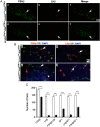
LHβ+ gonadotropes are notably ablated in ELHX3Cre/+/R26DTA/+ mice and LHβ cells colocalize with FSHβ and SF1. A, Frozen transverse pituitary sections of P0 animals were examined with antibodies to LHβ and FSHβ to determine the FSH+ and LH+ populations present in ELHX3Cre/+/R26DTA/+ mice. FSHβ and LHβ cells are predominantly located in the ventral area of the pituitary. Most LHβ+ and some FSHβ+ cells are ablated in ELHX3Cre/+/R26DTA/+ mice (panels a, b, d, and e). B, LHβ+ cells are predominantly FSHβ+, SF1+ bihormonal cells. The SF1 transcription factor is expressed in LHβ+ gonadotropes and is notably reduced in ELHX3Cre/+/R26DTA/+ mice. Frozen transverse pituitary sections at P0 were coimmunostained with antibodies recognizing gonadotrope hormones FSHβ (panels a and c) or LHβ (panels b and d) (red color); the SF1 transcription factor (green color), and 4′,6′-diamino-2-phenylindole stain to reveal nuclei (blue color). SF1+ cells colocalize with FSHβ and LHβ cells and are reduced in ELHX3Cre/+/R26DTA/+ mice (panels c and d). Example bihormonal (FSHβ+/LHβ+) cells are indicated by white arrows, example single hormone cells are indicated by white arrowhead, and SF1-only cells are indicated by orange arrowheads. C, LHβ-expressing cells are strikingly diminished in ELHX3Cre/+/R26DTA/+ mice. The graph shows the total cell number of αGSU-, FSHβ-, LHβ-, FSHβ/LHβ-, SF1-, LHβ/SF1-, and FSHβ/SF1-positive cells compared with controls. Experiments were carried out in control ELHX3+/+/R26DTA/+ littermates (n = 3) and ELHX3Cre/+/R26DTA/+ (n = 3). Data are expressed as means ± SEM. ***, Differences are significant at P < .001 as determined by a Student's t test.
Figure 6.
Figure 6.
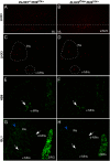
LHX3+ interneurons are reduced in the V2 interneuron region of the developing spinal cord in ELHX3Cre/+/R26DTA/+ mice, but motor neuron populations are unaffected. Sagittal (A and B) and transverse sections (C–H) of e14.5 spinal cords from ELHX3+/+/R26DTA/+ (as controls) and ELHX3Cre/+/R26DTA/+ mice were examined with antibodies against to LHX3 (A–D) or the motor neuron markers HB9 (E and F) and ISL1 (G and H). The dotted line in panels A and B indicates an arbitrary boundary between the intermediate and ventral zones. Large white dotted circles in panels C and D indicate the interneuron area, and small white dotted circles denote the motor neuron area. White arrows show HB9- or ISL1-positive cells. Blue arrows indicate the interneuron reduction in ELHX3Cre/+/R26DTA/+ mice. ML, Marginal layer of spinal cord; INs, interneurons; v-MNs, ventral-motor neurons; d-MNs, dorsal-motor neurons; DRG, dorsal root ganglion.
Figure 7.
Figure 7.
LHX3- and CHX10-expressing V2a interneuron cells are reduced in the spinal cord of ELHX3Cre/+/R26DTA/+ mice. Transverse sections from e14.5 ELHX3+/+/R26DTA/+ (A–C) and ELHX3Cre/+/R26DTA/+ (D–F) mouse spinal cords were colabeled with antibodies to CHX10 (A and D) and LHX3 (B and E). C and F, Merged channels. CHX10- and LHX3-colabeled cells are located in the intermediate ventral area (large white dotted circle). V2a interneuron CHX10- and LHX3-positive cells are reduced in ELHX3Cre/+/R26DTA/+ transgenic mice (panel F). INs, Interneurons; v-MNs, ventral motor neurons.
Figure 8.
Figure 8.
GATA2+ cells are reduced in the caudomedial region of the developing pituitary gland, but GATA2 expression is normal in the embryonic spinal cord. Frozen sagittal sections of the pituitary gland (A and B) and transverse sections of the spinal cord (C and D) from e14.5 ELHX3+/+/R26DTA/+ (controls) or ELHX3Cre/+/R26DTA/+ mice were stained with an antibody recognizing the GATA2 transcription factor. A and B, GATA2 expression is present in rostral tip area in both types of animal but is lost in the caudomedial region in ELHX3Cre/+/R26DTA/+ mice (B). White dotted circles indicate the caudomedial area of the pituitary. Arrows point to GATA2+ cells in the rostral tip area. White dotted rectangles indicate GATA2-positive cells in the spinal cord. R, Rostral tip region of the anterior lobe.

Similar articles

Cited by

References

    1. Kelberman D, Rizzoti K, Lovell-Badge R, Robinson IC, Dattani MT. Genetic regulation of pituitary gland development in human and mouse. Endocr Rev. 2009;30:790–829 - PMC - PubMed
    1. Davis SW, Castinetti F, Carvalho LR, et al. Molecular mechanisms of pituitary organogenesis: in search of novel regulatory genes. Mol Cell Endocrinol. 2010;323:4–19 - PMC - PubMed
    1. Mollard P, Hodson DJ, Lafont C, Rizzoti K, Drouin J. A tridimensional view of pituitary development and function. Trends Endocrinol Metab. 2012;23:261–269 - PubMed
    1. Prince KL, Walvoord EC, Rhodes SJ. The role of homeodomain transcription factors in heritable pituitary disease. Nat Rev Endocrinol. 2011;7:727–737 - PubMed
    1. Simmons DM, Voss JW, Ingraham HA, et al. Pituitary cell phenotypes involve cell-specific Pit-1 mRNA translation and synergistic interactions with other classes of transcription factors. Genes Dev. 1990;4:695–711 - PubMed

Publication types

MeSH terms