. 2013 Oct 14;203(1):115-28.
doi: 10.1083/jcb.201304188. Epub 2013 Oct 7.

Recruitment of the autophagic machinery to endosomes during infection is mediated by ubiquitin

Affiliations

Recruitment of the autophagic machinery to endosomes during infection is mediated by ubiquitin

Naonobu Fujita et al. J Cell Biol. .

Abstract

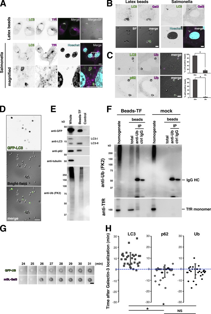
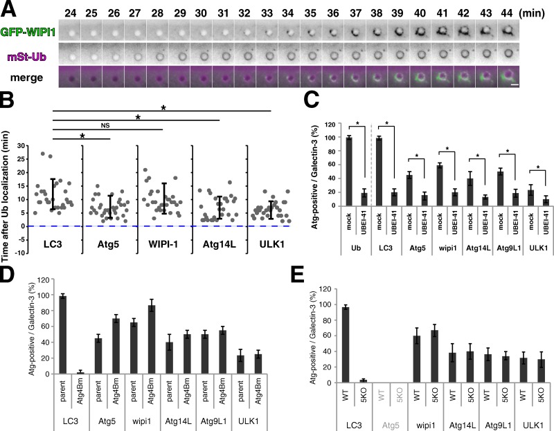
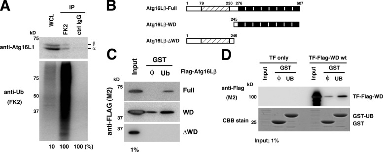
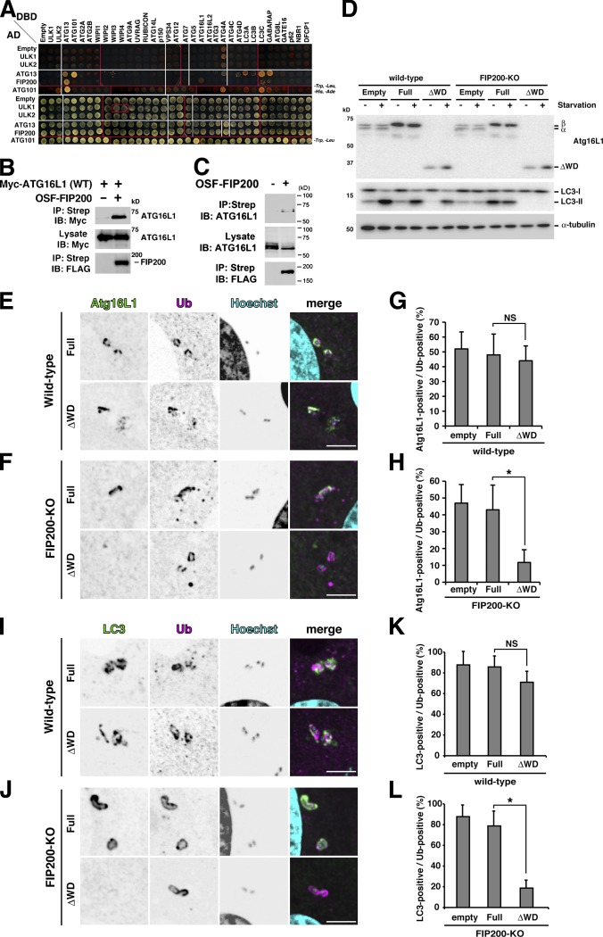
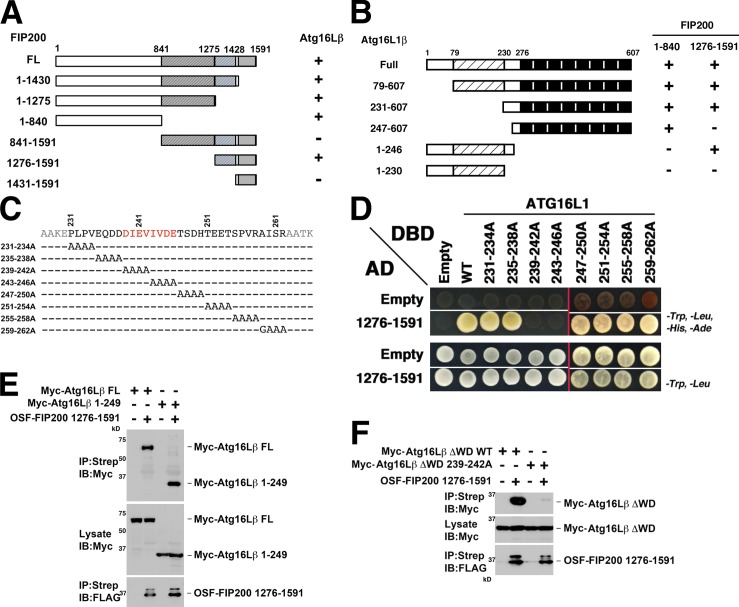
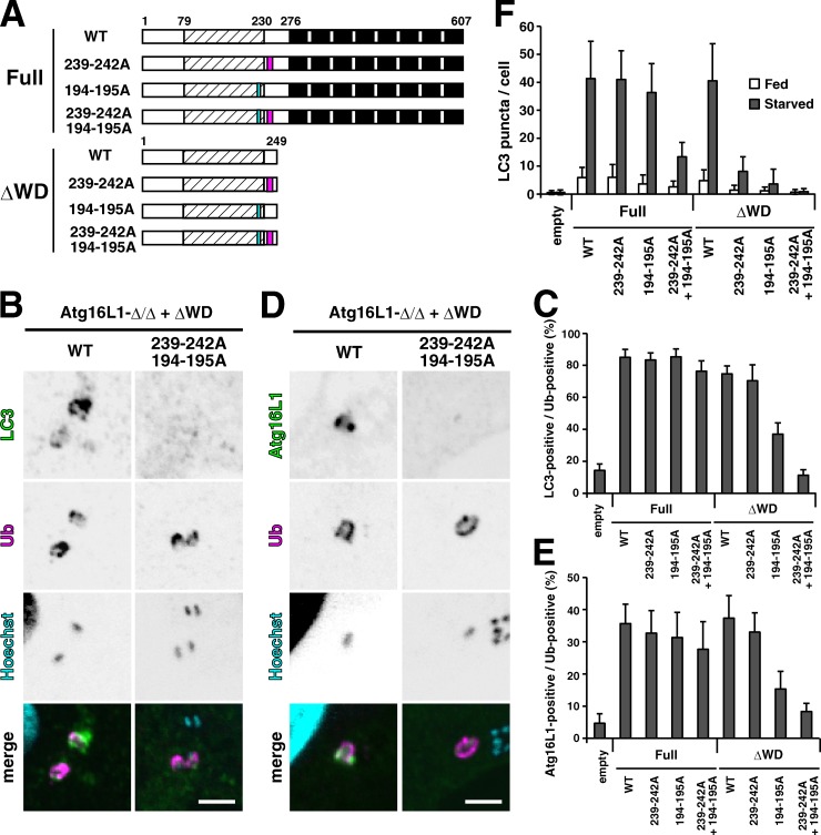
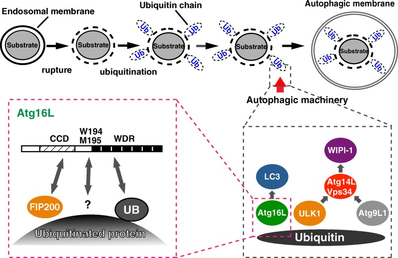
Although ubiquitin is thought to be important for the autophagic sequestration of invading bacteria (also called xenophagy), its precise role remains largely enigmatic. Here we determined how ubiquitin is involved in this process. After invasion, ubiquitin is conjugated to host cellular proteins in endosomes that contain Salmonella or transfection reagent-coated latex (polystyrene) beads, which mimic invading bacteria. Ubiquitin is recognized by the autophagic machinery independently of the LC3-ubiquitin interaction through adaptor proteins, including a direct interaction between ubiquitin and Atg16L1. To ensure that invading pathogens are captured and degraded, Atg16L1 targeting is secured by two backup systems that anchor Atg16L1 to ubiquitin-decorated endosomes. Thus, we reveal that ubiquitin is a pivotal molecule that connects bacteria-containing endosomes with the autophagic machinery upstream of LC3.


Figures

Figure 1.
Figure 1.
Ub-positive endosomes containing Salmonella or beads are targeted by autophagy. (A) HeLa cells were infected with S. Typhimurium (Salmonella) for 1 h or transfected with Effectene-coated latex beads for 3 h and then subjected to immunocytochemistry for LC3 and transferrin receptor (TfR). Bar, 10 µm. (B) HeLa cells were infected with Salmonella for 1 h or transfected with Effectene-coated latex beads for 3 h and then subjected to immunocytochemistry for LC3 and galectin3. Bar, 5 µm. (C) HeLa cells were transfected with Effectene-coated latex beads for 3 h and subjected to immunocytochemistry for LC3 and Ub (top) or LC3 and p62 (bottom). Bar, 5 µm. The percentages of LC3- or p62-positive beads per Ub-positive (Ub+) or Ub-negative (Ub−) beads were enumerated. Statistical analysis was performed by Student’s unpaired t test. *, P < 0.01. (D and E) HeLa cells stably expressing GFP-LC3 were transfected with Effectene-coated latex beads for 3 h. Bead–autophagosomes were fractionated as described in Materials and methods. The bead–autophagosome fraction was observed by confocal microscopy (D; bar, 10 µm) or lysed with RIPA buffer and subjected to Western blot analysis using the indicated antibodies (E). In a control sample, scraped cells were mixed with Effectene-coated beads and immediately homogenized. (F) The bead–autophagosome fraction was lysed and subjected to immunoprecipitation with an anti-Ub IgG antibody (FK2) or control IgG. Co-immunoprecipitated molecules were examined by Western blotting using the indicated antibodies. (G and H) NIH3T3 cells stably expressing mStrawberry (mStr)-Gal3 and GFP-LC3, mStr-Gal3, and GFP-p62, or mStr-Gal3 and GFP-Ub were transfected with Effectene-coated latex beads for 30 min and then washed. Live cells were observed at 1-min intervals by fluorescence microscopy. Bar, 3 µm. The time after galectin3 localization was measured for at least 30 cases for each combination (H). Statistical analysis was performed by Student’s unpaired t test. *, P < 0.05; NS, not significant.
Figure 2.
Figure 2.
Ubiquitination and recruitment of Atg proteins. (A and B) NIH3T3 cells stably expressing mStr-Ub and GFP-tagged LC3, Atg5, WIPI-1, Atg14L1, or ULK1 were transfected with Effectene-coated latex beads for 30 min. Then, live cells were observed at 1-min intervals by fluorescence microscopy. Bar, 3 µm. The time after Ub localization was measured for at least 30 cases for each combination. Each value represents the mean ± SD. Statistical analysis was performed by Student’s unpaired t test. *, P < 0.05; NS, not significant. (C) NIH3T3 cells stably expressing GFP-tagged Ub, LC3, Atg5, WIPI-1, Atg14L1, Atg9L1, or ULK1 were transfected with Effectene-coated beads for 3 h in the presence or absence (mock) of 30 µM UBEI-41 (a ubiquitin E1–specific inhibitor) and subjected to immunocytochemistry for galectin3. The percentages of GFP-positive per galectin3-positive beads were enumerated. At least 30 beads were counted (n = 3). The values are the mean ± SD. Statistical analysis was performed by Student’s unpaired t test. *, P < 0.05. (D and E) Parent NIH3T3 cells, Atg4B mutant overexpressing NIH3T3 cells (D), wild-type MEFs, and Atg5-KO MEFs (E) stably expressing GFP-tagged LC3, Atg5, WIPI-1, Atg14L1, Atg9L1, or ULK1 were transfected with Effectene-coated latex beads for 3 h and subjected to immunocytochemistry for galectin3. The percentages of Atg-positive per galectin3-positive beads were enumerated. At least 30 beads were counted (n = 3). The values are the mean ± SD.
Figure 3.
Figure 3.
The WD β-propellers of Atg16L1 directly interact with Ub. (A) HeLa cells were lysed with 1% Triton X-100 lysis buffer and subjected to immunoprecipitation with FK2- or control IgG-immobilized beads. The coimmunoprecipitated molecules were examined by Western blotting using the indicated antibodies. (B) Schematic diagram of Atg16L1 (Full) and its deletion constructs. (C) Purified recombinant GST or GST-Ub–immobilized glutathione Sepharose beads were incubated with lysates from HEK293T cells transiently expressing FLAG-tagged Atg16L1 constructs for 1 h at 25°C with gentle agitation. The beads were washed three times with ice-cold PBS and the bound complexes were eluted with 50 mM reduced glutathione and then subjected to Western blotting for FLAG. (D) Purified GST or GST-Ub–immobilized glutathione Sepharose beads were incubated with lysates from bacteria expressing trigger factor (TF) or TF-FLAG-WD β-propellers for 1 h at 25°C with gentle agitation. Washing and elution were performed as described in C. The eluted samples were subjected to SDS-PAGE and Western blotting for FLAG.
Figure 4.
Figure 4.
The WD β-propellers of Atg16L1 recruit LC3 in the absence of FIP200. (A) Yeast two-hybrid interactions of the ULK1 complexes with other human Atg proteins. ULK1, ULK2, Atg13, FIP200, and Atg101-AD fusions (rows 2–6) or control AD constructs (row 1, Empty) were coexpressed with control DBD constructs (column 1, Empty) or Atg protein–DBD fusions (columns 2–35) and tested for positive yeast two-hybrid interactions (top) or cotransformation (control, bottom). White lines divide images derived from the same plate, and red lines divide images from different plates. Atg16L1 positively interacted with FIP200 in this assay. (B) FIP200 binds to exogenously expressed Atg16L1. Myc-Atg16L1 coprecipitations with an empty vector control (lane 1) or One-STrEP-FLAG (OSF)-FIP200 (lane 2). (C) FIP200 binds endogenous Atg16L1. Atg16L1 coprecipitations with an empty vector control (lane 1) or OSF-FIP200 (lane 2). (D) Wild-type or FIP200-KO MEFs stably expressing the empty vector, full-length Atg16L1, or ΔWD mutant were incubated in growth medium (−) or Earle’s balanced salt solution (EBSS) (+) for 1 h and examined by Western blotting using the indicated antibodies. We previously reported that stable expression of an exogenous Atg16L1 construct can be used to replace the endogenous Atg16L1 protein with an exogenous Atg16L1 protein (Fujita et al., 2009) because free Atg16L1 molecules not complexed with the Atg12–Atg5 conjugate are preferentially degraded by the ubiquitin–proteasome system. In control cells (Empty), two Atg16L1 splicing variants, the α- and β-forms, were detected. In full-length β-form–replaced cells (Full), the α-form was not detected, whereas in ΔWD-replaced cells (ΔWD) both the α- and β-forms were minimally detected. Thus, we successfully obtained cells that express only endogenous levels of the full-length or ΔWD mutant. (E–L) Atg16L1-replaced wild-type or FIP200-KO MEFs were infected with Salmonella (MOI = 10) for 1 h and subjected to immunocytochemistry for Atg16L1 and Ub (E and F) or LC3 and Ub (I and J). The percentages of Atg16L1-positive (G and H) or LC3-positive per Ub-positive bacteria were enumerated (K and L). At least 50 Salmonella were counted. The average ± SD is shown for three independent experiments. Statistical analysis was performed by Student’s t test. *, P < 0.05; NS, not significant. Bar, 5 µm.
Figure 5.
Figure 5.
Atg16L1-FIP200 interacting domains. (A) Mapping of the Atg16L1-binding site on FIP200. Yeast two-hybrid mapping experiments showing that Atg16L1 binds to both the N-terminal and C-terminal coiled-coil regions of FIP200. (B) Mapping of the FIP200-binding site within Atg16L1. Yeast two-hybrid mapping experiments showing that the 1–840 fragment of FIP200 binds to the C-terminal 247–607 region, while the 1276–1591 fragment of FIP200 binds to the N-terminal 1–264 region of Atg16L1. (C and D) Additional yeast two-hybrid mapping experiments showing that the binding site of the FIP200 1276–1591 fragment maps to residues 239–246 of Atg16L1. White lines divide images derived from the same plate, and red lines divide images from different plates. (E) The N-terminal 1–249 fragment of Atg16L1 is sufficient to interact with FIP200. Full-length (lanes 1 and 2) or the N-terminal 1–249 fragments (lanes 3 and 4) of Myc-Atg16L1 coprecipitations with empty vector controls (lanes 1 and 3) or OSF-FIP200 1276–1591 (lanes 2 and 4). (F) Co-precipitation experiments confirming that OSF-FIP200 1276–1591 coprecipitates with wild-type (WT) Myc-Atg16L1 (1–249) fragments, but not with analogous fragments containing a mutant FIP200-binding site (Myc-Atg16L1 ΔWD 239–242A).
Figure 6.
Figure 6.
Effects of Atg16L1 mutations on autophagy against Salmonella. (A) Schematic diagram of Atg16L1 and various mutants. The magenta box indicates 239–242A and the cyan box indicates 194–195A. (B–E) Atg16L1-Δ/Δ MEFs stably expressing the indicated constructs were infected with Salmonella (MOI = 10) for 1 h and then analyzed by immunocytochemistry for LC3 (B) or Atg16L1 (D). Bar, 5 µm. The percentage of LC3- or Atg16L1-positive Salmonella per Ub-positive Salmonella was enumerated by fluorescence microscopy (C and E). At least 50 bacteria were counted. The average ± SD is shown for three independent experiments. (F) Atg16L1-Δ/Δ MEFs stably expressing the indicated constructs were cultured in growth medium (Fed) or EBSS (Starved) for 1 h and then subjected to immunocytochemistry using an anti-LC3 antibody. The number of LC3 puncta in each cell was counted for more than 50 cells. The average ± SD is shown for three independent experiments.
Figure 7.
Figure 7.
A possible mechanism of Ub-mediated autophagic sequestration of invading bacteria and transfection reagent–coated latex beads. After internalization and endosomal rupture, Ub is conjugated to host cellular proteins in endosomes that contain Salmonella or transfection reagent–coated latex beads (top). Ub would be recognized by three pivotal components of the autophagic machinery, notably the Atg16L1 complex, the ULK1 complex, and Atg9L1 (bottom right). Atg16L1 localizes to the ubiquitinated target through three mechanisms, the interaction of coiled-coil domain (CCD) with FIP200, the interaction of WD repeats (WDR) with Ub, and the 194–195 region-mediated mechanism (bottom left).

References

    1. Birmingham C.L., Brumell J.H. 2006. Autophagy recognizes intracellular Salmonella enterica serovar Typhimurium in damaged vacuoles. Autophagy. 2:156–158 - PubMed
    1. Desjardins M., Huber L.A., Parton R.G., Griffiths G. 1994. Biogenesis of phagolysosomes proceeds through a sequential series of interactions with the endocytic apparatus. J. Cell Biol. 124:677–688 10.1083/jcb.124.5.677 - DOI - PMC - PubMed
    1. Dupont N., Lacas-Gervais S., Bertout J., Paz I., Freche B., Van Nhieu G.T., van der Goot F.G., Sansonetti P.J., Lafont F. 2009. Shigella phagocytic vacuolar membrane remnants participate in the cellular response to pathogen invasion and are regulated by autophagy. Cell Host Microbe. 6:137–149 10.1016/j.chom.2009.07.005 - DOI - PubMed
    1. Durocher Y., Perret S., Kamen A. 2002. High-level and high-throughput recombinant protein production by transient transfection of suspension-growing human 293-EBNA1 cells. Nucleic Acids Res. 30:E9 10.1093/nar/30.2.e9 - DOI - PMC - PubMed
    1. Fujita N., Hayashi-Nishino M., Fukumoto H., Omori H., Yamamoto A., Noda T., Yoshimori T. 2008a. An Atg4B mutant hampers the lipidation of LC3 paralogues and causes defects in autophagosome closure. Mol. Biol. Cell. 19:4651–4659 10.1091/mbc.E08-03-0312 - DOI - PMC - PubMed

Publication types

MeSH terms