Review
. 2013 Nov;9(11):674-86.
doi: 10.1038/nrrheum.2013.147. Epub 2013 Oct 8.

Oxidative stress in the pathology and treatment of systemic lupus erythematosus

Affiliations
Review

Oxidative stress in the pathology and treatment of systemic lupus erythematosus

Andras Perl. Nat Rev Rheumatol. 2013 Nov.

Abstract

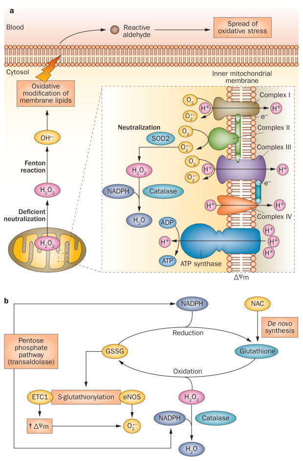
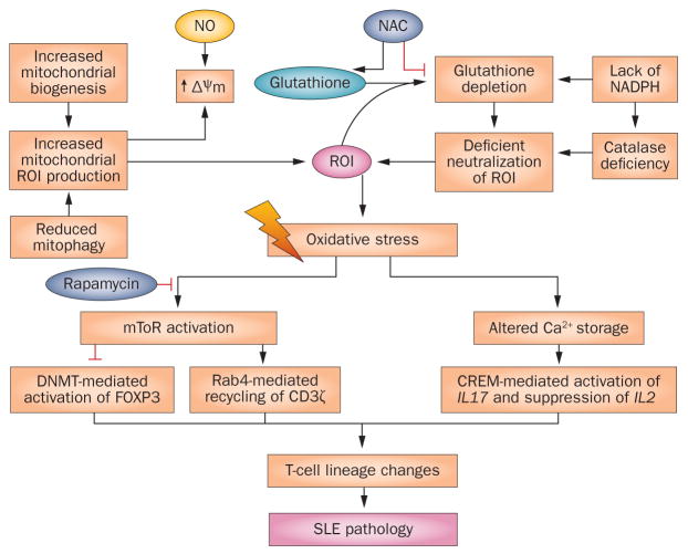
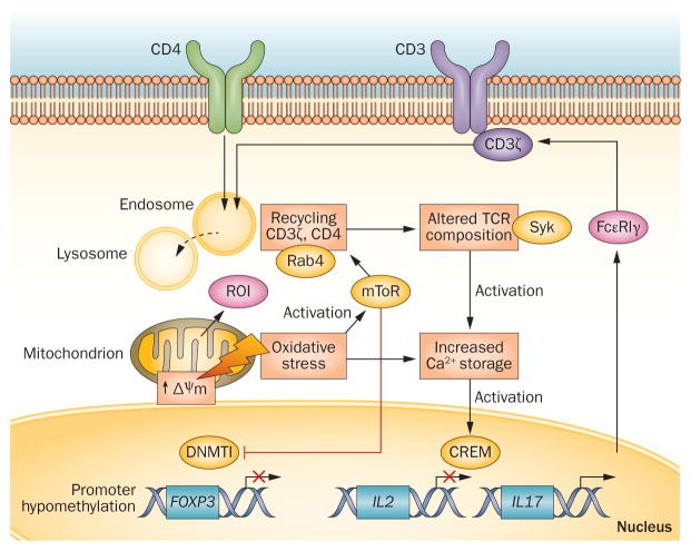
Oxidative stress is increased in systemic lupus erythematosus (SLE), and it contributes to immune system dysregulation, abnormal activation and processing of cell-death signals, autoantibody production and fatal comorbidities. Mitochondrial dysfunction in T cells promotes the release of highly diffusible inflammatory lipid hydroperoxides, which spread oxidative stress to other intracellular organelles and through the bloodstream. Oxidative modification of self antigens triggers autoimmunity, and the degree of such modification of serum proteins shows striking correlation with disease activity and organ damage in SLE. In T cells from patients with SLE and animal models of the disease, glutathione, the main intracellular antioxidant, is depleted and serine/threonine-protein kinase mTOR undergoes redox-dependent activation. In turn, reversal of glutathione depletion by application of its amino acid precursor, N-acetylcysteine, improves disease activity in lupus-prone mice; pilot studies in patients with SLE have yielded positive results that warrant further research. Blocking mTOR activation in T cells could conceivably provide a well-tolerated and inexpensive alternative approach to B-cell blockade and traditional immunosuppressive treatments. Nevertheless, compartmentalized oxidative stress in self-reactive T cells, B cells and phagocytic cells might serve to limit autoimmunity and its inhibition could be detrimental. Antioxidant therapy might also be useful in ameliorating damage caused by other treatments. This Review thus seeks to critically evaluate the complexity of oxidative stress and its relevance to the pathogenesis and treatment of SLE.


Conflict of interest statement

Competing interests

The author declares no competing interests.

Figures

Figure 1
Figure 1
Mitochondrial generation and systemic propagation of oxidative stress, and overview of redox balance mechanisms. a | Production of ROI and spread of oxidative stress. In all mammalian cells, ROI are generated by the ETC. Mitochondrial hyperpolarization, which occurs in T cells in SLE, increases this production by promoting transfer of electrons to molecular oxygen (generating O2 •−). O2 •− is not membrane permeable and is converted into H2O2 by SOD2 within the mitochondria. H2O2 is further neutralized into H2O by catalase at the expense of NADPH, but can also diffuse through membranes. Excess H2O2 is transformed into highly toxic OH•− through the Fenton reaction. OH•− damages lipids and other macromolecules in the immediate vicinity and generates diffusible and highly toxic lipid aldehydes, which spread oxidative stress from mitochondria to other intracellular organelles and through the bloodstream. b | Redox mechanisms that control oxidative stress. Glutathione metabolism regulates mitochondrial hyperpolarization, via S-glutathionylation of ETC complex 1, which increases production of O2•−. Reduced glutathione is regenerated at the expense of NADPH, which is primarily produced through the pentose phosphate pathway. Transaldolase activity, which is increased in T cells from patients with SLE, has been associated with depletion of NADPH and glutathione and with mitochondrial hyperpolarization. Abbreviations: ETC, electron transport chain; ROI, reactive oxygen intermediates; SLE, systemic lupus erythematosus; SOD2, superoxide dismutase [Mn], mitochondrial.
Figure 2
Figure 2
Overview of molecular pathways of oxidative stress and potential points of intervention in T cells in SLE. Upstream of oxidative stress, exposure to NO and/or depletion of glutathione generates ROI, which cause MHP, mitochondrial biogenesis and oxidative stress. In addition to biogenesis, an increase in the number of T-cell mitochondria in SLE has also been attributed to reduced mitophagy. NADPH is generated by the pentose phosphate pathway and is required both to generate NO and to regenerate glutathione. The glutathione precursor NAC is thought to work upstream of oxidative stress by replenishing glutathione and preventing MHP. mTOR is a sensor of MHP but precisely how it is activated by oxidative stress, and thus how NAC prevents its activation, is currently unclear. Downstream of oxidative stress, mTOR activates Rab4A and associated endocytic recycling of CD3ζ. These changes alter intracellular signal transduction and T-cell lineage specification, causing contraction of TH1-cell, TREG-cell and CD8+ T-cell subsets, and expansion of TH2-cell, TH17-cell and CD4CD8 T-cell subsets., Rapamycin prevents the activation of mTOR without affecting oxidative stress. Abbreviations: FcεRIγ, high affinity immunoglobulin ε receptor subunit β, γ chain; MHP, mitochondrial hyperpolarization; mTOR, serine/threonine-protein kinase mTOR; ROI, reactive oxygen intermediates; SLE, systemic lupus erythematosus; TREG cell, regulatory T cell.
Figure 3
Figure 3
Molecular targets of oxidative stress in T-cell signal transduction. The source of oxidative stress, ROI, primarily originate from mitochondria; small amounts can also be generated by NOX activity following TCR stimulation., MHP is induced by oxidative stress and activates mTOR, which in turn promotes T-cell activation via Rab4A-mediated downregulation of the TCR component CD3ζ and increased calcium flux. Activated mTOR inhibits DNMT1; subsequent promoter hypomethylation suppresses the transcription of FOXP3, which is required for TREG-cell development. Oxidative stress and associated changes to calcium storage also activate CREM, which suppresses IL-2 and enhances IL-17 promoter activity; these changes result in TH1-to-TH17 skewing in the T-cell compartment, as shown in Figure 4. Generation of ROI and MHP are depicted in Figure 1. Abbreviations: CREM, cAMP response element modulator; DNMT1, DNA methyltransferase 1; MHP, mitochondrial hyperpolarization; mTOR, serine/threonine-protein kinase mTOR; NAC, N-acetylcysteine; NOX, NADPH oxidase; ROI, reactive oxygen intermediates; TCR, T-cell receptor; TREG cell, regulatory T cell; TH1, type 1 T helper (cell); TH17, type 17 T helper (cell).
Figure 4
Figure 4
Consequences of compartmentalized oxidative stress in T cells and phagocytic cells for the proinflammatory intercellular signalling network in SLE. Oxidative stress is proposed to originate in CD4CD8 T cells, which are implicated in orchestrating dysfunction of the immune system in SLE. MHP, accumulation of mitochondria, and oxidative stress activate mTOR; in CD4CD8 T cells production of IL-17 and IL-4 is consequently increased, thus stimulating B cells and causing contraction of the CD4+CD25+FOXP3+ TREG-cell subset. mTOR activation also inhibits the development of TH1 cells and CD8+ T cells. Following activation, necrosis-prone T cells, marked by increased mitochondrial mass, release oxidized DNA and HMGB1, which stimulate B cells, macrophages, and DCs. In turn, macrophages and DCs produce NO, which stimulates MHP in T cells and production of BAFF, which further activates B cells and the production of ANA. GSH depletion and oxidative stress thus favour TH1 to TH2 polarization in the development of CD4+ T cells. Neutrophils extrude foreign DNA in NETs (by NETosis) that stimulate B cells via TLRs. NOX2-dependent oxidative stress facilitates destruction of infectious organisms in phagocytic cells, potentially limiting NETosis and TLR-mediated stimulation of B cells. Mitochondria are depicted in T cells that exhibit mitochondrial dysfunction. Abbreviations: ANA, antinuclear antibodies; BAFF, B cell activating factor (also known as BLyS and as TNF ligand superfamily, member 13b); DC, dendritic cell; GSH, glutathione; HMGB1, high mobility group protein-1; MHP, mitochondrial hyperpolarization; NET, neutrophil extracellular trap; NOX, NADPH oxidase; TH, helper T (cell); TLR, Toll-like receptor; TREG cell, regulatory T cell.

Similar articles

Cited by

References

    1. Francis L, Perl A. Pharmacotherapy of systemic lupus erythematosus. Expert Opin Pharmacother. 2009;10:1481–1494. - PubMed
    1. Tsokos GC. Systemic Lupus Erythematosus. N Engl J Med. 2011;365:2110–2121. - PubMed
    1. Perl A. Systems biology of lupus: Mapping the impact of genomic and environmental factors on gene expression signatures, cellular signaling, metabolic pathways, hormonal and cytokine imbalance, and selecting targets for treatment. Autoimmunity. 2010;43:32–47. - PMC - PubMed
    1. Perl A, Hanczko R, Telarico T, Oaks Z, Landas S. Oxidative stress, inflammation and carcinogenesis are controlled through the pentose phosphate pathway by transaldolase. Trends Mol Med. 2011;7:395–403. - PMC - PubMed
    1. Perl A, Gergely P, Jr, Nagy G, Koncz A, Banki K. Mitochondrial hyperpolarization: a checkpoint of T cell life, death, and autoimmunity. Trends Immunol. 2004;25:360–367. - PMC - PubMed

Publication types

MeSH terms