. 2013 Dec 1;19(23):6532-43.
doi: 10.1158/1078-0432.CCR-13-1305. Epub 2013 Oct 7.

Targeting SRC and tubulin in mucinous ovarian carcinoma

Affiliations

Targeting SRC and tubulin in mucinous ovarian carcinoma

Tao Liu et al. Clin Cancer Res. .

Abstract

Purpose: To investigate the antitumor effects of targeting Src and tubulin in mucinous ovarian carcinoma.

Experimental design: The in vitro and in vivo effects and molecular mechanisms of KX-01, which inhibits Src pathway and tubulin polymerization, were examined in mucinous ovarian cancer models.

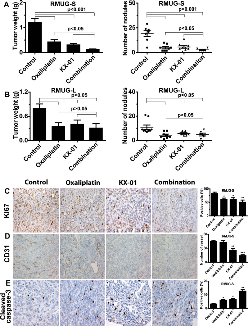
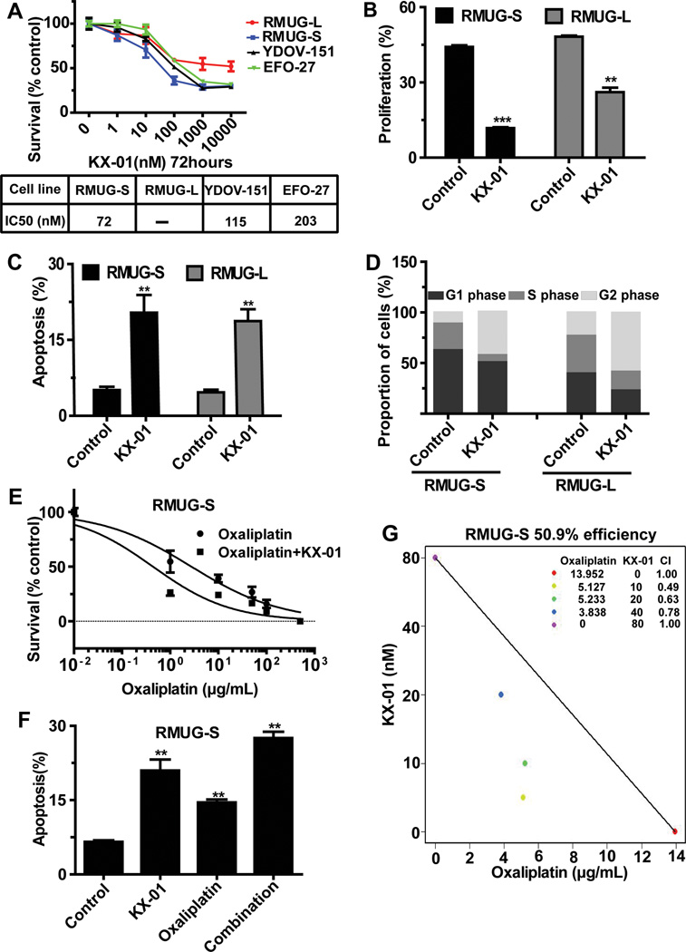
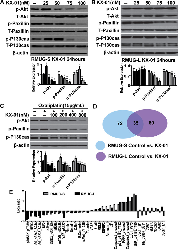
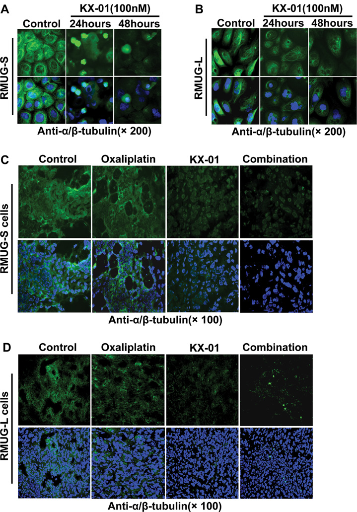
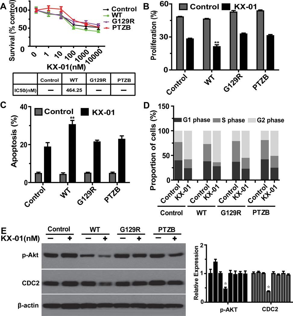
Results: In vitro studies using RMUG-S and RMUG-L cell lines showed that KX-01 inhibited cell proliferation, induced apoptosis, arrested the cell cycle at the G2-M phase, and enhanced the cytotoxicity of oxaliplatin in the KX-01-sensitive cell line, RMUG-S. In vivo studies showed that KX-01 significantly decreased tumor burden in RMUG-S and RMUG-L mouse models relative to untreated controls, and the effects were greater when KX-01 was combined with oxaliplatin. KX-01 alone and in combination with oxaliplatin significantly inhibited tumor growth by reducing cell proliferation and inducing apoptosis in vivo. PTEN knock-in experiments in RMUG-L cells showed improved response to KX-01. Reverse phase protein array analysis showed that in addition to blocking downstream molecules of Src family kinases, KX-01 also activated acute stress-inducing molecules.

Conclusion: Our results showed that targeting both the Src pathway and tubulin with KX-01 significantly inhibited tumor growth in preclinical mucinous ovarian cancer models, suggesting that this may be a promising therapeutic approach for patients with mucinous ovarian carcinoma.


Conflict of interest statement

Conflicts of interest: none to report

Figures

Figure 1
Figure 1
(A, B) Anti-tumor activity of KX-01 and oxaliplatin in (A) RMUG-S and (B) RMUG-L mouse models of mucinous ovarian carcinoma. Mice were randomized to four groups (n=10 mice per group) and underwent treatment as follows: (1) control group, (2) oxaliplatin group, (3) KX-01 group, and (4) combination group. Data are shown as mean ± standard deviation (error bars). (C, D, E) Effects of KX-01 and oxaliplatin on cell proliferation (Ki-67; C), angiogenesis (CD31; D), and apoptosis (Cleaved caspase-3; E) in the RMUG-S mouse model. Original magnification 200×. Bars in the graphs correspond sequentially to the labeled columns of images on the left. Data are shown as mean ± SEM (error bars). *p<0.05, **p<0.01, *** p<0.0001 compared with the control group.
Figure 2
Figure 2
Biological effects of KX-01 on mucinous ovarian carcinoma cells in vitro. (A) MTT assay results showing the effect of KX-01 on cell survival at various concentrations in a panel of mucinous ovarian carcinoma cells. Cell survival was calculated as the number of cells surviving relative to the number of cells surviving in the control group. IC50 indicates median inhibitory rate. (B) EdU assay results showing inhibition of cell proliferation in cells treated with KX-01 compared with control cells in RMUG-S and RMUG-L cells. (C) Annexin V and 7-aminoactinomycin D assay results showing cell apoptosis in cells treated with KX-01 compared with control cells in RMUG-S and RMUG-L cells. (D) Flow cytometry results showing induction of mitotic arrest in cells treated with KX-01 compared with control cells in RMUG-S and RMUG-L cells. (E) Cell cytotoxicity in RMUG-S cells treated with various concentrations of oxaliplatin with or without 100nM KX-01 for 72 hours. Results are presented as in A. (F) Isobologram analysis for the combination of KX-01 and oxaliplatin in RMUG-S cells. The interaction index was <1 at all examined points. (G) Apoptosis in RMUG-S cells after treatment with 15 µg/mL oxaliplatin, 100nM KX-01, or a combination of both treatments for 72 hours. All experiments were repeated at least 3 times, and bars represent means with standard errors. **p<0.01, ***p<0.001 compared with the control group.
Figure 3
Figure 3
Molecules involved in the mechanism of action in KX-01. (A, B) Western blot results showing the relative expression of Src downstream molecules (Akt, paxillin, and P130cas) in RMUG-S and RMUG-L cells treated with various concentrations of KX-01 for 24 hours. (C) Western blot results showing p-Akt, p-paxillin, and p-P130cas expression in cells treated with various concentrations of KX-01, followed by treatment with 15 µg/mL oxaliplatin. β-Actin was used as the loading control. Densitometry was used to calculate the ratio of each band to β-actin, normalized by control samples, each experiment was repeated three times, and bars represent means with standard errors. *p<0.05 compared with the control group. (D) Venn diagram showing the number of proteins involved in the mechanism of action in KX-01 in RMUG-S and RMUG-L cells according to reverse-phase protein array analysis. (E) Molecules whose expression differed significantly (p<0.05) between cells treated with KX-01 and control cells in both RMUG-S and RMUG-L cells.
Figure 4
Figure 4
The microtubule network was disrupted by KX-01. (A, B) α/β-tubulin immunofluorescent staining (green) in RMUG-S and RMUG-L cells treated with 100nM KX-01 for 24 or 48 hours and incubated with anti-α/β-tubulin antibody followed by Alexa Fluor 488–conjugated anti-rabbit antibody. Nuclear staining (blue) was with Hoechst 33342. Upper: tubulin stain only; Lower: tubulin and Hoechst 33342 nuclear stain. (C, D) α/β-tubulin immunofluorescent staining (green) in frozen tumor sections from the control, oxaliplatin, KX-01, and combination groups in the RMUG-S and RMUG-L mouse models. Nuclear staining (blue) was with Hoechst 33342. Upper: tubulin stain only; Lower: tubulin and Hoechst 33342 nuclear stain.
Figure 5
Figure 5
PTEN deletion is involved in resistance to KX-01. (A) MTT assay results showing cytotoxicity of KX-01 in RMUG-L cells transfected with wild type (WT) and domain mutation (G129R, PTZB) PTEN and exposed to various concentration of KX-01 for 72hours. Cell survival was calculated as the number of cells surviving relative to the number of cells surviving in the control group. IC50 indicates median inhibitory rate. (B) EdU assay results showing inhibition of cell proliferation in cells treated with 100nM KX-01 for 72 hours. (C) Annexin V and 7-aminoactinomycin D assay result showing cell apoptosis in cells treated with 100nM KX-01 for 72 hours. (D) Flow cytometry results showing induction of mitotic arrest in cells treated with 100nM KX-01 for 72hours. (E) Western blot results showing that p-Akt and CDC2 (cdk1) were inhibited in PTEN wild type cells. Cells were treated with 100nM KX-01 for 24 hours. β-actin was used as the loading control. All experiments were repeated at least 3 times, and bars represent means with standard errors. *p<0.05, **p<0.01 compared with control treatment (no KX-01).

Similar articles

Cited by

References

    1. Hess V, A'Hern R, Nasiri N, King DM, Blake PR, Barton DP, et al. Mucinous epithelial ovarian cancer: a separate entity requiring specific treatment. J Clin Oncol. 2004;22:1040–1044. - PubMed
    1. Frumovitz M, Schmeler KM, Malpica A, Sood AK, Gershenson DM. Unmasking the complexities of mucinous ovarian carcinoma. Gynecol Oncol. 2010;117:491–496. - PMC - PubMed
    1. Pectasides D, Fountzilas G, Aravantinos G, Kalofonos HP, Efstathiou E, Salamalekis E, et al. Advanced stage mucinous epithelial ovarian cancer: the Hellenic Cooperative Oncology Group experience. Gynecol Oncol. 2005;97:436–441. - PubMed
    1. Pisano C, Greggi S, Tambaro R, Losito S, Iodice F, Di Maio M, et al. Activity of chemotherapy in mucinous epithelial ovarian cancer: a retrospective study. Anticancer Res. 2005;25:3501–3505. - PubMed
    1. Bamias A, Psaltopoulou T, Sotiropoulou M, Haidopoulos D, Lianos E, Bournakis E, et al. Mucinous but not clear cell histology is associated with inferior survival in patients with advanced stage ovarian carcinoma treated with platinum-paclitaxel chemotherapy. Cancer. 2010;116:1462–1468. - PubMed

Publication types

MeSH terms