. 2014 Mar;22(3):554-566.
doi: 10.1038/mt.2013.237. Epub 2013 Oct 8.

Long-distance axonal transport of AAV9 is driven by dynein and kinesin-2 and is trafficked in a highly motile Rab7-positive compartment

Affiliations

Long-distance axonal transport of AAV9 is driven by dynein and kinesin-2 and is trafficked in a highly motile Rab7-positive compartment

Michael J Castle et al. Mol Ther. 2014 Mar.

Abstract

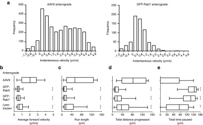
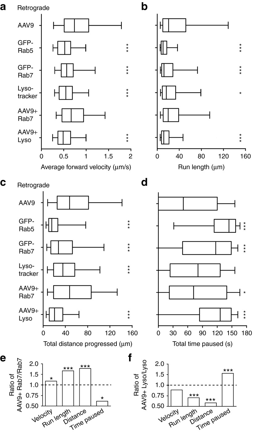
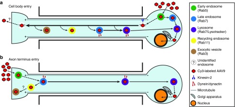
Adeno-associated virus (AAV) vectors can move along axonal pathways after brain injection, resulting in transduction of distal brain regions. This can enhance the spread of therapeutic gene transfer and improve treatment of neurogenetic disorders that require global correction. To better understand the underlying cellular mechanisms that drive AAV trafficking in neurons, we investigated the axonal transport of dye-conjugated AAV9, utilizing microfluidic primary neuron cultures that isolate cell bodies from axon termini and permit independent analysis of retrograde and anterograde axonal transport. After entry, AAV was trafficked into nonmotile early and recycling endosomes, exocytic vesicles, and a retrograde-directed late endosome/lysosome compartment. Rab7-positive late endosomes/lysosomes that contained AAV were highly motile, exhibiting faster retrograde velocities and less pausing than Rab7-positive endosomes without virus. Inhibitor experiments indicated that the retrograde transport of AAV within these endosomes is driven by cytoplasmic dynein and requires Rab7 function, whereas anterograde transport of AAV is driven by kinesin-2 and exhibits unusually rapid velocities. Furthermore, increasing AAV9 uptake by neuraminidase treatment significantly enhanced virus transport in both directions. These findings provide novel insights into AAV trafficking within neurons, which should enhance progress toward the utilization of AAV for improved distribution of transgene delivery within the brain.


Figures

Figure 1
Figure 1
AAV9 particles undergo axonal transport and induce transgene expression following axon- or cell body–specific application. (a) Rat E18 cortical neurons cultured in the microfluidic chamber are healthy and extend axons through the grooves after 5 days in vitro (arrows). (b) An 80 µl difference in media volume across the grooves isolates 5 ng AF488-Dextran within the cell channel 4 hours after application. (c,d) Specific application of AAV9 to the axon termini of identical cultures for 2.5 hours resulted in retrograde transduction and expression of the eGFP transgene 13 days after application of virus. (c) Neurons that extended axons across the grooves to the isolated channel (arrow), to which AAV9 was applied, exhibited strong eGFP expression. (d) Cells that did not extend axons into the isolated channel were not transduced. Thresholds were adjusted to improve visibility of axonal eGFP. (e,f) AAV9 axonal transport was tracked in the groove by sequential image acquisition (3 seconds/frame, 63×). The leftmost panel of each sequence displays the imaged axon in phase. Thresholds were adjusted to improve visibility of Cy3-AAV9. (e) 1 hour after application of mCy3-AAV9 to the axon side of the chamber, an AAV9 punctum (arrows) makes a representative retrograde run of 124 µm at 1.01 µm/second. See also Supplementary Movie S1. (f) 1 hour after application of bCy3-AAV9 to the cell body side of the chamber, an AAV9 punctum (arrows) makes a representative anterograde run of 143 µm at 2.99 µm/second. See also Supplementary Movie S2. AAV, adeno-associated virus.
Figure 2
Figure 2
AAV9 retrograde transport is mediated by dynein/dynactin, and anterograde transport is mediated by kinesin-2. In mass cultures of rat E18 cortical neurons treated with 1 × 109 mCy3- or bCy3-AAV9, the number of AAV9 puncta progressing more than 50 μm retrograde or anterograde was counted and then divided by the total length of axon imaged. Stars depict the statistical comparison of each plasmid transfection against mock transfection. **P < 0.01; ***P < 0.001. Error bars represent one standard deviation from the mean among n = 4 cultures for each condition. Transfection of CC1, a dominant negative inhibitor of dynein/dynactin, resulted in a 59.1% decrease in retrograde-directed puncta, indicating that the dynein/dynactin complex mediates retrograde AAV9 transport. Transfection of Kif5C Tail, a dominant negative inhibitor of kinesin-1, had no effect on AAV9 transport, whereas Kif3A-HL, a dominant negative inhibitor of kinesin-2, decreased anterograde-directed puncta by 76.2%. This indicates that kinesin-2, and not kinesin-1, mediates anterograde AAV9 transport. AAV, adeno-associated virus.
Figure 3
Figure 3
AAV9 distributes widely throughout the endosomal system regardless of the site of entry. (a) Images of a representative microfluidic immunocytochemistry (ICC) stain, in which mCy3-AAV9 was applied to the cell body side of the chamber for 4 hours, followed by fixation and staining with an anti-Rab11 primary antibody and AF488 secondary antibody. Colocalization of Rab11 and AAV9 can be observed in the grooves (arrow). All axons are stained with anti-Rab11 (green), although only a small percentage contain AAV9 (red). Similar punctate staining was observed with all other Rab antibodies. (b) ICC was conducted using antibodies against Rab5, 7, 11, or 3, 1 or 4 hours after application of AAV9 to the axon side of the chamber, or 4 hours after application to the cell body side. A minimum of n = 146 AAV9 puncta were counted for each time point, with a total n = 7828 counted. Stars depict the significance of a logistic regression among all time points for each antibody. ***P < 0.001; *P < 0.05. Error bars represent one standard deviation from the mean among a minimum of n = 3 cultures. Colocalization with the exocytic vesicle increased from 1 to 4 hours after AAV9 application, and colocalization with the recycling endosome was greater at 4 hours after axon terminus than after cell body application. (c) The colocalization of AAV9 with Lysotracker during microfluidic live imaging was calculated in 1 hour blocks. Each 1 hour block represents a minimum of n = 4 cultures and n = 172 puncta. Stars depict the statistical significance of a logistic regression among all three 1 hour blocks. ***P < 0.001. Error bars represent one standard deviation from the mean among cultures. For both application sites, colocalization with Lysotracker was observed to increase over time, peaking at ˜26% from 3 to 4 hours after AAV9 application. AAV, adeno-associated virus.
Figure 4
Figure 4
AAV9 undergoes axonal transport in the late endosome/lysosome but not in early or recycling endosomes. (a,b) Colocalized movement was tracked by sequential image acquisition (4.5 seconds/frame, 63×). Displacement of the green punctum ahead of the red punctum occurs due to sequential acquisition (250 ms delay between channels). The leftmost panel of each sequence shows the imaged axon in phase. Thresholds were adjusted to improve visibility. Retrograde movement of AAV9 was observed in both GFP-Rab7- and Lysotracker-labeled endosomes. (a) 4 hours after application of mCy3-AAV9 (red) to the axon side of a chamber treated with Lysotracker (green), a colocalized punctum (arrows) makes a representative retrograde run of 33 µm at 0.42 µm/second, then pauses. See also Supplementary Movie S5. (b) 1 hour after application of bCy3-AAV9 (red) to the axon side of a chamber transfected with GFP-Rab7 (green), a colocalized punctum (arrows) makes a representative retrograde run of 86 µm at 1.1 µm/second. See also Supplementary Movie S6. (c) Colocalized puncta were counted from 1 to 4 hours after application of AAV9 and classified as retrograde-directed or anterograde-directed (progressing more than 10 µm in the respective direction over 3 minutes) or bidirectional/stationary (not progressing at least 10 µm in either direction). This graph summarizes categorical observations from all live imaging experiments that were performed, and thus the number of replicates is larger for groups that were statistically analyzed (Figure 7). Minimal movement was observed in colocalization with GFP-Rab5 after application of AAV9 to the axon channel (n = 41 puncta from three cultures), or with GFP-Rab11 after application to the axon (n = 45, 2) or cell body channel (n = 91, 6). By contrast, ˜40% of AAV9 puncta moved retrograde when colocalized with GFP-Rab7 (n = 182, 17), or with Lysotracker after application to the axon (n = 162, 4) or cell body channel (n = 301, 4). AAV, adeno-associated virus.
Figure 5
Figure 5
AAV9 is delivered to the Golgi and nucleus after retrograde transport to the cell body. Following specific application of mCy3-AAV9 for 1 or 4 hours to either the axon or the cell body side of microfluidic chambers, cells were fixed and stained with anti-Giantin (green, Golgi) and DAPI (blue, nucleus). (a) Representative images (63×) of cell bodies within the microfluidic channels demonstrate colocalization with both the Golgi and the nucleus. Thresholds were adjusted for improved visibility of colocalization. (b) The percentage of AAV9 puncta that colocalized with the Golgi, the nucleus, or neither (uncolocalized) was counted 1 hour after axon application (n = 38 cells from n = 2 cultures), 4 hours after axon application (n = 45, 2), 1 hour after cell body application (n = 45, 3), and 4 hour after cell body application (n = 45, 3). Stars depict post hoc significance following an ANOVA comparing all time points. ***P < 0.001; **P < 0.01; *P < 0.05. While all conditions demonstrated colocalization with both the Golgi and the nucleus, a greater percentage of cellular AAV9 was found in the Golgi following retrograde transport from the axon terminus, and less AAV9 was associated with the nucleus 1 hour after entry at the axon terminus. AAV, adeno-associated virus.
Figure 6
Figure 6
Comparison against endosomes indicates that AAV9 undergoes high-speed anterograde axonal transport. Anterograde-directed AAV9 puncta were tracked within the microfluidic groove from 1 to 5 h after specific application to the cell body side of the chamber. (a) Histograms of instantaneous velocity, defined as the speed (µm/second) of movement between two sequential images during a run, depict the speeds at which each population is capable of moving, as well as the relative frequency of each speed. Each value represents the velocity of a single step between two frames, with negative values representing steps in reverse of the overall direction of movement (e.g., a retrograde-directed punctum stepping backwards in the anterograde direction). Anterograde AAV9 movement is bimodal, making frequent high-velocity steps, whereas all other populations observed in this study were unimodal and showed little-to-no movement beyond 2.0 µm/second (see also Supplementary Figure S2). (be) Box plots portray the quantified axonal transport of anterograde-directed AAV9 and intracellular compartments. Stars depict the significance of the comparison between the anterograde AAV9 population and each of the GFP-Rab5, GFP-Rab7, or Lysotracker populations. ***P < 0.001; **P < 0.01. Whiskers represent the 5–95th percentile, with outliers omitted due to large sample sizes. (b) Anterograde-directed AAV9 puncta move at higher average velocities, (c) make runs of longer distance, (d) progress further total distance, and (e) spend less time paused than any of the examined intracellular compartments, suggesting that AAV9 is targeted for high speed anterograde axonal transport. AAV, adeno-associated virus.
Figure 7
Figure 7
AAV9 undergoes retrograde transport within highly motile Rab7-positive late endosomes/lysosomes. Retrograde-directed AAV9 puncta were tracked within the microfluidic groove from 1 to 5 hours after specific application to the axon side of the chamber. (ad) Box plots portray quantified axonal transport parameters. Stars depict the statistical comparison of retrograde-directed AAV9 against each intracellular compartment, as well as against GFP-Rab7- or Lysotracker-colocalized AAV9 subpopulations. ***P < 0.001; **P < 0.01; *P < 0.05. Whiskers represent the 5–95th percentile, with outliers omitted due to large sample sizes. Retrograde-directed AAV9 puncta (a) move at higher average velocities, (b) make longer runs, and (c) progress further total distance than any of the examined endosomal compartments, and also (d) spend less time paused than GFP-Rab5- and GFP-Rab7-labeled endosomes. No significant differences were detected between GFP-Rab7-colocalized AAV9 and AAV9 alone, except for a small increase in time spent paused when colocalized with GFP-Rab7. Lysotracker-colocalized AAV9 moved more slowly, made shorter runs, progressed less total distance, and spent more time paused than the AAV9 retrograde population as a whole. (ef) Ratio comparisons of data from ad indicate that late endosomes/lysosomes move differently when carrying AAV9. Stars represent a statistical comparison between those labeled endosomes which colocalized with AAV9 and the labeled endosome population as a whole (i.e., the two populations compared within each bar). ***P < 0.001; *P < 0.05. (e) GFP-Rab7-labeled late endosomes/lysosomes moved faster, made longer runs, progressed further distance, and spent less time paused when colocalized with AAV9. (f) By contrast, Lysotracker-labeled late endosomes/lysosomes made shorter runs, progressed less total distance, and spent more time paused when colocalized with AAV9. AAV, adeno-associated virus.
Figure 8
Figure 8
AAV9 axonal transport and endosomal trafficking after entry at the cell body or axon terminus. (a) After endocytosis at the plasma membrane of the cell body, AAV9 moves by the late endosome/lysosome to the perinuclear region, where it is released and trafficked into the Golgi and the nucleus. AAV9 is also trafficked into an unknown compartment that moves anterograde at high velocity by the motor kinesin-2. AAV9 in the axon can be transferred from this unknown compartment into recycling endosomes, exocytic vesicles, and late endosomes/lysosomes, which move retrogradely to the cell body by the motor dynein in complex with dynactin. In vivo, AAV9 is released at the synapse and endocytosed by second-order neurons. (b) After endocytosis at the plasma membrane of the axon terminus, AAV9 is trafficked into the early endosome, the late endosome, the lysosome, the recycling endosome, and the exocytic vesicle. AAV9 in the late endosome/lysosome is transported retrogradely to the cell body by the motor dynein in complex with dynactin. AAV9 in the Rab7-labeled late endosome/lysosome moves at higher velocities and spends less time paused than AAV9 in the later, more acidic Lysotracker-labeled late endosome/lysosome, indicating that movement slows as this compartment matures. Retrograde transport of AAV9 by the late endosome/lysosome requires both Rab7 function and endosomal acidification. Once the acidified late endosome/lysosome reaches the cell body, AAV9 is efficiently delivered to the Golgi and the nucleus. AAV, adeno-associated virus.

References

    1. Atchison RW, Casto BC, Hammon WM. Adenovirus-associated defective virus particles. Science. 1965;149:754–756. - PubMed
    1. Hoggan MD, Blacklow NR, Rowe WP. Studies of small DNA viruses found in various adenovirus preparations: physical, biological, and immunological characteristics. Proc Natl Acad Sci USA. 1966;55:1467–1474. - PMC - PubMed
    1. Samulski RJ, Chang LS, Shenk T. A recombinant plasmid from which an infectious adeno-associated virus genome can be excised in vitro and its use to study viral replication. J Virol. 1987;61:3096–3101. - PMC - PubMed
    1. Grieger JC, Choi VW, Samulski RJ. Production and characterization of adeno-associated viral vectors. Nat Protoc. 2006;1:1412–1428. - PubMed
    1. Rivera VM, Gao GP, Grant RL, Schnell MA, Zoltick PW, Rozamus LW, et al. Long-term pharmacologically regulated expression of erythropoietin in primates following AAV-mediated gene transfer. Blood. 2005;105:1424–1430. - PubMed

Publication types